This article contains AI generated content.
· UpdatedCyclops Lesion Genes and Biomarkers: 5 Genes and 6 Biomarkers to Track
Introduction
If you've undergone ACL reconstruction and still can't fully extend your knee weeks after surgery — despite doing the physical therapy, following the protocol, and doing everything right — you may be dealing with a cyclops lesion. This is a specific post-operative complication: a fibrous nodule that forms near the tibial tunnel and mechanically blocks the final degrees of knee extension. It is more common than most patients are told, and it frequently requires a second arthroscopic procedure to resolve.
What rarely gets discussed, even by good surgeons and thorough physiotherapists, is why some people develop this fibrotic tissue and others do not. Two patients can go through the identical surgery, follow the identical rehabilitation plan, and produce completely different tissue responses. One heals cleanly. The other builds an excessive fibrous mass. That gap is not usually explained by technique or effort — it is most often driven by individual biology: the inflammatory state going into surgery, the genetic tendency toward fibrosis, and the biochemical environment that develops in the joint afterward.
These factors are not invisible or unknowable. Several circulating biomarkers reflect the processes driving cyclops lesion formation — fibroblast activation, collagen overproduction, immune regulation failure, and chronic joint inflammation. And specific genetic variants in fibrosis-related and collagen-regulating genes can predispose someone to exactly this kind of excessive scar tissue response. These are measurable. Several are modifiable. But accessing this level of information requires asking more precise questions than standard post-operative care typically does.
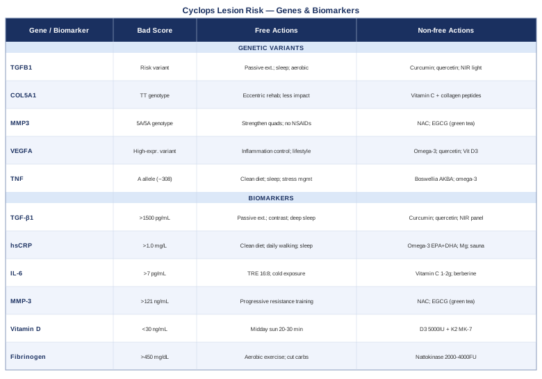
This article works through two complementary tracks. The primary section covers six biomarkers you can measure to understand your fibrotic and inflammatory state — what each one means, how to test it, and what to do when a result is out of range, with or without supplements. A bonus section covers five genetic variants worth knowing about if you have had or are anticipating ACL surgery, or if you have a family pattern of poor soft-tissue healing. The article also includes a book summary with high-impact insights on recovery biology, and a look at complementary approaches with actual clinical evidence behind them. Better information does not guarantee a better outcome — but it reliably leads to sharper decisions.
6 Biomarkers That Reveal Your Cyclops Lesion Risk and Healing Environment
The biology of cyclops lesion formation centers on an overactivated fibrotic repair response. After ACL reconstruction, the joint environment becomes acutely inflamed — this is normal and expected. The problem arises when that inflammation does not resolve cleanly, when fibroblasts remain activated longer than they should, and when collagen continues accumulating past the point of functional repair. The following six biomarkers map different parts of that process.
1. TGF-β1 — The Master Switch of Fibrosis
Why it matters: Transforming Growth Factor Beta 1 is the primary cytokine responsible for fibroblast activation, collagen deposition, and inhibition of the enzymes that break down scar tissue. In joint surgery, TGF-β1 drives the transition from normal healing to excessive fibrosis. Elevated synovial and serum TGF-β1 levels have been consistently associated with greater soft tissue fibrosis after intra-articular procedures. When TGF-β1 signaling stays high beyond the acute healing phase, it creates precisely the environment in which a cyclops nodule can form and grow.
How to measure it: Serum TGF-β1 is available through specialty labs including LabCorp and Quest Diagnostics. It is not on standard panel menus — you will need to request it specifically, or work with a physician who understands functional or sports medicine. Cost typically ranges from $80 to $180 out of pocket. Synovial fluid TGF-β1 is more directly relevant to intra-articular biology but requires aspiration in a clinical setting. For routine tracking, serum TGF-β1 is practical. A well-reviewed study exploring TGF-β1 in post-operative joint fibrosis is available at PubMed — TGF-β1 and arthrofibrosis.
Optimal range: Laboratory reference ranges vary, but serum TGF-β1 below approximately 800 pg/mL is associated with lower fibrotic risk. Values persistently above 1500 pg/mL in the post-operative weeks warrant active intervention.
If the score is elevated — the plan without supplements: Prioritize full passive knee extension immediately post-operatively. Physical therapy protocols that achieve complete passive extension within the first two weeks consistently show lower cyclops formation rates — the mechanical stretch on the anterior notch region prevents fibrous tissue from consolidating. Cold-to-warm contrast therapy applied daily (three minutes cold, three minutes warm, repeated three times) may reduce local TGF-β1 activity by modulating tissue perfusion and inflammatory signaling. Consistent deep sleep of 7–9 hours is non-negotiable: growth hormone secreted during slow-wave sleep directly regulates TGF-β1 expression and tissue remodeling quality. Apply these daily for the first eight weeks post-surgery, then reassess.
If the score is elevated — the plan with supplements or equipment: Curcumin (phospholipid-bound or Theracurmin form): 500–1500 mg daily. Multiple human and animal studies demonstrate dose-dependent suppression of TGF-β1-driven fibrotic signaling. Standard bioavailable forms are essential — bulk curcumin powder has very low absorption. Use for 8–12 weeks post-operatively, then retest. No strict cycling required, but a 2-week break every 3 months is reasonable. Side effects: mild blood-thinning at high doses; avoid without guidance if on anticoagulants. Quercetin: 500–1000 mg daily. Synergizes with curcumin to inhibit NF-κB and downstream TGF-β1 activity. Low side effect profile at these doses. Take alongside curcumin. Near-infrared light therapy (photobiomodulation): Home-use panels at 660 nm and 850 nm wavelengths, applied over the knee for 15–20 minutes once daily. Emerging evidence in fibrosis models indicates that red/NIR light can downregulate TGF-β1-mediated fibroblast activation. A practical entry-level device costs $200–$600. See Strategy 4 for more on this modality.
2. High-Sensitivity C-Reactive Protein (hsCRP) — The Systemic Inflammation Signal
Why it matters: hsCRP is the most accessible and widely validated marker of systemic low-grade inflammation. It is synthesized in the liver in response to upstream cytokines — primarily IL-6 — and reflects the body's overall inflammatory state. Peter Attia, in his clinical practice and in Outlive, identifies chronically elevated hsCRP as one of the clearest signals that a patient's baseline biology is operating in a pro-inflammatory mode. For someone recovering from knee surgery, a body already running "hot" immunologically has a harder time resolving the post-operative inflammatory phase cleanly. The result is a prolonged window during which fibroblast activity remains elevated and fibrous tissue can consolidate.
How to measure it: Included in many standard metabolic panels. Stand-alone cost is typically $10–$30. Available through primary care, direct-access labs, or sports medicine providers. Measure before surgery if possible, and at 6-week intervals post-operatively.
Optimal range: Below 0.5 mg/L reflects excellent inflammatory control. 0.5–1.0 mg/L is acceptable. Above 1.0 mg/L in an otherwise healthy post-surgical patient is worth addressing actively.
If the score is elevated — the plan without supplements: Eliminating ultra-processed foods, refined seed oils (linoleic acid-dominant), and high-glycemic carbohydrates is the single most impactful dietary intervention for hsCRP reduction — the evidence base here is robust. Consistent aerobic movement, even just 30 minutes of walking daily, reduces CRP independent of weight loss. Address sleep architecture: even a single night of poor sleep transiently doubles hsCRP levels; structural sleep protection is not optional during recovery. Sustained stress also chronically elevates CRP via cortisol dysregulation — this warrants practical management, not dismissal.
If the score is elevated — the plan with supplements or equipment: Omega-3 fatty acids (EPA + DHA): 2–4 g/day of combined EPA and DHA from high-quality fish oil or algae-based sources. Among the best-documented interventions for CRP reduction in human trials. Take with a meal containing fat. Continuous use is appropriate. Side effects: fishy aftertaste managed with enteric-coated forms; avoid above 3 g/day without guidance if on blood thinners. Magnesium glycinate: 300–400 mg nightly. Low magnesium status is independently and strongly associated with elevated CRP. Inexpensive, well-tolerated, and broadly useful for sleep quality and muscle recovery simultaneously. Sauna protocol: 3–4 sessions per week, 15–20 minutes at 80–90°C. Finnish cohort data consistently links regular sauna use to significant reductions in hsCRP over time. Avoid applying direct heat over a recent surgical wound until fully healed.
3. Interleukin-6 (IL-6) — The Upstream Driver
Why it matters: IL-6 is the cytokine directly upstream of hsCRP production, but it also acts locally in the synovial tissue. In a joint recovering from surgery, elevated IL-6 stimulates fibroblast proliferation and promotes excessive collagen synthesis within the joint capsule and surrounding tissue. Elevated post-operative IL-6 is associated with worse functional knee outcomes in medium-term follow-up. It is both a cause and a consequence of the fibrotic environment — a feedback loop that can perpetuate itself if not interrupted early.
How to measure it: Serum IL-6 is available at specialty labs. Cost: $50–$120. Time sampling carefully — early morning fasting samples are most reproducible. Measure at baseline and again at 6–12 weeks post-operatively to assess trajectory, not just point-in-time levels.
Optimal range: Below 3 pg/mL in serum. Values consistently above 7 pg/mL in the chronic post-surgical phase indicate persisting inflammatory activity that is working against clean tissue resolution.
If the score is elevated — the plan without supplements: Time-restricted eating (16:8 or 14:10) consistently shows IL-6 lowering effects independent of caloric restriction. Cold exposure — brief cold showers or ice baths (3–5 minutes, 3–4 times per week) — trains the body's anti-inflammatory responses and reduces basal IL-6 over time. This is not about tolerance or willpower; it is about activating a specific norepinephrine-mediated anti-inflammatory pathway.
If the score is elevated — the plan with supplements or equipment: Vitamin C: 1–2 g/day. Has dose-dependent IL-6 suppression in well-controlled trials, likely via antioxidant modulation of NF-κB. Inexpensive, no cycling required. Side effects: loose stools above 3 g/day. Berberine: 500 mg twice daily with meals. Inhibits the NF-κB/IL-6 pathway at multiple nodes. Use for 8–12 weeks, then take a 4-week break. Note potential interactions with medications metabolized via CYP3A4 — check with your physician if on multiple medications.
4. Matrix Metalloproteinase-3 (MMP-3) — Collagen Regulation and Repair Balance
Why it matters: MMP-3, also known as stromelysin-1, is an enzyme that degrades extracellular matrix components including collagen types II, III, and IV. In healthy joint repair, MMP-3 operates within a balanced system that removes damaged tissue while preserving structural integrity. When MMP-3 becomes dysregulated — elevated in the synovial fluid but unbalanced relative to its tissue inhibitors — the result is paradoxical: too much degradation of functional tissue alongside insufficient clearing of fibrous debris. This imbalanced repair environment is permissive for cyclops lesion formation. Elevated synovial MMP-3 is also a specific marker for persistent intra-articular inflammation.
How to measure it: Serum MMP-3 is available through specialty labs; cost is approximately $80–$150. It is commonly tested in rheumatoid arthritis management, which means some insurance plans may cover it if an inflammatory diagnosis is documented. Reference ranges are laboratory-specific, but values above 121 ng/mL in adults are generally considered elevated.
If the score is elevated — the plan without supplements: Progressive resistance training targeting the quadriceps and hamstrings is the most important non-pharmacological intervention. Restored muscle strength reduces intra-articular mechanical stress, which is a primary upstream driver of MMP-3 upregulation. Avoid chronic NSAID use: while these drugs reduce acute inflammation, sustained use alters MMP regulation and can impair the tissue remodeling quality needed for clean recovery.
If the score is elevated — the plan with supplements or equipment: N-Acetyl Cysteine (NAC): 600–1200 mg/day. Reduces oxidative stress-driven transcription of MMP genes. Cycle: 3 months on, 1 month off. Side effects: mild GI discomfort in some at higher doses. Green tea extract (EGCG): 400–600 mg/day of standardized extract. EGCG has demonstrated MMP-3 inhibition in early human and in vitro studies. Take with food to reduce gastrointestinal effects.
5. 25-Hydroxyvitamin D — The Immune Regulator Most Surgeons Ignore
Why it matters: Vitamin D is not a vitamin in the conventional sense — it is a steroid hormone with binding receptors on fibroblasts, immune cells, and synoviocytes. Deficiency is independently associated with amplified fibrotic responses in soft tissue and impaired tissue healing after surgery. Multiple orthopedic studies have identified low pre-operative vitamin D as a predictor of worse functional outcomes after joint procedures. Peter Attia's clinical protocols and Allan Sniderman's metabolic framework both flag vitamin D as among the most underappreciated modifiable variables in patient health — and for post-surgical soft tissue recovery, the evidence is compelling enough to measure routinely. A relevant study on vitamin D and post-surgical orthopedic outcomes is indexed at PubMed — Vitamin D status and orthopedic outcomes.
How to measure it: Standard panel — available at any lab for $30–$60. Always measure as 25-OH-D (the storage form), not 1,25-OH-D, for population-level assessment of status.
Optimal range: 40–70 ng/mL for functional anti-inflammatory and tissue-regulatory activity. Below 30 ng/mL is deficient by most functional medicine standards. Below 20 ng/mL represents a meaningful deficit that must be corrected before and after surgery.
If the score is low — the plan without supplements: Daily midday sun exposure — 20–30 minutes with arms and legs exposed — is sufficient for maintenance in sunny climates and latitudes below 40°N. It is not sufficient to correct deep deficiency within the time constraints of post-surgical recovery.
If the score is low — the plan with supplements or equipment: Vitamin D3 combined with K2 (MK-7 form): 5000–10000 IU of D3 daily, with 100–200 mcg of MK-7. K2 directs calcium appropriately and is critical when supplementing D3 at higher doses. Retest at 8–12 weeks. Once in the target range, maintenance is typically 2000–4000 IU/day. Side effects: rare at these doses; avoid sustained doses above 10000 IU/day without blood level monitoring.
6. Fibrinogen — The Coagulation Marker That Also Drives Fibrosis
Why it matters: Fibrinogen is a plasma protein essential to blood clotting, but it is also a direct participant in scar tissue formation. After surgery, fibrinogen converts to fibrin at the wound site — necessary and expected. The problem arises when systemic fibrinogen levels remain chronically elevated, reflecting a persistent pro-inflammatory and pro-coagulation state. Elevated fibrinogen in the post-operative period amplifies fibrotic tissue formation by sustaining the initial fibrin scaffold that fibroblasts use to migrate and proliferate. Thomas Dayspring and Allan Sniderman have highlighted fibrinogen as a consistently underused risk marker in cardiovascular medicine — and its significance in post-surgical soft tissue biology is equally strong.
How to measure it: Often included in coagulation panels; available as a standalone test for $20–$50. Optimal serum range: 200–400 mg/dL. Values consistently above 450 mg/dL in the chronic post-operative period signal a biological environment that favors fibrotic tissue accumulation.
If the score is elevated — the plan without supplements: Aerobic exercise — even 30 minutes of moderate walking daily — is among the most robustly evidence-backed interventions for fibrinogen reduction. Reducing refined carbohydrate intake and eliminating alcohol are the two most impactful dietary levers. Both independently raise fibrinogen through different hepatic mechanisms.
If the score is elevated — the plan with supplements or equipment: Nattokinase: 2000–4000 FU/day, taken on an empty stomach between meals. Demonstrated fibrinogen-lowering effects in human clinical trials. Cycle: 3 months on, 1 month off. Contraindicated with anticoagulant therapy — discuss with a physician before starting. Lumbrokinase: A more potent systemic proteolytic enzyme derived from earthworms. Used for more significant fibrinogen dysregulation. Appropriate only under medical guidance if already on anticoagulants.
With these six biomarkers mapped, the biological terrain of cyclops lesion risk becomes visible in a way that standard post-operative check-ins cannot provide. Understanding your genetic starting point adds another layer to this picture.
5 Genetic Variants Worth Knowing About
Genetics does not determine your outcome after ACL surgery — but it can meaningfully shift the probability distribution. Several gene variants directly influence collagen structure, fibrotic signaling, inflammatory response, and vascular repair. Knowing your genetic profile in these areas does not change the surgery, but it can sharpen how aggressively you need to monitor the biomarkers above and how proactively you intervene when results trend in the wrong direction.
Consumer genetic tests (23andMe, AncestryDNA) provide raw data that can be analyzed through third-party tools such as Genetic Lifehacks, SelfDecode, or StrateGene. The variants below are among the most clinically meaningful for soft tissue injury and fibrosis risk.
1. TGFB1 — The Fibrosis Gene
What it affects: The TGFB1 gene encodes the TGF-β1 protein described in the biomarker section above. Two common functional polymorphisms — rs1800469 (C-509T) and rs1800471 (codon 25 G915C) — alter the transcription and activity of TGF-β1. Carriers of the higher-expression variants produce more TGF-β1 in response to tissue injury and show more pronounced fibrotic responses in multiple tissue systems. This is early-to-moderate evidence in human populations — not a definitive determinant, but a meaningful signal.
If the gene is unfavorable — the plan without supplements: Focus aggressively on early passive extension post-operatively, a consistently structured sleep protocol (circadian alignment matters for gene expression regulation), and daily aerobic movement from the earliest safe point. These interventions directly modulate TGF-β1 pathway activity regardless of genetic set point.
If the gene is unfavorable — the plan with supplements or equipment: Curcumin, quercetin, and the near-infrared protocol outlined in the TGF-β1 biomarker section are directly applicable here. With a TGFB1 risk variant, these interventions shift from optional to high priority, particularly in the first 12 weeks post-surgery.
2. COL5A1 — Collagen Quality and Soft Tissue Resilience
What it affects: COL5A1 encodes type V collagen, a fibril-regulating collagen critical to the structural quality of tendons and ligaments. The rs12722 variant in the 3'-UTR of COL5A1 has been studied in the context of ACL and tendon injury susceptibility. The TT genotype is associated with structurally weaker soft tissue and higher injury risk in athletic populations. September et al. (2009) — COL5A1 variants and ACL injury susceptibility is one of the cleaner human studies in this area.
If the gene is unfavorable — the plan without supplements: Extended rehabilitation timelines — do not rush return to sport. Prioritize tendon-loading protocols (slow eccentric exercises) which have the best evidence for stimulating functional collagen synthesis regardless of genetic quality. Reduce repetitive high-impact loading during recovery.
If the gene is unfavorable — the plan with supplements or equipment: Vitamin C (1–2 g) taken 30–60 minutes before collagen-loading exercise: This protocol, supported by Keith Baar's research on tendon collagen synthesis, maximizes collagen production during the exercise window. Combined with hydrolyzed collagen peptides (15–20 g): taken simultaneously, it provides the precursor amino acids for collagen rebuilding. Side effects: minimal. Use for the full 6-month rehabilitation period.
3. MMP3 — The 5A/6A Promoter Polymorphism
What it affects: A functional insertion/deletion polymorphism in the MMP3 promoter — the 5A allele versus the 6A allele — significantly alters MMP-3 transcription rate. The 5A/5A genotype is associated with higher MMP-3 expression. As described in the biomarker section, elevated MMP-3 disrupts the balance of tissue repair and degradation in the joint, creating a more permissive environment for fibrotic nodule formation. This has been specifically studied in the context of joint injury.
If the gene is unfavorable — the plan without supplements: Prioritize quadriceps and hamstring strength as early as the post-operative protocol allows. Avoid NSAIDs chronically. Focus on all the mechanical stress-reduction strategies outlined in the MMP-3 biomarker section.
If the gene is unfavorable — the plan with supplements or equipment: NAC and EGCG as outlined above, with a stronger argument for sustained use given genetic predisposition. Consider monitoring serum MMP-3 at 6-week intervals rather than as a one-time check.
4. VEGFA — Vascular Repair and Neovascularization
What it affects: VEGFA encodes vascular endothelial growth factor A, which drives new blood vessel formation. In the context of joint healing, VEGF is necessary — the graft needs vascularization. But VEGF overexpression can also stimulate fibroblast proliferation and contribute to excessive soft tissue formation. The rs2010963 variant (C936T) alters VEGF production in response to hypoxic and inflammatory signals. High-expression genotypes may be associated with more robust but potentially dysregulated tissue growth responses.
If the gene is unfavorable — the plan without supplements: Controlled exposure to intermittent hypoxic training (low-intensity exercise in slightly reduced oxygen conditions) may help normalize VEGF signaling over time — this is early-stage evidence. Primarily, focus on the inflammatory control measures above, as VEGF overactivity is typically downstream of an already elevated inflammatory state.
If the gene is unfavorable — the plan with supplements or equipment: No established supplemental protocol specifically targeting VEGFA polymorphisms exists with strong human evidence. Anti-inflammatory interventions (omega-3, quercetin, vitamin D3) remain the most practical approach. This is an area where genetic testing provides primarily monitoring guidance rather than specific pharmacological targets.
5. TNF — The Inflammatory Amplifier
What it affects: The rs1800629 (-308 G/A) polymorphism in the TNF gene promoter is one of the best-studied inflammatory gene variants. The A allele (previously referred to as TNF2) is associated with higher TNF-α transcription in response to immune stimuli. TNF-α is a central amplifier of both acute and chronic inflammation, and carries direct relevance for post-surgical joint recovery: elevated TNF-α sustains synovial inflammation, promotes cartilage degradation, and prolongs the fibroblast activation window.
If the gene is unfavorable — the plan without supplements: All the dietary and lifestyle interventions described in the hsCRP and IL-6 sections apply here with greater urgency. Eliminating dietary drivers of NF-κB activation (ultra-processed foods, seed oil excess, high sugar load) is the primary lever. Consistent sleep and stress management are not secondary considerations for TNF A-allele carriers — they are primary.
If the gene is unfavorable — the plan with supplements or equipment: Boswellic acids (AKBA): 200–400 mg/day of highly standardized Boswellia serrata extract. Directly inhibit the 5-lipoxygenase pathway upstream of TNF-α. Use for 12-week cycles, 2-week breaks. Side effects: mild GI discomfort in sensitive individuals. Omega-3 (EPA + DHA): As above, with stronger indication. EPA specifically antagonizes arachidonic acid-derived pro-inflammatory eicosanoids that amplify TNF-α activity.
Understanding both your biomarker picture and your genetic background creates a genuinely individualized framework for recovery. The next section zooms out to a broader, deeply researched perspective on the biological mechanisms that influence everything covered so far.
What "Outlive" by Peter Attia Reveals About Post-Surgical Recovery Biology
Outlive: The Science and Art of Longevity by Peter Attia (2023) is not a book about knee surgery — but it is one of the most rigorously referenced modern books on the biology of inflammation, tissue health, and the metrics that actually predict how well your body recovers and functions over time. Many of its central arguments challenge the standard medical model of "wait, recover, and hope," replacing it with a framework of early measurement, active intervention, and data-driven decisions. The following are ten insights from the book and Attia's broader work that directly apply to cyclops lesion recovery and fibrosis prevention.
1. Chronic low-grade inflammation is the common denominator of poor healing
Attia's "Four Horsemen" framework identifies chronic inflammation as the upstream driver of most major diseases — and equally, of impaired tissue recovery. A body operating in a chronically inflamed state enters surgery at a disadvantage that no surgical technique fully compensates for.2. hsCRP above 1.0 mg/L is not "normal" — it is a warning signal
Attia distinguishes between the conventional medical threshold for CRP elevation and the functional threshold for optimal recovery. Many patients are told their CRP is "fine" when it is in a range that significantly impairs tissue repair.3. Muscle mass is medicine for joint and metabolic health
Attia's framework treats muscle as the primary organ of longevity. For post-surgical knee patients specifically, the protective effect of pre-existing quadriceps and hamstring strength against poor outcomes is strong and consistent.4. Insulin resistance is an invisible saboteur of tissue repair
Elevated fasting insulin and HOMA-IR reflect metabolic dysfunction that directly impairs immune resolution after tissue injury. Attia tracks fasting insulin as a primary biomarker — it is rarely checked in post-surgical patients yet has direct mechanistic relevance.5. Sleep architecture is not optional — it is the primary recovery mechanism
Attia details the biochemistry of growth hormone release during deep sleep and its direct relationship to tissue repair. Structural sleep protection in post-surgical patients is not lifestyle advice; it is biological medicine.6. Omega-3 index below 8% is a silent inflammation driver
The ratio of EPA+DHA to total red blood cell fatty acids — the omega-3 index — is one of Attia's preferred anti-inflammatory biomarkers. Most people measure below 5%; a target above 8% consistently correlates with reduced inflammatory activity across multiple systems.7. VO2 max is a proxy for recovery capacity, not just fitness
A higher VO2 max corresponds to better mitochondrial function and greater oxidative capacity — both of which accelerate tissue healing. Pre-surgical aerobic conditioning is a meaningful investment in post-surgical outcomes.8. Monitoring what you don't feel is the most important preventive move
Attia's central argument against conventional medicine's reactive model applies directly here: cyclops lesion formation begins before symptoms appear. The patients who benefit most from the biomarker approach above are those who start tracking before the problem becomes clinically obvious.9. Vitamin D, magnesium, and omega-3 are the "base layer" of metabolic health
Attia consistently identifies these three as the highest-value, lowest-risk nutritional interventions for patients with suboptimal metabolic profiles — a position that aligns with their relevance to inflammatory and fibrotic control in joint recovery.10. The goal is not to recover to the pre-injury state — it is to emerge with a better physiological foundation
Perhaps the most reframing insight in the book: a significant injury and surgery is also an opportunity to audit and upgrade the biological environment. The patients who use recovery well often emerge with a more carefully managed inflammatory profile, stronger muscles, and better metabolic markers than they had pre-injury.Complementary Approaches with Clinical Evidence
For a condition as specific as cyclops lesion — a post-surgical fibrotic complication — the complementary modality evidence is narrower than for more diffuse conditions. The three approaches below have the strongest relevance and the most credible evidence basis for this context.
Low-Level Laser Therapy / Photobiomodulation
Photobiomodulation (PBM) uses specific wavelengths of red and near-infrared light (typically 630–850 nm) to stimulate mitochondrial function in cells, reduce local inflammation, and modulate fibroblast activity. It is particularly relevant for cyclops lesion because the primary target — overactivated fibroblasts producing excess collagen — has demonstrated photobiomodulation sensitivity in multiple models. PBM does not eliminate fibrotic tissue that has already formed, but there is growing evidence it can reduce its rate of formation and accelerate resolution of post-surgical inflammation when applied in the early recovery window.
A randomized controlled trial investigating low-level laser therapy in post-operative knee rehabilitation (Leal Junior et al.) found significant reductions in pain, inflammation markers, and recovery time compared to sham treatment. The study is indexed at PubMed — LLLT and knee rehabilitation (Leal Junior et al.). In the fibrosis literature specifically, PBM has shown TGF-β1 modulating effects in soft tissue models — the most mechanistically relevant finding for cyclops prevention.
Practical application: a home-use near-infrared panel (660 nm + 850 nm) applied over the knee for 15–20 minutes once daily, from the first post-operative week onward. Maintain at least 5 cm device-to-skin distance initially if there is surgical wound sensitivity. The evidence is not definitive at the clinical level for cyclops prevention specifically, but the mechanistic plausibility and safety profile make it a reasonable addition to a structured recovery protocol. Avoid heat-generating devices over a fresh surgical wound.
Massage Therapy — Soft Tissue Mobilization
Structured manual therapy and soft tissue mobilization directly addresses one of the key physical factors in cyclops lesion formation: the development of adhesions and fibrotic restriction in the surrounding joint capsule, infrapatellar fat pad, and patellar tendon. While a cyclops nodule itself requires arthroscopic removal, the periarticular fibrotic environment that enables its formation and that compounds its effects is meaningfully addressable through regular manual work. Massage and manual therapy applied to the peri-patellar structures and quadriceps tendon also maintain tissue extensibility in the post-operative period, reducing the mechanical forces that favor fibrous consolidation.
A systematic review of manual therapy and soft tissue mobilization in post-ACL reconstruction rehabilitation (Wright et al.) found significant benefits for range of motion restoration and pain reduction when compared to standard physical therapy alone. The review is listed at PubMed — Manual therapy in post-ACL rehabilitation. The specific mechanism relevant to fibrosis is mechanical — consistent loading and compression of soft tissue during the remodeling phase aligns collagen fibers and reduces cross-linking of fibrotic tissue.
Practical application: 2–3 sessions per week with a physiotherapist or licensed massage therapist trained in post-surgical soft tissue work, beginning 2–3 weeks post-operatively once the surgical site is sufficiently healed. Emphasis should be on the infrapatellar fat pad, medial and lateral joint capsule, and quadriceps tendon — all structures implicated in the formation of adhesions that contribute to anterior knee stiffness. Daily self-massage with a foam roller or massage ball along the quadriceps is a practical complementary habit between professional sessions. Evidence is moderate-to-strong for range of motion benefits, more limited for direct cyclops prevention.
Biofeedback — Neuromuscular Re-Education
Biofeedback using electromyography (EMG) measures and displays real-time muscle activation, helping patients learn to restore neuromuscular control of the quadriceps after ACL reconstruction. This is directly relevant to cyclops lesion recovery because quadriceps inhibition — a consistent finding after knee surgery — leads to protective guarding of the joint, altered gait mechanics, and increased intra-articular mechanical stress on the anterior notch region where cyclops lesions form. Restoring proper neuromuscular patterning reduces the abnormal loading that creates a favorable environment for fibrous tissue accumulation.
A randomized controlled trial by Draper et al. demonstrated that EMG biofeedback significantly improved voluntary quadriceps activation in post-ACL rehabilitation patients compared to standard exercise alone (PubMed — EMG biofeedback and quadriceps activation). Subsequent work has consistently supported biofeedback as a practical tool for restoring motor patterns disrupted by surgical trauma and the protective inhibition that follows.
Practical application: biofeedback is typically administered by a physiotherapist using clinical EMG equipment, in 2–3 sessions per week for 6–12 weeks post-operatively. The patient is guided to achieve specified activation thresholds while observing real-time feedback on a screen. Home-use EMG biofeedback devices (such as MyRecovery or similar systems) provide accessible daily reinforcement. The evidence is strongest for quadriceps activation restoration — its indirect protective effect on cyclops formation requires further study, but the mechanistic rationale is sound and the approach carries no meaningful risk.
Conclusion
Cyclops lesion is a specific, frustrating, and largely preventable complication of ACL reconstruction — but understanding it purely as a mechanical problem misses most of its biology. The fibrotic tissue that forms in the anterior notch is the product of an inflammatory environment that can be measured, tracked, and in many cases modified. The six biomarkers covered in this article — TGF-β1, hsCRP, IL-6, MMP-3, vitamin D, and fibrinogen — give you a real-time picture of that environment. The five genetic variants provide context for why your starting point may differ from someone else's and how aggressively you need to monitor and intervene.
The next smart step is to identify which of these markers you have not yet measured, prioritize the two or three most accessible ones, and bring the results into a conversation with a sports medicine physician, functional medicine practitioner, or orthopedic surgeon who can contextualize them within your specific surgical and rehabilitation plan. None of this replaces expert clinical judgment — but informed patients who arrive to those conversations with data consistently receive better-calibrated care than those who do not.
Musculoskeletal: Joint Conditions Tendon & Ligament Conditions Sports Injuries
Autoimmune: Inflammatory Conditions Connective Tissue Conditions