This article contains AI generated content.
· UpdatedCysticercosis Arthritis Genes Biomarkers — 5 Genes and 6 Biomarkers to Track
Introduction
Joint pain that resists every standard treatment, inflammation markers that never quite normalize, a diagnosis that took months or years to arrive — if any of this sounds familiar, you already know how isolating cysticercosis-related arthritis can be. This is a condition that mimics rheumatoid arthritis, reactive arthritis, and spondyloarthropathy well enough to fool experienced clinicians. The underlying cause — larvae of the tapeworm Taenia solium lodging in muscles, soft tissue, or joint spaces, and the immune system's attempt to eliminate them — creates a pattern of inflammation that conventional rheumatology approaches only partially address.
What makes this particularly frustrating is that generic anti-inflammatory advice does not account for the parasitic origin of the inflammation. Treating the joint symptoms without confirming and managing the underlying infection leaves the root cause intact. And once the infection is confirmed, most clinical guidance stops at antiparasitic drugs and corticosteroids, with little attention to the individual biological variables that determine how severe the joint damage becomes and how long recovery takes.
That is where biomarkers and genetics become genuinely useful. Not every person exposed to T. solium develops the same degree of joint inflammation. Some experience mild reactive arthritis; others develop chronic, erosive joint disease. The difference often comes down to specific immune gene variants, baseline inflammatory capacity, and how well the immune system regulates its own response during and after treatment. Understanding these variables does not replace medical care, but it does make that care more targeted and more likely to actually work.
This article covers two complementary approaches. The first and primary one focuses on six measurable biomarkers that give a real-time picture of infection status, joint inflammation, and immune activity — each with practical guidance on what to do when a result is abnormal. The second covers five genetic variants that explain differences in susceptibility and inflammatory severity, with strategies calibrated to each variant. Together, they offer a more precise framework than "reduce inflammation" — a foundation for decisions, not just symptoms to suppress.
Summary
This article takes a layered approach to cysticercosis-related arthritis — starting with the six most actionable biomarkers to track (anti-T. solium antibodies, hsCRP, ESR, eosinophil count, IL-6, and complement C3/C4), each explained with measurement costs, target values, and specific plans with and without supplementation. It then examines five genes that shape how severely the body responds to the parasite and its antigens — HLA-B27, TNF-α rs1800629, IL-10 rs1800896, TLR4 Asp299Gly, and HLA-DRB1 — with practical compensatory strategies for unfavorable variants. A summary of the key insights from Peter Attia's Outlive follows, reframed around the inflammatory biology of parasitic arthritis. Finally, five evidence-supported complementary approaches are examined: mindfulness-based stress reduction, low-level laser therapy, microbiome-directed interventions, the autoimmune protocol, and yoga — selected for their relevance to inflammation modulation and joint recovery in this specific context. Whether you are early in the diagnostic process or managing an established case, the information here is built around one goal: giving you better leverage over a condition that is rarely explained in full.
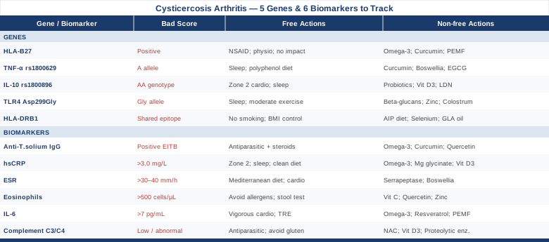
6 Biomarkers to Monitor When Cysticercosis Affects Your Joints
Biomarker tracking in cysticercosis-related arthritis serves two overlapping purposes: confirming the diagnosis and its activity, and monitoring the inflammatory damage to joints. These are not the same thing. A person can have serologically confirmed cysticercosis with no joint involvement, or joint inflammation that persists well after the antiparasitic treatment has cleared the bulk of the infection. Tracking the right set of markers in sequence tells you where you are in that process and which mechanisms still need attention.
The six markers below cover the spectrum from infection confirmation to downstream inflammatory activity, and are selected for their clinical relevance, measurement availability, and interpretability in the context of this condition specifically.
Anti-Taenia solium Antibodies: The Diagnostic Anchor
The single most important biomarker in any suspected cysticercosis case is the serology that confirms the infection. Without this confirmation, joint inflammation attributed to T. solium remains a clinical hypothesis. The Enzyme-Linked Immunoelectrotransfer Blot (EITB) test, developed at the CDC, remains the gold standard for this purpose. It detects IgG antibodies against specific lentil-lectin purified glycoprotein antigens from T. solium cysticerci, with sensitivity around 94–98% and specificity approaching 100% in individuals with two or more viable cysts. Standard ELISA assays are more widely available but less specific, with higher rates of cross-reactivity against other helminths.
In the context of arthritis, a positive EITB confirms that the immune system has been — or is currently — responding to T. solium. It does not by itself confirm articular involvement; that requires clinical evaluation and, in some cases, synovial biopsy. But it is the prerequisite for attributing joint inflammation to this infection rather than a coincidental autoimmune process.
How to measure it
EITB is available through reference laboratories such as the CDC-associated labs and specialty parasitology centers. Standard ELISA for anti-cysticercus antibodies is available through most major commercial labs (Quest, LabCorp). Cost ranges from $75 to $220 depending on the specific test and laboratory. In practice, ELISA is often ordered first; EITB is ordered for confirmation when clinical suspicion remains high despite a negative or borderline ELISA. Both require a standard venous blood draw.
If the result is positive, the plan without supplements
A confirmed positive serology in someone with active joint symptoms should trigger an immediate referral to both an infectious disease specialist and a rheumatologist experienced with reactive or parasite-driven arthritis. Antiparasitic treatment — typically albendazole (400 mg twice daily for 8–30 days depending on burden and location) or praziquantel — is the foundation of treatment, but must be managed carefully because cyst die-off triggers a secondary inflammatory spike. This is why albendazole is almost always co-administered with corticosteroids during the treatment course. Joint rest, physiotherapy, and non-steroidal anti-inflammatory drugs (NSAIDs) address joint symptoms. Follow-up serology at 3, 6, and 12 months tracks whether antibody titers are declining — which suggests treatment success — or persisting, which may indicate continued antigen exposure. CDC cysticercosis overview
If the result is positive, the plan with supplements or equipment
During and after antiparasitic treatment, several evidence-supported adjuncts can reduce the secondary inflammatory spike and support joint tissue recovery. Omega-3 fatty acids (EPA+DHA, 2–4 g/day with food) have well-established anti-inflammatory effects mediated through specialized pro-resolving mediators (SPMs) that promote the resolution — rather than just suppression — of inflammation; cycle on continuously during the treatment and recovery phase. Curcumin with piperine (500–1000 mg standardized curcumin with 5–10 mg piperine, twice daily with meals) inhibits NF-κB signaling and reduces IL-6 and TNF-α; cycle 8 weeks on, 2 weeks off. Avoid high-dose curcumin alongside antiparasitic drugs without physician oversight due to potential CYP450 interactions. Quercetin (500 mg twice daily) acts as a natural mast cell stabilizer relevant to the eosinophilic component of parasitic arthritis; best cycled 6 weeks on, 2 weeks off. Infrared heating pads or far-infrared sauna (20–30 minutes, 3–4x per week) may support joint circulation and recovery during the subacute phase but should be avoided during active fever or high systemic inflammation.
High-Sensitivity C-Reactive Protein: Reading the Systemic Flame
High-sensitivity CRP (hsCRP) is the most widely used marker of systemic low-grade to moderate inflammation, and it responds sensitively to both active infection and joint inflammation. In cysticercosis-related arthritis, hsCRP tends to spike sharply during antiparasitic treatment — when dying cysts release antigens that trigger a brisk immune response — and should gradually trend downward over the 3–6 months following successful treatment. A persistently elevated hsCRP after treatment suggests ongoing inflammatory activity, either from residual antigenic burden, secondary joint damage, or a coexisting autoimmune process triggered by molecular mimicry.
Peter Attia, in his clinical framework for longevity medicine, considers hsCRP one of the three most actionable inflammatory biomarkers alongside IL-6 and fibrinogen. His target is below 1.0 mg/L for optimal metabolic and cardiovascular protection. In the context of parasitic arthritis, a more realistic acute target is below 3.0 mg/L within 3 months post-treatment, trending toward sub-1.0 mg/L at 6–12 months.
How to measure it
hsCRP is available at virtually every commercial laboratory and is often included in standard metabolic panels. It is important to specify high-sensitivity CRP, not standard CRP, which lacks precision at the lower end of the range. Cost ranges from $15 to $50. Measure at baseline before treatment, at the peak of treatment (week 2–3, where it may be highest), and then at 3, 6, and 12 months post-treatment to track the trend.
If the score is elevated, the plan without supplements
Sustained hsCRP above 3.0 mg/L outside of an active treatment window warrants investigation into whether the inflammatory trigger has been adequately addressed. This means confirming adequate antiparasitic treatment completion, ruling out residual viable cysts through imaging (brain MRI, soft tissue ultrasound), and evaluating for secondary autoimmune arthritis that may now be self-sustaining. Lifestyle factors that reliably lower hsCRP include Zone 2 aerobic exercise (3–4 sessions per week at a pace where conversation is possible without gasping), elimination of ultra-processed foods, improving sleep duration to 7–9 hours, and addressing visceral adiposity — all of which reduce the baseline inflammatory load independent of the infection.
If the score is elevated, the plan with supplements or equipment
Omega-3 fatty acids (EPA+DHA, 3–4 g/day) consistently lower hsCRP in multiple randomized trials; this is a continuous intervention with no cycling needed at therapeutic doses. Magnesium glycinate (300–400 mg at night) supports anti-inflammatory signaling and reduces NF-κB activity; safe for long-term use with a break every 3 months to assess need. Vitamin D3 + K2 (2,000–5,000 IU D3 with 100–200 mcg K2 MK-7 daily) supports immune regulation; dose to achieve serum 25-OH-D of 50–70 ng/mL. High-intensity infrared sauna therapy (20–30 minutes, 3–4x per week) has demonstrated reductions in inflammatory cytokines in several small studies and may provide an additive benefit during the post-treatment recovery phase.
Erythrocyte Sedimentation Rate: The Slow-Moving But Reliable Signal
ESR measures how quickly red blood cells settle in a test tube — a process accelerated by inflammatory proteins, especially fibrinogen and immunoglobulins, that coat the cells. Unlike CRP, which rises and falls quickly (within hours to days), ESR lags by several days and falls more slowly. This makes it a better marker for tracking the overall trajectory of chronic inflammation rather than acute spikes. In cysticercosis-related arthritis, ESR above 30–40 mm/h in men or 40–50 mm/h in women (adjusting for age) during the post-treatment phase suggests that the inflammatory process has not resolved and that joint tissue may continue to sustain damage.
ESR also correlates with disease activity in reactive arthritis, which is one of the clinical patterns cysticercosis-related joint inflammation most closely resembles. Tracking ESR alongside CRP gives a more complete picture — a CRP that normalizes while ESR remains elevated is a sign worth pursuing.
How to measure it
ESR is included in many basic lab panels and costs $10 to $30 as a standalone test. It requires no special preparation. Measure alongside hsCRP at each follow-up interval.
If the score is elevated, the plan without supplements
Persistently elevated ESR beyond 3 months post-treatment should prompt a reassessment of the antiparasitic treatment completeness, a review of joint function to rule out established structural damage needing orthopedic management, and consideration of whether a low-grade autoimmune process has been initiated. Anti-inflammatory dietary patterns — specifically a Mediterranean-style diet with high polyphenol intake, fatty fish twice weekly, and minimized refined carbohydrates — reduce fibrinogen and other acute-phase reactants that drive ESR elevation. Structured aerobic exercise remains one of the most evidence-supported interventions for lowering ESR in inflammatory arthritis.
If the score is elevated, the plan with supplements or equipment
Serrapeptase (80,000–120,000 SPU on an empty stomach) is a proteolytic enzyme that breaks down fibrinogen and other inflammatory proteins and may contribute to lowering ESR; cycle 4 weeks on, 2 weeks off and monitor for GI tolerance. Boswellia serrata extract (300–500 mg standardized to 65% boswellic acids, twice daily with food) reduces leukotriene synthesis and has RCT support for inflammatory arthritis; cycle 8 weeks on, 2 weeks off. Monitor liver enzymes with prolonged use. Research on boswellia and arthritis
Eosinophil Count: The Parasite Burden Signal
Peripheral blood eosinophilia — an elevated eosinophil count above 500 cells/μL, or above 5% of the white cell differential — is the immune system's signature response to helminth (worm) infections, including cysticercosis. Eosinophils are recruited to combat parasites too large for phagocytosis, and their activation also contributes to tissue inflammation in joints and other sites. The degree of eosinophilia correlates roughly with the magnitude of the parasitic burden and the intensity of the host immune response, though individual variation is significant.
Crucially, eosinophil counts that normalize after treatment and then rise again may signal re-infection or an incomplete treatment response. Tracking this marker provides a biological readout of the ongoing parasitic immune engagement that other inflammatory markers do not capture as specifically.
How to measure it
Eosinophil count is a standard component of the complete blood count (CBC) with differential, available at any lab for $10 to $40. Request the full differential, not just the total white count. Baseline measurement before treatment, re-measurement at treatment completion (4–6 weeks), and then at 3 and 6 months post-treatment is a reasonable tracking schedule.
If the count is elevated, the plan without supplements
Persistent eosinophilia beyond 4–6 weeks post-treatment is a significant finding that should prompt reassessment of treatment adequacy with an infectious disease specialist. In the interim, avoiding dietary lectins, gluten, and known allergenic foods can reduce the environmental eosinophil trigger load and support resolution. A comprehensive stool analysis to rule out concurrent gastrointestinal helminths (which could independently sustain eosinophilia) is also warranted.
If the count is elevated, the plan with supplements or equipment
Vitamin C (1,000–2,000 mg/day in divided doses) supports immune regulatory function and may help modulate eosinophil activity; safe for continuous use. Quercetin (500 mg twice daily) stabilizes mast cells and reduces eosinophil activation — particularly relevant in the context of parasitic immune responses; cycle 6 weeks on, 2 weeks off. Zinc picolinate (25–30 mg/day with food) supports T-regulatory cell function and immune balance; do not exceed 40 mg/day long-term; take copper 2 mg/day alongside zinc if using for more than 8 weeks.
Interleukin-6: The Joint Destruction Driver
Interleukin-6 (IL-6) is a pro-inflammatory cytokine that plays a central role in joint damage in inflammatory arthritis. It drives the production of acute-phase proteins (including CRP), stimulates osteoclast activity leading to bone erosion, promotes synovial fibroblast proliferation, and activates immune cell recruitment to the joint. In parasite-driven arthritis, the dying cysticerci release antigens that trigger persistent IL-6 production in the synovium, particularly in genetically susceptible individuals.
This is one of the biomarkers Peter Attia tracks most closely in his patients, alongside hsCRP, precisely because IL-6 represents the signaling upstream of many inflammatory processes — it is not just a reflection of inflammation but an active driver of it. Elevated IL-6 after treatment resolution of cysticercosis may indicate that the immune system has developed a self-sustaining joint inflammatory loop that requires active intervention beyond antiparasitic therapy.
How to measure it
IL-6 is a specialty test available through Quest Diagnostics, LabCorp, and hospital-based specialty labs. It requires a standard blood draw but must be processed carefully — the sample must be kept cold and processed quickly, or IL-6 degrades and gives falsely low readings. Cost ranges from $50 to $150. The reference range is typically below 7 pg/mL, with values above 10 pg/mL considered clearly elevated. Measure at baseline and at 3 and 6 months post-treatment. IL-6 research in inflammatory arthritis
If the score is elevated, the plan without supplements
Persistently elevated IL-6 after successful antiparasitic treatment is a signal that inflammation has become partially autonomous and requires active management. Vigorous aerobic exercise — not just gentle walking, but sessions that raise heart rate to 70–85% of maximum — has among the best evidence for lowering IL-6 through mechanisms that include increased skeletal muscle production of anti-inflammatory myokines and improved IL-6 receptor sensitivity. Time-restricted eating (compressing the eating window to 8–10 hours) has modest evidence for reducing basal IL-6. In more severe cases, rheumatology consultation about IL-6 receptor antagonists (such as tocilizumab) is appropriate.
If the score is elevated, the plan with supplements or equipment
Omega-3 fatty acids (EPA+DHA, 3–4 g/day) directly reduce IL-6 synthesis; continuous use. Resveratrol (500 mg/day with food, trans-resveratrol form) inhibits NF-κB and STAT3 pathways that drive IL-6 production; cycle 8 weeks on, 4 weeks off — avoid with blood thinners. PEMF (Pulsed Electromagnetic Field) therapy devices (home units, 20 minutes daily on affected joints) have RCT evidence for reducing inflammatory cytokines including IL-6 in osteoarthritis and rheumatoid arthritis; continuous use is safe with no significant side effects documented in standard protocols.
Complement C3 and C4: The Immune Complex Window
When antibodies bind to T. solium antigens — or to self-antigens in cases of molecular mimicry — they form immune complexes that can deposit in joint synovium, triggering an intense local inflammatory response. The complement system is activated by these complexes, and its proteins are consumed in the process. Monitoring complement C3 and C4 levels provides a window into whether immune complex-mediated joint damage is occurring. Low or low-normal C3/C4 in the context of active arthritis suggests active immune complex formation and complement consumption, which is both diagnostically informative and prognostically important.
Conversely, elevated C3 and C4 in the absence of joint improvement may indicate ongoing antigenic stimulation without adequate immune resolution — a pattern seen in persistent low-grade cysticercosis.
How to measure it
C3 and C4 complement levels are available through all major commercial laboratories as part of a complement panel or as individual tests. Cost ranges from $30 to $85 for both tests. Normal C3 is 90–180 mg/dL; normal C4 is 16–47 mg/dL. Values below the lower limit of the range in the context of active joint inflammation are clinically significant. Test at baseline and at 3 and 6 months post-treatment.
If the score is low or abnormal, the plan without supplements
Low complement in the context of active parasitic arthritis most directly signals the need for aggressive antiparasitic treatment to reduce the antigenic burden driving immune complex formation. Dietary reduction of pro-inflammatory foods that drive immune complex-mediated reactions — particularly dairy, gluten, and high-AGE foods produced by high-temperature cooking — can reduce the background immune activation load. Avoidance of alcohol and NSAIDs long-term (which can impair complement function) is also advisable.
If the score is low or abnormal, the plan with supplements or equipment
N-Acetylcysteine (NAC) (600 mg twice daily) supports glutathione production and has been shown to modulate complement activation in inflammatory conditions; cycle 8 weeks on, 4 weeks off. Vitamin D3 at adequate repletion levels (achieving 50–70 ng/mL serum 25-OH-D) supports immune complex clearance and complement regulation. Proteolytic enzymes (nattokinase, serrapeptase, or a combination enzyme product taken between meals) support degradation of immune complexes and fibrin deposits in joint tissue; safe for cycle use of 4–6 weeks with periodic reassessment.
The six biomarkers above, tracked together on a 3-month schedule, give a far more complete picture of where cysticercosis-related joint inflammation stands than any single marker alone. Recognizing that genetics significantly shapes how these markers behave — and how aggressively the inflammation responds — is the next layer of this framework.
5 Genes That Shape Your Body's Response to Cysticercosis
Not everyone who ingests T. solium eggs develops the same course of disease. Some clear the infection with minimal symptoms; others develop extensive cysticercosis with severe joint involvement. Individual differences in immune gene architecture explain much of this variation. The five variants below are among the most clinically relevant — each supported by human genetic data, each with practical implications for how joint inflammation should be managed if the variant is present.
Genetic testing for these variants is available through companies such as 23andMe, AncestryDNA, or specialty clinical genetics panels. Raw data from 23andMe can be analyzed through platforms like Genetic Genie or StrateGene to identify most of these SNPs. Clinical HLA typing requires a dedicated blood test.
HLA-B27: The Reactive Arthritis Risk Gene
HLA-B27 is one of the most clinically well-established genetic markers in all of inflammatory arthritis medicine. It encodes a cell surface protein that presents peptide antigens to cytotoxic T cells. When an infection — whether bacterial, viral, or parasitic — triggers an immune response in an HLA-B27 positive individual, the risk of developing reactive arthritis (sterile joint inflammation remote from the infection site) is dramatically elevated — approximately 20-fold compared to HLA-B27 negative individuals.
In the context of cysticercosis, HLA-B27 positive individuals who mount an immune response to T. solium antigens are at significantly higher risk of developing a reactive arthritis pattern — including asymmetric oligoarthritis, enthesitis (inflammation at tendon insertion sites), and potentially sacroiliitis. The mechanism involves molecular mimicry: T. solium antigens share structural similarities with self-proteins, and HLA-B27 may present these cross-reactive peptides in a way that directs immune attack toward joint tissues. Early genetic data on HLA associations in cysticercosis is reviewed in several parasitology immunology studies.
If the gene is unfavorable (HLA-B27 positive), the plan without supplements
In HLA-B27 positive individuals with cysticercosis-related joint inflammation, the priority is completing antiparasitic treatment promptly and completely, since the antigenic trigger needs to be eliminated before the reactive arthritis loop can resolve. Extended NSAID use (with gastroprotection) is a first-line joint strategy in spondyloarthropathy-pattern reactive arthritis, which is more likely in B27-positive individuals. Physiotherapy focused on maintaining spinal mobility and preventing enthesitis-related stiffness is important — loss of spinal motion in this group is a real long-term risk. Avoid high-impact loading of the affected joints during the active phase.
If the gene is unfavorable, the plan with supplements or equipment
Omega-3 fatty acids (EPA+DHA, 3–4 g/day) reduce leukotriene-mediated joint inflammation particularly relevant in enthesitis; continuous use. Curcumin (500–1000 mg standardized, twice daily with food) inhibits NF-κB, which is centrally involved in HLA-B27-related inflammatory signaling; cycle 8 weeks on, 2 weeks off. PEMF mat or focused PEMF device applied to sacroiliac or axial joints (20 minutes daily) has evidence in ankylosing spondylitis-related conditions, which share pathophysiology with HLA-B27-associated reactive arthritis. Monitor D3 levels and optimize to 60–70 ng/mL, as vitamin D regulates HLA-B27-related inflammatory cascades.
TNF-α -308 Polymorphism: Your Inflammatory Thermostat
TNF-alpha (Tumor Necrosis Factor alpha) is one of the master regulators of the inflammatory response. The rs1800629 variant at position -308 of the TNF-α promoter region controls how much TNF-α the immune system produces in response to an inflammatory stimulus. The A allele at this position (the -308A variant) is associated with higher TNF-α transcription — meaning the inflammatory response is turned up louder and for longer. Individuals with the AA genotype are high TNF-α producers; GA is intermediate; GG is lower.
In cysticercosis, higher TNF-α production is a double-edged sword. It supports parasite killing, which is beneficial. But during the die-off phase of antiparasitic treatment and in the post-infection joint inflammation period, high TNF-α accelerates synovial inflammation, cartilage degradation, and bone erosion. Several studies have found the -308A allele overrepresented in individuals with more severe inflammatory manifestations of parasitic infections. TNF-α polymorphism research in infectious disease
If the gene is unfavorable (A allele present), the plan without supplements
High TNF-α producers benefit most from an actively anti-inflammatory lifestyle during and after antiparasitic treatment. This means prioritizing sleep (since TNF-α rises sharply with sleep deprivation), minimizing chronic stress (cortisol dysregulation amplifies TNF-α-driven inflammation), and following a polyphenol-rich diet that broadly suppresses NF-κB and AP-1 transcription factors driving TNF-α production. Cold exposure (cold showers, cold water immersion) has modest evidence for transiently reducing TNF-α, though this should be avoided during acute infection phases. In severe cases, biological TNF-α inhibitors (etanercept, adalimumab) may be considered by a rheumatologist if the post-infectious arthritis is destructive and unresponsive to standard therapy.
If the gene is unfavorable, the plan with supplements or equipment
Curcumin with piperine (500–1000 mg twice daily) directly inhibits TNF-α gene expression — one of the best-studied natural TNF-α modulators; cycle 8 weeks on, 2 weeks off. Boswellia serrata (300–400 mg, 65% boswellic acids, twice daily) inhibits 5-lipoxygenase and reduces TNF-α-driven leukotriene production in joints; cycle 6 weeks on, 2 weeks off. Green tea extract / EGCG (400–500 mg/day, decaffeinated) is an NF-κB inhibitor with clinical evidence in RA for reducing TNF-α activity; monitor for caffeine sensitivity even with decaf preparations; cycle 8 weeks on, 4 weeks off.
IL-10 Promoter Polymorphism: The Anti-Inflammatory Capacity Gene
If TNF-α is the accelerator of inflammation, Interleukin-10 (IL-10) is one of the primary braking mechanisms. IL-10 is an anti-inflammatory cytokine produced by regulatory T cells and macrophages that dampens excessive immune responses and promotes tissue repair. The rs1800896 polymorphism at position -1082 of the IL-10 promoter controls basal IL-10 output. The AA genotype at this position is associated with low IL-10 production — meaning the immune system has a weaker anti-inflammatory brake. GG is the high-producer genotype; GA is intermediate.
In cysticercosis, low IL-10 producers have greater difficulty resolving the inflammatory cascade triggered by parasite antigens. The immune response may overshoot and persist, contributing to chronic synovitis and structural joint damage even after the infection itself is cleared. Several studies have linked low IL-10-producing genotypes to more severe outcomes in helminth infections. IL-10 polymorphism in helminth infection studies
If the gene is unfavorable (AA genotype), the plan without supplements
Low IL-10 producers need to actively support the regulatory immune arm. Aerobic exercise at moderate intensity (Zone 2 cardio, 3–5 sessions per week) consistently upregulates IL-10 production from skeletal muscle-derived myokines — one of the most reliable non-pharmacological IL-10 inducers. Adequate sleep (7–9 hours, with attention to sleep architecture including deep sleep) supports regulatory T cell function, which is the primary cellular source of IL-10. Chronic stress suppresses IL-10 production, so stress management becomes directly relevant to immune balance in this genotype.
If the gene is unfavorable, the plan with supplements or equipment
Probiotics (specifically Lactobacillus rhamnosus, Bifidobacterium longum strains, 10–50 billion CFU/day) have evidence for upregulating IL-10 through gut-immune axis signaling; cycle 8–12 weeks with reassessment. Vitamin D3 (dosed to serum level 60–70 ng/mL) is one of the most potent natural inducers of IL-10 from macrophages and T-regulatory cells; continuous use with monitoring. Low-dose naltrexone (LDN) (1.5–4.5 mg at bedtime, prescribed) transiently blocks opioid receptors overnight, leading to a rebound upregulation of endorphins that promote IL-10 production and regulatory T cell activity — increasingly used off-label in autoimmune and post-infectious inflammatory conditions. Requires a physician prescription and periodic liver function monitoring.
TLR4 Asp299Gly Variant: Innate Immunity and Parasite Recognition
Toll-Like Receptor 4 (TLR4) is a pattern recognition receptor of the innate immune system that detects molecular patterns associated with pathogens, including glycolipids from helminth parasites. The Asp299Gly variant (rs4986790) alters the extracellular domain of TLR4, reducing its signaling capacity. Individuals carrying this variant have a blunted innate immune response to TLR4 ligands — which means a slower and weaker initial recognition of T. solium antigens.
This blunted innate immunity has complex consequences in cysticercosis. On one hand, it may reduce the intensity of the acute inflammatory response during initial infection — potentially explaining why some TLR4-variant carriers have atypical or delayed presentations. On the other hand, inefficient initial parasite detection may allow more extensive dissemination before the immune response is fully engaged, ultimately leading to a larger antigenic burden and more protracted joint inflammation during treatment.
If the gene is unfavorable (Gly allele present), the plan without supplements
The clinical implication of this variant in cysticercosis management is primarily about vigilance — ensuring that diagnostic workup is thorough even when initial serological response appears mild, and not assuming that a lower-intensity presentation means lower parasite burden. Optimizing innate immune function through consistent sleep, regular moderate exercise, and minimizing immunosuppressive factors (alcohol, chronic stress, poor diet) is the baseline approach.
If the gene is unfavorable, the plan with supplements or equipment
Beta-glucans (from Saccharomyces cerevisiae or oats, 250–500 mg/day) are well-established TLR2 and Dectin-1 agonists that strengthen innate immune readiness through pathways adjacent to TLR4, potentially compensating for TLR4 insufficiency; safe for long-term use. Zinc picolinate (25 mg/day with food) supports TLR-mediated signaling broadly and innate immune cell activity; take with 2 mg copper for extended use. Colostrum supplements (2–4 g/day) provide immunoglobulins and innate immune proteins including lactoferrin that support pattern recognition independent of TLR4; cycle 8 weeks on, 4 weeks off.
HLA-DRB1: The Autoimmune Susceptibility Factor
HLA-DRB1 alleles shape how the adaptive immune system recognizes and responds to antigens. Specific alleles — particularly those carrying the "shared epitope" (HLA-DRB1*01, *04, and *10 groups) — are the strongest genetic risk factor for rheumatoid arthritis and are also implicated in autoimmune articular responses triggered by infectious antigens through molecular mimicry.
In cysticercosis, parasite antigens may be processed and presented by HLA-DRB1 molecules in ways that trigger cross-reactive T cell responses against joint self-antigens. This mechanism has been documented in other infectious arthritides — Lyme arthritis being perhaps the best-characterized example — and the shared epitope architecture of HLA-DRB1 makes it a plausible contributor to post-cysticercosis autoimmune joint disease. In populations where cysticercosis is endemic, HLA-DRB1 typing may help identify individuals at risk for a more chronic, autoimmune-patterned arthritis after infection.
If the gene is unfavorable (shared epitope alleles present), the plan without supplements
For shared epitope-positive individuals with post-cysticercosis arthritis that persists beyond 6 months, the clinical question becomes whether autoimmune RA has been triggered and whether disease-modifying antirheumatic drugs (DMARDs) are warranted. This decision belongs with a rheumatologist. From a lifestyle standpoint, aggressive management of metabolic risk factors — especially BMI and insulin resistance, which amplify autoimmune joint disease — is particularly important in this genotype. Smoking, which dramatically worsens shared epitope-associated RA, must be eliminated. Avoiding mechanical joint stress during active disease prevents secondary damage.
If the gene is unfavorable, the plan with supplements or equipment
The Autoimmune Protocol (AIP) as described by Sarah Ballantyne provides a systematic elimination and reintroduction framework for identifying dietary triggers that amplify autoimmune joint responses in genetically susceptible individuals; this approach is detailed further below in the complementary strategies section. Selenium (100–200 mcg/day as selenomethionine) supports regulatory T cell function and has some evidence in autoimmune conditions for modulating disease activity; do not exceed 400 mcg/day. Evening primrose oil or GLA-rich oils (3–6 g/day) shift prostaglandin balance toward anti-inflammatory PGE1 in a way particularly relevant to T-cell-mediated joint inflammation; cycle 8 weeks on, 4 weeks off.
What "Outlive" by Peter Attia Reveals About Tracking Inflammation Smarter
Peter Attia's Outlive: The Science and Art of Longevity (2023) is not specifically about parasitic diseases, but it is arguably the most practically rigorous book available about the biomarker approach to managing chronic disease — and the inflammatory biology it describes maps directly onto what drives joint damage in post-cysticercosis arthritis. Here are ten of the book's most relevant insights.
Chronic disease begins with metabolic dysfunction long before symptoms appear
Attia argues that the major killers of our era — cardiovascular disease, cancer, metabolic disease, and neurodegeneration — share a root in metabolic and inflammatory dysregulation that is measurable years before clinical disease. The same principle applies to post-infectious arthritis: the inflammatory ground is set by metabolic health well before the infectious trigger arrives.
hsCRP and IL-6 together are more informative than either alone
Attia tracks both markers routinely because they occupy different positions in the inflammatory cascade. CRP reflects the downstream production of acute-phase proteins driven by IL-6, so a normal CRP with elevated IL-6 can indicate smoldering upstream inflammation not yet visible in the CRP result. This combination tracking is exactly what is needed in post-cysticercosis joint disease monitoring.
ApoB matters more than LDL-C for cardiovascular risk — and chronic inflammation elevates both
While the primary application in cysticercosis arthritis is inflammatory rather than cardiometabolic, Attia's point that inflammation fundamentally alters lipid metabolism is relevant here. Chronic joint inflammation raises cardiovascular risk through shared pathways — a reason to take inflammatory resolution seriously beyond joint protection alone.
Zone 2 aerobic exercise is the single most powerful tool for reducing systemic inflammation
Attia is emphatic that long-duration, moderate-intensity aerobic exercise (the zone where lactate stays below the first threshold — approximately 65–75% max heart rate for most people) consistently reduces IL-6, CRP, and insulin resistance better than any supplement protocol. For cysticercosis arthritis patients, incorporating Zone 2 training as joint symptoms allow is a non-negotiable foundational intervention.
Muscle mass is a metabolic organ — not just structural tissue
Skeletal muscle produces myokines — anti-inflammatory signaling molecules including IL-10 and IL-15 — during contraction. Attia frames resistance training as essential for this reason. In the context of post-infectious arthritis, building and maintaining muscle mass around affected joints also protects them mechanically while simultaneously reducing the systemic inflammatory burden.
Sleep is a biological necessity, not a lifestyle preference — it regulates every inflammatory marker covered in this article
Attia dedicates substantial space to sleep architecture and its impact on inflammation. A single night of poor sleep raises IL-6 and CRP measurably the following morning. In chronic inflammatory joint conditions, sleep disruption creates a vicious cycle — pain disrupts sleep, and sleep disruption amplifies pain and inflammation. Addressing sleep quality is therefore not adjunctive but foundational.
Protein intake is systematically underestimated in recovery and immune function
Attia recommends 1.6–2.2 g of protein per kg of body weight per day for most adults, particularly during periods of recovery or inflammation. Immune cell synthesis, joint tissue repair, and anti-inflammatory protein production all depend on adequate amino acid availability. Most people in chronic disease states eat half this amount without realizing the deficit.
Time-restricted eating reduces basal inflammation — but context matters
Attia is carefully nuanced about time-restricted eating: it reduces inflammatory signaling in metabolically unhealthy individuals but may not add benefit in those who already maintain lean muscle and good metabolic health. For cysticercosis arthritis patients who are metabolically compromised, compressing the eating window to 8–10 hours can reduce overnight inflammatory signaling.
Emotional health and stress regulation are quantitatively linked to inflammatory marker levels
Attia frames psychological health as inseparable from physical biomarkers. Elevated cortisol from chronic psychological stress directly upregulates TNF-α and IL-6 expression — particularly relevant for carriers of the TNF-α -308A and low IL-10 genotypes described above. Structured stress management is therefore part of the biomarker protocol, not a soft add-on.
Medicine 3.0 is about acting early on probabilistic risk, not waiting for disease thresholds
The core philosophical shift Attia advocates is acting on rising inflammatory markers before they produce irreversible joint damage — not waiting for radiographic erosions or disability before intervening. This principle is the intellectual foundation of the biomarker tracking approach described throughout this article: measure early, act while the system is still responsive.
Complementary Approaches With Real Relevance to Parasitic Arthritis
The strategies below are not alternatives to antiparasitic treatment or rheumatological care. They are evidence-supported adjuncts that address the inflammatory, immune, and physical dimensions of cysticercosis-related joint disease through mechanisms that standard pharmacology does not cover.
Mindfulness Meditation and MBSR
Mindfulness-based stress reduction is relevant to cysticercosis arthritis not because it treats the infection, but because it addresses a biological mechanism that amplifies and perpetuates joint inflammation — the stress-inflammation axis. Chronic psychological stress upregulates NF-κB and raises circulating levels of IL-6 and TNF-α through glucocorticoid resistance pathways. This is particularly significant for carriers of the TNF-α -308A allele and low IL-10 genotypes described above.
The MBSR evidence base in inflammatory arthritis includes multiple randomized trials showing reductions in self-reported joint pain and fatigue, with some studies documenting reductions in CRP and IL-6. A well-designed RCT in patients with RA (Pradhan et al., Arthritis Care and Research) found that MBSR significantly reduced disease activity scores and psychological distress at 6 months compared to controls.
In practice, the standard 8-week MBSR program — taught by a certified instructor or through validated digital platforms — is the most studied format. For someone managing active joint disease, 20–30 minutes of body scan or breath-focused meditation daily is the starting protocol. The evidence is strongest for pain perception, psychological well-being, and reduction of fatigue, with modest direct effects on biomarkers. The signal is real, but it is adjunctive rather than primary.
Low-Level Laser Therapy and Photobiomodulation
Low-level laser therapy (LLLT) and photobiomodulation (PBM) use specific wavelengths of near-infrared and red light (typically 630–1000 nm) to stimulate mitochondrial function in cells, reduce oxidative stress, and modulate inflammatory cytokine production in tissues including synovium. The mechanism involves activation of cytochrome c oxidase in the mitochondrial electron transport chain, which increases ATP production and reduces reactive oxygen species — both of which are relevant to the energy deficits and oxidative damage that characterize inflamed joint tissue.
In inflammatory arthritis, multiple meta-analyses support LLLT for reducing joint pain and morning stiffness. A Cochrane-level systematic review on LLLT in RA found statistically significant reductions in pain and morning stiffness when using wavelengths in the 630–860 nm range applied directly to affected joints, with no serious adverse effects reported. While specific cysticercosis arthritis trials do not exist, the underlying inflammatory mechanisms are shared with other reactive arthritides.
In practical terms, LLLT can be applied through a qualified physiotherapy clinic (5–10 sessions) or via an evidence-quality home device (Class II/III near-infrared devices, 660 nm + 850 nm). Applied to affected joints for 10–15 minutes per session, 3–4 times per week for 8–12 weeks, it serves as a safe adjunct during the post-treatment inflammatory recovery phase. Avoid direct application over active cellulitis, open wounds, or areas of malignancy.
Microbiome-Directed Therapies
The gut microbiome is an active participant in immune regulation, and its relevance to parasitic arthritis is more direct than it might initially appear. T. solium infection is almost always acquired through contaminated food or water, environments that simultaneously disrupt the gut microbiome. More importantly, the gut immune axis is central to tolerance versus reactivity decisions — whether the immune system damping down or amplifying responses to antigen exposure. Dysbiosis reduces the population of IL-10-producing regulatory immune cells that depend on microbiome-derived short-chain fatty acids for their function.
Human data supporting microbiome interventions in inflammatory arthritis is growing. A clinical trial of Lactobacillus casei in RA demonstrated reductions in inflammatory cytokines and disease activity scores. Post-infectious gut dysbiosis after any systemic infection is well-documented and provides an additional rationale for targeted microbiome support in cysticercosis recovery.
Practically, a fermented food protocol (1–2 servings daily of yogurt, kefir, sauerkraut, or kimchi — human trial data from the Sonnenburg lab found that increased fermented food intake raised microbial diversity and lowered 19 inflammatory proteins including IL-6) represents a low-cost, accessible starting point. For a more targeted approach, a multi-strain probiotic containing Lactobacillus rhamnosus GG, L. acidophilus, and Bifidobacterium species at 25–50 billion CFU/day, taken for 8–12 weeks, can help rebuild regulatory immune tone. Prebiotic foods (chicory root, Jerusalem artichoke, garlic, leek) feed these organisms and should accompany probiotic supplementation.
The Autoimmune Protocol by Sarah Ballantyne
The Autoimmune Protocol (AIP), developed by Dr. Sarah Ballantyne and detailed in her book The Paleo Approach, is a dietary and lifestyle framework designed to reduce the dietary drivers of autoimmune-pattern inflammation. While not specifically designed for post-infectious arthritis, the AIP is directly relevant when cysticercosis has triggered a reactive or autoimmune-pattern joint disease — particularly in HLA-DRB1 shared epitope carriers or HLA-B27 positive individuals as described above.
The AIP eliminates all grains, legumes, dairy, eggs, nightshade vegetables, nuts, seeds, alcohol, and NSAIDs during its elimination phase (typically 30–90 days), then systematically reintroduces food groups to identify triggers. The rationale is that gut barrier dysfunction — exacerbated by both the infection and the antiparasitic treatment course — allows dietary antigens to reach the immune system in ways that amplify inflammatory joint responses. Ballantyne cites multiple human studies on intestinal permeability, immune activation, and autoimmune disease in the scientific foundation of the protocol.
For cysticercosis arthritis specifically, the most relevant aspect of the AIP is the elimination of dietary triggers during the inflammatory recovery phase, combined with a nutrient-dense focus on organ meats, fatty fish, fermented foods, and vegetables that support immune regulatory function. The protocol is demanding and should be entered at a point of nutritional stability, ideally with guidance from a nutritionist familiar with the approach. It is not a permanent elimination diet — the reintroduction phase is essential for identifying individual triggers rather than maintaining blanket restriction indefinitely.
Yoga
Yoga's relevance to cysticercosis-related arthritis lies in its combined effect on joint mobility, inflammation-relevant physiology, and the stress-inflammation axis. Restorative and gentle yoga styles — Iyengar, restorative yoga, and Viniyoga — are the most appropriate for individuals with active or recently resolved joint inflammation, as they emphasize supported postures that reduce mechanical stress while improving circulation and range of motion.
A randomized trial published in the Journal of Rheumatology found that 8 weeks of Iyengar yoga significantly reduced pain, fatigue, and depression in RA patients compared to a wait-list control, with improvements maintained at 9-month follow-up. Yoga's anti-inflammatory effects are mediated through reductions in cortisol, downregulation of NF-κB activity, and improvements in sleep quality — all relevant to the biomarker panel described above.
In practice, beginning with a 20–30 minute restorative yoga session 3–4 times per week during the subacute recovery phase is appropriate. Avoid high-demand standing balances, deep squats, or positions that load acutely inflamed joint spaces. As joint symptoms resolve, transitioning to a more active practice (Hatha, Vinyasa) further supports muscle mass, coordination, and the myokine-mediated anti-inflammatory benefits that Attia identifies as central to long-term inflammatory control.
Conclusion
Cysticercosis-related arthritis sits at an intersection that most clinical guidelines do not fully address: it is simultaneously an infectious disease, an inflammatory joint condition, and in some individuals, an autoimmune-pattern disease — all shaped by genetic factors that vary meaningfully between individuals. Managing it well requires more than completing an antiparasitic course and waiting. It requires tracking the right biomarkers at the right intervals, understanding which genetic variants increase your vulnerability, and making targeted interventions — lifestyle, dietary, and where appropriate, supplemental — that match the specific mechanisms driving inflammation in your case.
The six biomarkers covered here — anti-T. solium IgG, hsCRP, ESR, eosinophil count, IL-6, and complement C3/C4 — give you a complete enough picture to know whether treatment is working, whether inflammation is resolving, and whether additional intervention is needed. The five genes — HLA-B27, TNF-α -308, IL-10 -1082, TLR4 Asp299Gly, and HLA-DRB1 — explain much of the variance in why identical diagnoses lead to different outcomes. Neither set of information replaces a qualified clinician; both make conversations with clinicians far more productive.
The best next step is a practical one: if you have confirmed or suspected cysticercosis with joint involvement, request the baseline biomarker panel from your physician, note which ones are already included in standard orders and which need to be specifically requested, and begin tracking them on a 3-month schedule. If you have access to genetic data, review the variants listed above. Then bring both sets of information — your biomarker trajectory and your genetic profile — to your rheumatologist and infectious disease specialist together. Better data leads to better decisions. That is the only realistic form of hope this condition allows, and it is real.
Musculoskeletal: Joint Conditions Muscle Conditions
Autoimmune: Inflammatory Conditions
Infectious: Parasitic Infections