This article contains AI generated content.
· UpdatedDermatomyositis — 6 Genes And 7 Biomarkers To Track
Introduction
Dermatomyositis is not a condition that reveals itself in a straightforward way. It can begin quietly — unusual muscle weakness climbing stairs, a purplish rash on the eyelids or knuckles, fatigue that feels disproportionate to effort. By the time a diagnosis lands, months or even years may have passed, and most people are left with a stack of prescriptions and very few tools for understanding what is actually driving their disease. That gap between diagnosis and true understanding is where this article begins.
The frustrating reality of dermatomyositis is that standard care addresses the inflammation broadly — corticosteroids, immunosuppressants, routine enzyme tests — without distinguishing between the highly different subtypes that drive different risks and trajectories. Someone with anti-MDA5-positive amyopathic dermatomyositis faces a completely different threat profile than someone with anti-TIF1-gamma-positive disease, yet both may receive almost identical management protocols early on. Generic advice rarely moves the needle when the underlying mechanisms are this heterogeneous.
What does help is precision: knowing which biomarkers actually reflect disease activity and tissue damage in your case, understanding which genetic variants make you more susceptible to flares, to interstitial lung disease, or to cancer-associated disease, and then having a concrete plan that goes beyond waiting for the next rheumatology appointment. This is not about replacing medical care — it is about building a sharper picture alongside it.
The sections that follow cover two structured approaches. The first maps out the seven most clinically meaningful biomarkers for dermatomyositis — what each one reveals, how to measure it, and what you can do when a number comes back outside the healthy range. The second covers six key genes linked to dermatomyositis risk and subtype, with practical frameworks for addressing each. Together, they offer something generic advice never could: a personalized starting point grounded in the biology of this specific disease.
7 Biomarkers to Track in Dermatomyositis
Biomarker tracking in dermatomyositis serves two distinct purposes. The first is disease monitoring — gauging whether inflammation and muscle damage are worsening, stable, or improving. The second is subtype clarification — identifying which immunological pathway is dominant in your case, which directly shapes risk stratification and management priorities. The seven biomarkers below cover both functions.
1. Creatine Kinase (CK)
Why it matters: Creatine kinase is the front-line enzyme for detecting muscle damage. When muscle fibers are inflamed or destroyed, CK leaks into the bloodstream. In dermatomyositis, CK can run anywhere from mildly elevated (two to three times the upper limit of normal) to fifty times normal during a severe flare. It remains one of the most direct signals of active myositis and responds fairly quickly to effective treatment.
What it may reveal: A persistently elevated CK despite immunosuppressive therapy suggests that inflammation is not adequately controlled. Importantly, CK can sometimes be normal even in active dermatomyositis — particularly in amyopathic or hypomyopathic subtypes — so it should always be read alongside clinical symptoms and other markers.
How to measure it: Standard serum CK is available through any routine blood panel. Cost typically ranges from $10 to $30 in the United States. Reference ranges vary by sex and age, but most labs flag anything above 200 U/L as elevated. Isoenzyme fractionation (CK-MM, CK-MB) is rarely needed for dermatomyositis monitoring but can occasionally help distinguish cardiac from skeletal muscle involvement.
If the score is bad — the plan without supplements: Graded activity modification is the most immediate non-pharmaceutical tool. Complete rest counterproductively leads to further muscle atrophy, but high-intensity exercise during active disease accelerates damage. A gentle program of range-of-motion work and low-resistance movement, supervised by a physiotherapist familiar with inflammatory myopathies, helps preserve function without driving CK higher. Anti-inflammatory dietary principles — reducing ultra-processed foods, refined carbohydrates, and seed oils — reduce systemic inflammatory load that amplifies muscle breakdown.
If the score is bad — the plan with supplements or equipment: Curcumin (with piperine for bioavailability) at 500–1000 mg daily has human evidence for reducing markers of muscle damage and oxidative stress in inflammatory conditions. Cycling is recommended: eight weeks on, two weeks off. Omega-3 fatty acids (EPA/DHA, 2–3 g daily) have anti-inflammatory effects well-documented at the cellular level. Vitamin D deficiency is common in dermatomyositis and independently associated with worse muscle function; supplementing to maintain serum 25(OH)D between 50–70 ng/mL is a reasonable target. Side effects of curcumin are mild (occasional GI upset); omega-3 at this dose is well-tolerated but may require monitoring in those on anticoagulants.
2. Aldolase
Why it matters: Aldolase is a glycolytic enzyme released from damaged muscle tissue. Its key advantage over CK in dermatomyositis is that it can remain elevated even when CK normalizes — which happens in a subset of patients who still have active disease. This makes aldolase a useful complementary marker rather than a redundant one.
What it may reveal: Isolated aldolase elevation with normal CK is particularly associated with active skin disease and subtle ongoing muscle injury. In practical terms, if CK looks reassuring but symptoms persist, aldolase often tells the fuller story. Some clinicians also use aldolase trajectories to assess treatment response over weeks.
How to measure it: Serum aldolase is a standard lab test, typically costing $20–50. Normal range is approximately 1.0–7.5 U/L for adults. It is not included in standard metabolic panels, so must be specifically ordered.
If the score is bad — the plan without supplements: Since aldolase elevation often reflects continued underlying inflammation rather than a specific deficiency, the non-supplement approach mirrors CK management: meticulous sleep hygiene (seven to nine hours, consistent schedule), stress reduction (chronic cortisol elevates inflammatory cytokines), and dietary elimination of known pro-inflammatory triggers. An elimination trial removing gluten and dairy for eight weeks can be informative in autoimmune conditions where gut permeability is a contributing factor.
If the score is bad — the plan with supplements or equipment: N-acetylcysteine (NAC) at 600 mg twice daily addresses oxidative stress that contributes to ongoing muscle fiber damage. It is well-tolerated and inexpensive. Magnesium glycinate (300–400 mg at night) supports muscle recovery and often corrects deficiencies common in autoimmune patients on long-term medications. Photobiomodulation therapy (red light, 630–850 nm, 10–20 minutes daily over affected muscle groups) has emerging evidence for reducing muscle cell damage markers, though dermatomyositis-specific trials remain limited.
3. Anti-MDA5 Antibody
Why it matters: Anti-MDA5 (anti-melanoma differentiation-associated gene 5, also called anti-CADM-140) is one of the most clinically significant myositis-specific antibodies. It defines a subtype of dermatomyositis characterized by prominent skin disease, often mild or absent muscle weakness (amyopathic or hypomyopathic DM), and a high risk of rapidly progressive interstitial lung disease (RP-ILD). RP-ILD associated with anti-MDA5 can be fatal within months of onset, making early identification critical.
What it may reveal: A positive anti-MDA5 result fundamentally reshapes the management approach. Pulmonary function testing, high-resolution CT of the chest, and more aggressive immunosuppression are all triggered by this finding. Serial antibody titers can also track treatment response — falling titers generally correlate with clinical improvement and reduced ILD activity.
How to measure it: Anti-MDA5 is measured via enzyme-linked immunosorbent assay (ELISA) or line blot assay, typically as part of a myositis-specific autoantibody panel. Cost ranges from $150 to $350 depending on the panel breadth and the laboratory. It is not part of standard autoimmune screens and must be specifically requested.
If the score is bad — the plan without supplements: Anti-MDA5 positivity with ILD requires immediate escalation of medical management — this is not a context where lifestyle interventions replace pharmaceutical care. However, avoiding smoking (which worsens ILD dramatically), reducing air pollution exposure, and using a HEPA air purifier indoors are concrete environmental actions. Gentle breathing exercises (not forced breathing against resistance, but diaphragmatic breathing training) help maintain respiratory muscle function and oxygen exchange in early-to-moderate ILD.
If the score is bad — the plan with supplements or equipment: NAC has specific evidence in pulmonary fibrosis, reducing oxidative injury in the lung parenchyma. At 600 mg three times daily, it has been studied in ILD-related oxidative stress contexts. A home pulse oximeter (SpO2 monitor, cost $15–30) is an essential and underused tool for MDA5-positive patients — tracking resting and post-exertion oxygen saturation at home allows early detection of functional decline between appointments.
4. Anti-Jo-1 and Anti-ARS Antibodies
Why it matters: Anti-Jo-1 is the most common myositis-specific antibody overall, found in approximately 20–30% of dermatomyositis and polymyositis patients. It is the founding member of the anti-synthetase syndrome antibody family (also including anti-PL-7, anti-PL-12, anti-EJ, anti-OJ, anti-KS, and others). The anti-synthetase syndrome presents as a triad: inflammatory myopathy, interstitial lung disease, and arthritis — often with mechanic's hands (hyperkeratotic fissuring of the fingertips) as a distinctive finding.
What it may reveal: Positive anti-Jo-1 identifies a specific risk profile: ILD (more common and typically more manageable than MDA5-associated ILD), inflammatory polyarthritis, and Raynaud's phenomenon. Anti-Jo-1 titers can correlate with disease activity, though less consistently than MDA5. Testing the full ARS panel (not just anti-Jo-1) is important because other anti-synthetase antibodies carry overlapping but not identical risk profiles.
How to measure it: Full anti-synthetase panel testing costs $200–400 as part of a comprehensive myositis autoantibody panel. Individual anti-Jo-1 testing is less expensive ($50–100) but misses the broader picture. Annual pulmonary function tests (PFTs) are recommended for all anti-Jo-1-positive patients regardless of current respiratory symptoms.
If the score is bad — the plan without supplements: Joint-protective movement strategies are central here. Water-based exercise (aquatic therapy) is particularly effective for anti-synthetase syndrome because it provides resistance without high joint loading, preserving muscle mass while minimizing joint inflammation. Occupational therapy assessment for hand function is valuable when mechanic's hands interfere with daily tasks.
If the score is bad — the plan with supplements or equipment: Omega-3 fatty acids (2–3 g EPA/DHA daily) have consistent evidence for reducing inflammatory arthritis activity and may benefit the joint component of anti-synthetase syndrome. Evening primrose oil (GLA 500–1000 mg daily) has modest human evidence for inflammatory joint conditions. Spirometry home device ($30–80) allows tracking of forced vital capacity trends between clinic visits — a simple and empowering monitoring tool for patients with ILD risk.
5. Anti-TIF1-Gamma (Anti-p155/140)
Why it matters: Anti-TIF1-gamma is a myositis-specific antibody strongly associated with cancer-associated dermatomyositis. In adults over 40 with dermatomyositis, anti-TIF1-gamma positivity carries approximately a 30–40% risk of an underlying malignancy, making this one of the most clinically urgent autoantibody findings in the field. The cancer is typically detected within three years of dermatomyositis diagnosis, though occasionally the tumor precedes it.
What it may reveal: A positive anti-TIF1-gamma result is a trigger for aggressive cancer surveillance — typically including CT of chest/abdomen/pelvis, mammography, colonoscopy, and CA-125 depending on sex and age. In younger patients and children, the cancer association is much weaker, and anti-TIF1-gamma in pediatric dermatomyositis more often predicts calcinosis and severe skin involvement.
How to measure it: Anti-TIF1-gamma is included in extended myositis autoantibody panels, cost $150–350 depending on panel configuration. It requires a specialized laboratory and cannot be reliably detected by general ANA panels.
If the score is bad — the plan without supplements: In the context of anti-TIF1-gamma positivity, the primary action is medical — thorough cancer screening as guided by a rheumatologist or oncologist. Beyond that, maintaining a healthy weight, limiting alcohol, and pursuing regular physical activity reduce background cancer risk in a meaningful and evidence-based way.
If the score is bad — the plan with supplements or equipment: Melatonin (1–10 mg at night) has emerging oncological research suggesting anti-tumor properties and antioxidant effects. It is generally safe for long-term use at physiological doses. Vitamin D optimization (maintaining 50–70 ng/mL) has epidemiological associations with reduced cancer incidence. Both should be discussed with the treating physician but carry a favorable risk-benefit profile.
6. Ferritin
Why it matters: Serum ferritin is an acute-phase reactant that rises steeply during inflammatory flares in dermatomyositis. In MDA5-positive disease specifically, hyperferritinemia (ferritin above 500–1000 ng/mL) is one of the strongest prognostic indicators of rapidly progressive ILD and poor outcome. Some research groups have proposed ferritin as a serial monitoring tool to detect flares and guide treatment intensification before clinical deterioration becomes apparent.
What it may reveal: Beyond general inflammation, very high ferritin in dermatomyositis can signal macrophage activation syndrome (MAS), a rare but life-threatening complication requiring urgent medical escalation. Tracking ferritin trends over time — not just single values — provides a more meaningful signal than any individual reading.
How to measure it: Ferritin is a standard blood test ($15–40), available through any lab. The conventional normal range (12–300 ng/mL) is not nuanced enough for dermatomyositis monitoring — context-specific interpretation by a specialist is important. Iron studies should be checked alongside ferritin to distinguish true inflammation-driven elevation from iron overload.
If the score is bad — the plan without supplements: Consistent sleep (seven to nine hours), elimination of alcohol, and resolving any concurrent infection reduce secondary contributions to ferritin elevation. An anti-inflammatory dietary pattern centered on colorful vegetables, lean protein, and healthy fats (Mediterranean-style eating) reduces the inflammatory cytokine burden that drives ferritin production.
If the score is bad — the plan with supplements or equipment: Curcumin inhibits the NF-κB inflammatory cascade that drives ferritin synthesis and has been studied specifically in autoimmune inflammatory conditions. Lactoferrin supplementation (200–300 mg daily) modulates iron metabolism and has some evidence for down-regulating inflammatory ferritin responses. Both are well-tolerated; cycle curcumin eight weeks on, two weeks off to avoid desensitization.
7. C-Reactive Protein (CRP) and Erythrocyte Sedimentation Rate (ESR)
Why it matters: CRP and ESR are general-purpose inflammation markers that serve as baseline context for interpreting more specific dermatomyositis biomarkers. Their limitation in this disease is notable: dermatomyositis often drives significant muscle and lung inflammation while producing only modestly elevated or even normal CRP. This disconnects from the true inflammatory burden can mislead clinicians and patients alike.
What it may reveal: A markedly elevated CRP in dermatomyositis should raise suspicion for superimposed infection (a major risk in immunosuppressed patients) or overlap connective tissue disease. ESR elevation, while nonspecific, correlates broadly with overall autoimmune activity and can help contextualize trends. High-sensitivity CRP (hsCRP) at levels above 3 mg/L also predicts cardiovascular risk, which is elevated in chronic autoimmune conditions.
How to measure it: CRP and ESR are standard, inexpensive tests ($10–20 each). High-sensitivity CRP is preferred for cardiovascular risk assessment and is available through most labs for approximately $20–40.
If the score is bad — the plan without supplements: A low-glycemic, high-fiber diet consistently reduces CRP at population level. Time-restricted eating (16:8 or 14:10 schedule) has emerging evidence for reducing systemic inflammatory markers. Regular moderate exercise, when disease activity permits, is one of the strongest evidence-based reducers of hsCRP.
If the score is bad — the plan with supplements or equipment: Omega-3 fatty acids at 2–3 g EPA/DHA daily reduce CRP and ESR with strong human evidence. Magnesium deficiency independently elevates CRP — correcting to optimal levels (serum magnesium 0.85–1.0 mmol/L) is a simple intervention. Berberine (500 mg twice daily with meals) has anti-inflammatory and metabolic effects relevant to systemic inflammatory reduction; cycle it twelve weeks on, four weeks off.
The seven biomarkers above — CK, aldolase, anti-MDA5, anti-Jo-1/ARS, anti-TIF1-gamma, ferritin, and CRP/ESR — form a practical monitoring panel that covers muscle damage, autoantibody subtype, organ risk, and systemic inflammation. No single marker tells the complete story, but together they give a picture precise enough to act on.
Dermatomyositis Genetics: 6 Key Genes
While biomarkers reflect what is happening now, genetics reveals the architectural susceptibilities that shaped why dermatomyositis developed in the first place — and which pathways are most likely to need support. The research in this area is younger and less clinically actionable than biomarker science, but it is advancing quickly and offers meaningful insight.
1. HLA-DRB1*03:01 — The Immune Presentation Gene
What it affects: HLA-DRB1 encodes a major histocompatibility complex (MHC) class II molecule that presents antigens to T helper cells. The DRB1*03:01 allele is the single strongest genetic risk factor for dermatomyositis and polymyositis identified to date, with an odds ratio of approximately 2–4 in European ancestry populations. It is particularly associated with anti-Jo-1-positive disease and anti-synthetase syndrome. Evidence comes from genome-wide association studies (GWAS) and is robust across multiple populations.
If the gene is bad — the plan without supplements: HLA alleles cannot be changed, but downstream effects can be modulated. Reducing the antigen load that an overactive immune system encounters is the underlying logic. This means minimizing occult infections, dental disease, and gut dysbiosis — all of which generate immunostimulatory signals. The specific elimination of smoking is critical: tobacco-derived compounds can trigger citrullination of self-proteins, generating neoantigens that DRB1*03:01-carrying immune systems may then attack.
If the gene is bad — the plan with supplements or equipment: Vitamin D at 4000–6000 IU daily (with K2 100–200 mcg) modulates MHC class II antigen presentation and has specific immunomodulatory effects in T helper cell polarization. Maintaining optimal serum 25(OH)D is arguably the highest-impact supplement intervention for any HLA-associated autoimmune disease. Consider serum testing every six months to titrate dose.
2. HLA-DQA1 — The Antibody-Specific Risk Gene
What it affects: HLA-DQA1 alleles work synergistically with DRB1 variants to define specific autoantibody susceptibility patterns in dermatomyositis. Certain DQA1 alleles are particularly associated with anti-MDA5 antibody generation and anti-NXP2 (associated with calcinosis in juvenile and adult DM). The mechanistic pathway runs through altered peptide presentation in the thymus and periphery, shaping which self-antigens escape tolerance.
If the gene is bad — the plan without supplements: Central tolerance cannot be restored pharmacologically, but peripheral tolerance can be supported through gut microbiome optimization. A high-fiber, diverse plant-based diet increases regulatory T-cell induction via short-chain fatty acid production — a direct counter to the regulatory T-cell deficits seen in HLA-DQA1-associated autoimmunity.
If the gene is bad — the plan with supplements or equipment: Probiotics containing Lactobacillus rhamnosus and Bifidobacterium longum strains have the strongest evidence for promoting regulatory immune responses. A clinically dosed multi-strain formulation (10–50 billion CFU daily) for three months, followed by reassessment, is a reasonable protocol. Cycling is not strictly necessary but rotating strains every three months may broaden microbiome diversity.
3. PTPN22 R620W — The Autoimmune Rheostat Gene
What it affects: PTPN22 encodes Lymphoid Tyrosine Phosphatase (LYP), which regulates T-cell and B-cell activation thresholds. The R620W variant (rs2476601) lowers the activation threshold, making immune cells easier to trigger — a shared risk factor across type 1 diabetes, rheumatoid arthritis, lupus, and dermatomyositis. Note that PTPN22 evidence in dermatomyositis specifically is more preliminary than in other autoimmune conditions; it should be interpreted cautiously.
If the gene is bad — the plan without supplements: Caloric restriction and time-restricted eating reduce T-cell hyperactivation through mTOR pathway inhibition and improved autophagy. These are not cures but represent meaningful modulation of the immune signaling milieu. Cold exposure (cold showers, 2–3 minutes, three times weekly) activates anti-inflammatory norepinephrine-mediated pathways and has increasing research attention in autoimmune modulation.
If the gene is bad — the plan with supplements or equipment: Spermidine (found in wheat germ and as a supplement, 1–5 mg daily) is gaining attention for its autophagy-inducing effects, which appear to regulate B and T cell activation in aged immune systems. Evidence is early and largely from cell and animal studies, but the safety profile is favorable. Quercetin (500–1000 mg daily) modulates T-cell signaling and LYP-related pathways; cycle eight weeks on, two weeks off.
4. STAT4 — The Interferon Amplifier
What it affects: STAT4 mediates signaling downstream of interleukin-12 and type I interferons, promoting Th1 and Th17 immune responses. The rs7574865 risk variant in STAT4 has been associated with dermatomyositis, lupus, rheumatoid arthritis, and Sjögren's syndrome. In dermatomyositis, type I interferon overactivation is a central disease mechanism — the interferon signature is detectable in the majority of patients — making STAT4 variants pathophysiologically relevant.
If the gene is bad — the plan without supplements: Reducing UV light exposure during flares (UV triggers interferon production in skin) through physical sun protection (UPF clothing, broad-spectrum SPF 50+ sunscreen) directly addresses one of the most reproducible environmental triggers for dermatomyositis activity. Blue light reduction in the evening (blue-light-blocking glasses, screen time limits after 8 PM) modulates circadian immune rhythms that amplify interferon signaling.
If the gene is bad — the plan with supplements or equipment: Palmitoylethanolamide (PEA, 600 mg twice daily) has specific anti-inflammatory and neuroprotective effects via PPAR-alpha activation, modulating type I interferon downstream signaling. Evidence for PEA in autoimmune conditions is growing. Melatonin (0.5–3 mg at night) has direct interferon-modulating effects in addition to sleep support. These are generally safe long-term.
5. IRF5 — The Interferon Switch
What it affects: Interferon Regulatory Factor 5 (IRF5) controls transcription of type I interferon genes. Risk variants in IRF5 (particularly rs2004640 and rs10954213) are associated with elevated interferon-alpha production — exactly the cytokine overproduction central to dermatomyositis pathogenesis. IRF5 variants have also been linked to anti-Jo-1 and anti-Ro antibody production. The evidence for IRF5 in dermatomyositis comes from association studies; functional human trials of IRF5-targeting interventions are not yet available.
If the gene is bad — the plan without supplements: Stress management has a direct and underappreciated connection here: psychological stress activates plasmacytoid dendritic cells (the primary IRF5-expressing cells that produce massive amounts of interferon-alpha). MBSR programs with eight weeks of practice have been shown to measurably reduce type I interferon-associated gene expression in lupus, a condition with overlapping IRF5 involvement.
If the gene is bad — the plan with supplements or equipment: Hydroxychloroquine (a prescription medication but worth noting here) directly inhibits the endosomal TLR-IRF5 signaling cascade that drives interferon production, and is used clinically in dermatomyositis. For non-prescription options, resveratrol (250–500 mg daily, with a meal) has modest IRF pathway modulating effects in cellular research. Green tea catechins (EGCG, 400 mg daily) also influence TLR-IRF signaling. Cycling of resveratrol is recommended: three months on, one month off.
6. TYK2 — The JAK Pathway Entry Point
What it affects: Tyrosine Kinase 2 (TYK2) is a JAK family kinase that transduces interferon-alpha, interferon-beta, and interleukin-12 signaling. Its relevance for dermatomyositis has increased dramatically with the emergence of JAK inhibitor therapy (ruxolitinib, baricitinib, tofacitinib) as a promising treatment approach for refractory disease. TYK2 variants (notably the protective P1104A variant in rs34536443) modulate autoimmune susceptibility across multiple conditions including lupus, psoriasis, and inflammatory bowel disease. Early data suggest relevance to myositis, though dermatomyositis-specific studies are still preliminary.
If the gene is bad — the plan without supplements: High-intensity interval training (HIIT), when disease activity permits, has been shown to downregulate JAK-STAT inflammatory signaling in peripheral blood mononuclear cells in autoimmune populations. Even two sessions per week of near-maximal effort intervals (supervised and graded carefully in DM) appear sufficient to generate this effect. Work with a physiotherapist familiar with myositis to calibrate appropriately.
If the gene is bad — the plan with supplements or equipment: Barberry extract (berberine, 500 mg twice daily) has TYK2/JAK pathway modulation effects in early research. EGCG and quercetin have overlapping JAK-relevant anti-inflammatory actions. The prescription JAK inhibitor landscape (baricitinib, tofacitinib) is evolving rapidly for dermatomyositis and represents the most potent pharmacological intervention targeting this pathway — worth discussing with a rheumatologist for refractory cases.
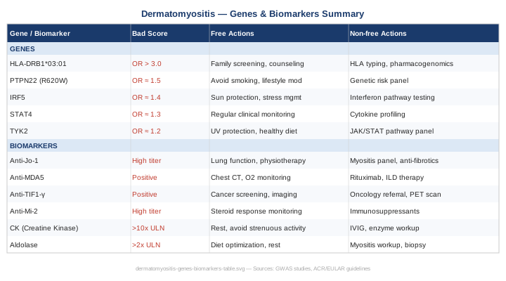
Building on this genetic picture, it helps to see genes and biomarkers side-by-side as an action matrix.
A Research Summary That May Change Your Approach: The Wahls Protocol and Autoimmune Mitochondrial Science
Terry Wahls, MD, published The Wahls Protocol after reversing her own secondary progressive multiple sclerosis — a severe autoimmune condition — through a rigorously designed nutritional intervention rooted in mitochondrial biology and immunology. While dermatomyositis is not the primary studied condition, the mechanistic underpinnings overlap substantially, and Wahls' research is among the most robust dietary autoimmune work available with actual clinical trial data.
1. Mitochondria as the Upstream Target
Wahls argues — backed by cell biology — that inflamed, autoimmune-damaged tissues have universally impaired mitochondria. Rather than targeting inflammation directly (as drugs do), her protocol supplies the raw materials mitochondria need to function: sulfur amino acids, B vitamins, CoQ10 cofactors, heme iron, and omega-3s. In dermatomyositis, where mitochondrial dysfunction in muscle fibers is well-documented, this framing is directly relevant.
2. Nine Cups per Day as a Therapeutic Dose
The protocol prescribes nine cups of produce daily: three cups of leafy greens (kale, chard, beet greens — rich in B vitamins and vitamins K), three cups of sulfur-rich vegetables (cabbage family, onions, mushrooms), and three cups of deeply pigmented vegetables and fruits (beets, berries, peppers — rich in antioxidants). Each category maps to specific mitochondrial cofactor pathways, not just general "healthy eating."
3. The Neurological and Muscular Case for Coenzyme Q10
Wahls consistently highlights CoQ10 as foundational to mitochondrial electron transport chain function. In dermatomyositis specifically, CoQ10 deficiency has been identified in muscle biopsies and CoQ10 supplementation (200–300 mg daily of ubiquinol form) is one of the few supplements with disease-specific rationale. The Wahls Protocol provides this through offal consumption but supplementation achieves equivalent coenzyme repletion.
4. Myelin and Muscle Membrane Repair Through Dietary Fat
Medium-chain triglycerides (MCT), from coconut oil or dedicated MCT oil, provide alternative fuel for mitochondria-impaired cells that cannot efficiently oxidize long-chain fatty acids. This is particularly relevant for dermatomyositis muscle tissue during active inflammation. Wahls uses one to three tablespoons of coconut oil daily as a metabolic support tool.
5. Sulfur Amino Acids for Glutathione Synthesis
Cysteine, methionine, and glycine from sulfur-rich foods (eggs, meat, onions, garlic) are rate-limiting precursors for glutathione — the cell's master antioxidant. Dermatomyositis tissue shows profound oxidative stress; glutathione depletion amplifies the inflammatory cycle. The dietary strategy is fundamentally different from taking antioxidant supplements: it supplies the building blocks rather than the finished product.
6. Elimination of Dietary Lectins
Wahls (and the overlapping autoimmune protocol literature) recommends eliminating dietary lectins (legumes, grains, nightshades, dairy) during the first phase of recovery. The mechanistic argument centers on gut permeability: lectins disrupt tight junctions, increase intestinal permeability, and allow bacterial antigens to enter circulation where they can trigger or sustain autoimmune activation. This claim has variable evidence quality — some is well-supported, some extrapolated from animal models.
7. Time-Restricted Eating as an Immune Regulator
Wahls incorporates a twelve-to-sixteen hour overnight fast as standard practice. Emerging research on time-restricted eating and autoimmunity suggests fasting intervals reduce dendritic cell activation, lower systemic cytokines, and improve the Treg-to-Teffector ratio — directly relevant to dermatomyositis pathophysiology.
8. Heavy Metal Detoxification Through Cruciferous Vegetables
Sulforaphane from broccoli sprouts and other crucifers activates the Nrf2 pathway, upregulating phase II detoxification enzymes that clear reactive oxygen species and environmental toxins. Silica and heavy metal accumulation have been explored as potential environmental triggers for dermatomyositis in occupational studies. Crucifer-rich diets provide a practical mitigation strategy regardless of individual exposure history.
9. The Clinical Trial Evidence
Wahls published a pilot clinical trial of her protocol in multiple sclerosis (published in PLOS One, 2014), showing statistically significant improvements in fatigue, cognitive function, and quality of life. While this was not a dermatomyositis trial, fatigue and quality of life are dominant symptom burdens in both conditions, and the metabolic mechanisms are conserved. A full dermatomyositis-specific Wahls trial has not been conducted; extrapolation requires appropriate epistemic humility.
10. The Progressive Intensity Levels as a Practical Pathway
The protocol has three tiers — Basic Wahls, Wahls Paleo, and Wahls Paleo Plus — allowing gradual dietary change rather than an abrupt overhaul. This tiered approach makes it practically sustainable for patients managing fatigue and disability alongside a complex medical regimen. Starting with Wahls Basic (primarily the nine cups framework) and progressing only if well-tolerated is a reasonable and cautious application for dermatomyositis.
Complementary Approaches with Clinical Relevance
The following modalities have meaningful human clinical evidence in autoimmune or closely related inflammatory muscle conditions. They are not substitutes for rheumatological care but represent valuable adjuncts when used thoughtfully.
The Autoimmune Protocol (AIP) from Sarah Ballantyne
The Autoimmune Protocol, developed and systematized by Sarah Ballantyne, PhD, is a structured nutritional and lifestyle program specifically designed for autoimmune conditions. It functions by removing dietary inputs that promote intestinal permeability and immune activation — including grains, legumes, nightshades, eggs, dairy, nuts, seeds, alcohol, and processed foods — while simultaneously increasing nutrient density through organ meats, diverse vegetables, bone broth, and fermented foods. The framework also incorporates sleep optimization, stress reduction, and gentle movement as essential non-negotiables alongside the dietary component.
For dermatomyositis specifically, the AIP is relevant because gut permeability and dysbiosis are increasingly recognized as contributors to autoimmune disease maintenance, not just initiation. A 2017 pilot study published in Inflammatory Bowel Diseases demonstrated clinically meaningful improvements in inflammatory bowel disease using the AIP, providing human proof-of-concept for the dietary framework's anti-inflammatory efficacy. While no dermatomyositis-specific AIP trial exists, the mechanistic overlap — intestinal permeability, T-cell dysregulation, systemic cytokine elevation — justifies its consideration.
In practical application for dermatomyositis: begin with a strict six-week elimination phase, tracking muscle enzyme levels (CK, aldolase) and symptom severity with a daily journal. Reintroduce eliminated foods systematically, one at a time, every five to seven days, recording any symptom exacerbation. Most people discover one to three specific food triggers that meaningfully worsen their inflammatory profile. Work with a registered dietitian experienced in AIP to ensure nutritional adequacy during elimination, particularly for calcium, vitamin D, and adequate protein for muscle preservation.
Mindfulness-Based Stress Reduction (MBSR)
Chronic psychological stress elevates cortisol, disrupts regulatory T-cell populations, and amplifies proinflammatory cytokine production — all of which directly worsen dermatomyositis disease activity. MBSR, developed by Jon Kabat-Zinn at the University of Massachusetts, is an eight-week structured program combining body scan meditation, sitting meditation, and mindful yoga, typically delivered in two-and-a-half-hour weekly sessions plus a full-day retreat.
Human evidence for MBSR in autoimmune and inflammatory conditions includes a well-conducted randomized trial in rheumatoid arthritis demonstrating significant reductions in disease activity scores and inflammatory markers after eight weeks, published in Rheumatology. A systematic review of mind-body interventions in inflammatory conditions found consistent reductions in perceived pain, fatigue, and quality-of-life impairment — outcomes that map directly onto the dermatomyositis symptom burden.
For dermatomyositis, the practical recommendation is to join a formally certified MBSR program (available through hospitals, community health centers, and online platforms) rather than self-directed meditation apps, particularly at first. Eight weeks of consistent practice (forty-five minutes daily) appears to be the minimum dose for measurable neuroimmune effects. Those with active disease should choose the gentler yoga component variants and communicate with the instructor about physical limitations.
Tai Chi
Tai chi is a low-impact, mind-body movement practice involving slow, deliberate movements, weight shifting, and coordinated breathing. Its relevance for dermatomyositis lies in its ability to preserve and gently restore muscle function, balance, and neuromuscular coordination without the inflammatory stimulus of conventional exercise. For patients with proximal muscle weakness — the hallmark of dermatomyositis — activities requiring minimal eccentric loading (like tai chi) are preferable to conventional resistance training during active disease.
A randomized controlled trial of tai chi in systemic lupus erythematosus — a closely related autoimmune condition — published in Arthritis Care and Research found significant improvements in fatigue, depression, and physical function versus a control group. A meta-analysis of tai chi in fibromyalgia and chronic inflammatory conditions found consistent improvements in pain, stiffness, and functional capacity. Evidence specifically in dermatomyositis remains limited to expert recommendations rather than controlled trials.
Begin with a beginner-level tai chi class (Yang style is the most accessible), two to three times weekly, each session thirty to forty-five minutes. Physiotherapist supervision or a class taught by an instructor familiar with chronic illness is preferable. Advance pace and duration slowly over three to six months; track CK and functional strength markers to ensure activity is not triggering flares.
Low-Level Laser Therapy / Photobiomodulation
Photobiomodulation (PBM) uses red and near-infrared light (typically 630–850 nm) at low intensities to stimulate cellular mitochondrial function, reduce oxidative stress, and modulate inflammatory pathways. The proposed mechanism involves cytochrome c oxidase activation in mitochondria, increasing ATP production and reducing reactive oxygen species — directly relevant to the mitochondrial dysfunction documented in dermatomyositis muscle fibers.
Human evidence for PBM in inflammatory muscle conditions includes trials in rheumatoid arthritis (showing reduced joint inflammation and pain) and in delayed-onset muscle soreness (showing faster recovery and lower CK elevation). A systematic review of PBM in musculoskeletal conditions found consistent anti-inflammatory and analgesic effects. Dermatomyositis-specific clinical trials do not yet exist; this is an emerging application requiring cautious extrapolation.
Practical application: a home red light therapy device (cost $200–600) emitting 630–850 nm at 50–150 mW/cm² applied to major proximal muscle groups (thighs, shoulders) for ten to twenty minutes daily. Avoid applying over skin with active rash (heliotrope, Gottron's papules) as photosensitivity is a feature of dermatomyositis and light exposure to affected skin may exacerbate it. Begin with every-other-day sessions and monitor skin tolerance carefully.
Microbiome-Directed Therapies
The gut microbiome's role in autoimmune disease has moved from hypothesis to well-supported biology over the past decade. Studies in multiple autoimmune conditions — including lupus, rheumatoid arthritis, and multiple sclerosis — consistently identify characteristic gut dysbiosis patterns, with reduced microbial diversity, depleted butyrate-producing bacteria, and expanded pro-inflammatory species. For dermatomyositis, direct microbiome data are emerging: a 2021 study in Frontiers in Immunology identified specific gut microbiome alterations in dermatomyositis patients compared to healthy controls, including reduced Faecalibacterium prausnitzii — a key anti-inflammatory butyrate producer.
The interventional logic is to restore microbial diversity and butyrate-producing capacity through combined dietary and probiotic strategies. High-fiber intake (25–35 g daily from diverse plant sources), fermented foods (kefir, kimchi, sauerkraut, yogurt), and targeted probiotic supplementation collectively shift the microbiome toward an anti-inflammatory configuration. Prebiotic fibers (inulin, FOS, partially hydrolyzed guar gum) specifically feed butyrate producers.
In practice: introduce three to four daily servings of fermented foods, targeting variety over quantity. Add a clinically dosed multi-strain probiotic (at least four to six strains, 20–50 billion CFU). Rotate probiotic products every three months to broaden exposure. Track digestive symptoms and inflammatory markers over twelve weeks. Those on immunosuppressive therapy should consult their rheumatologist before beginning probiotic supplementation, as there are theoretical (though practically uncommon) considerations regarding immunomodulation.
Conclusion
Dermatomyositis is a disease that rewards precision. Vague approaches — generalized anti-inflammatory advice, broad immunosuppression without subtype stratification — miss too much of what makes this condition individual. The seven biomarkers covered here give you a concrete monitoring framework: CK and aldolase track muscle damage in real time; anti-MDA5, anti-Jo-1/ARS, and anti-TIF1-gamma define your subtype and risk profile; ferritin tracks systemic severity; and CRP/ESR provide inflammatory context. Together they create a picture no single appointment can capture. The six genes — particularly HLA-DRB1, IRF5, STAT4, and TYK2 — explain why your immune system behaves as it does and identify the specific pathways most worth supporting.
The practical next step is not to overhaul everything at once. Choose one domain — run the full autoantibody panel if you have not yet, or bring ferritin and aldolase tracking to your next appointment, or begin the AIP elimination phase, or start MBSR. Small, targeted actions, built on accurate information, accumulate into meaningful change. Discuss the biomarker panel, the genetic context, and any supplement protocols with your rheumatologist or an integrative physician familiar with inflammatory myopathies — they are your most important partner in applying what this article covers.
Cancer & Oncology Skin Autoimmune
Musculoskeletal: Muscle Conditions
Respiratory: Lung Conditions
Autoimmune: Inflammatory Conditions Autoimmune Skin Conditions