This article contains AI generated content.
· UpdatedDevelopmental Dysplasia of the Hip - 5 Genes And 5 Biomarkers To Track
Introduction
For many adults, discovering they have developmental dysplasia of the hip (DDH) comes as a quiet but life-altering shock. It usually starts as an occasional ache deep in the groin, a clicking sensation during a normal walk, or a feeling of instability when standing up. Too often, the diagnosis is delayed until early joint wear has already begun, leaving patients to navigate a complex medical landscape of physical therapy, pelvic osteotomies, or the prospect of an early total hip replacement.
Standard advice for managing hip dysplasia is frequently frustrating in its simplicity. Being told to "do low-impact exercise," "lose weight," or "take a generic joint supplement" does not address the mechanical and biological realities of a shallow hip socket that fails to adequately support the femoral head. These broad recommendations ignore the unique cellular environment of your joint capsule and fail to provide a personalized blueprint for protecting your cartilage.
This article takes a deeper, science-aware approach to managing the condition. By exploring the molecular biomarkers of joint degradation and the genetic pathways that govern joint structure and repair, we can look past generic advice. Understanding how your specific cartilage matrix is reacting to mechanical stress enables you to make informed decisions that target the root biological mechanisms of joint wear.
There is grounded hope in this approach. Better information leads to better decisions, and understanding the specific biological levers of your condition can help you preserve joint function. By tracking key biomarkers, understanding genetic predispositions, and applying targeted neuromuscular and complementary therapies, you can work alongside your clinical team to build a proactive, highly effective hip preservation plan.
Summary
This article provides a comprehensive, science-driven guide to managing developmental dysplasia of the hip (DDH) by examining the structural, biological, and genetic factors that dictate joint health. We explore five critical biomarkers—such as Cartilage Oligomeric Matrix Protein (COMP) and urinary CTX-II—that allow you to measure the active rate of joint wear and cartilage degradation. Additionally, we investigate five key susceptibility genes, including GDF5 and ASPN, explaining how genetic variations influence your joint structure and detailing precise lifestyle and supplemental strategies to compensate for these underlying vulnerabilities.
Further in the article, we outline a specialized joint-preservation protocol inspired by recent neurobiology and exercise science, followed by an objective evaluation of five complementary modalities with clinical evidence for managing chronic joint discomfort. The schematic below illustrates the mechanical relationship between a dysplastic hip joint and the corresponding biomarkers of joint wear.
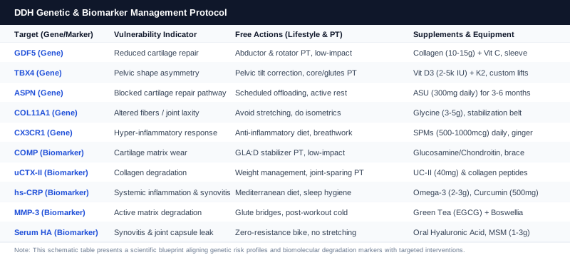
Reverse Developmental Dysplasia of the Hip: 5 Biomarkers to Track
To manage hip dysplasia effectively, we must look beyond static imaging. While X-rays and MRIs show the physical structure of your hip joint, they only reveal damage that has already occurred. Biological markers, on the other hand, provide a real-time window into the rate of joint degradation and cartilage turnover. By measuring these markers, you and your medical team can track whether your joint-preservation strategies are working or if mechanical wear is outstripping your body's ability to repair.
1. Cartilage Oligomeric Matrix Protein (COMP)
Cartilage Oligomeric Matrix Protein (COMP) is a non-collagenous glycoprotein found abundantly in the extracellular matrix of articular cartilage. When joint cartilage is subjected to abnormal mechanical shear forces—a hallmark of the shallow socket in hip dysplasia—chondrocytes undergo cellular stress, and the cartilage matrix begins to break down. This structural breakdown releases COMP fragments directly into the synovial fluid, where they eventually enter the bloodstream. Measuring serum COMP provides a highly sensitive gauge of active cartilage remodeling and joint wear.
The clinical utility of tracking COMP is supported by the Johnston County Osteoarthritis Project, which demonstrated that elevated serum COMP levels directly correlate with the presence and radiographic severity of joint degeneration. For individuals with hip dysplasia, an elevated COMP score indicates that the structural instability of the hip is causing active, ongoing cartilage wear, even before significant joint space narrowing is visible on a standard X-ray.
How to measure it
Serum COMP is measured via a standard venous blood draw analyzed using an enzyme-linked immunosorbent assay (ELISA). The test is available through specialized functional laboratories and joint health panels. The cost range for a standalone serum COMP test typically runs between $100 and $250.
If the score is bad, the plan without supplements
If your COMP levels are elevated, the immediate goal is to reduce mechanical shear stress on the hip joint while enhancing muscular stability. You must prioritize neuromuscular stabilization training, such as the GLA:D protocol, focusing on strengthening the gluteus medius, gluteus minimus, and deep hip rotators (including the piriformis and obturators). Strengthening these stabilizers helps pull the femoral head deeper into the shallow acetabular socket, improving joint congruency. Avoid activities that place high shear forces on the joint, such as deep loaded hip flexion (squats past parallel) and high-impact running, opting instead for low-impact resistance training.
If the score is bad, the plan with supplements or equipment
To support the cartilage matrix under mechanical stress, incorporate a combination of Glucosamine Sulfate (1500 mg daily) and Chondroitin Sulfate (800 mg daily). These compounds serve as crucial structural precursors for glycosaminoglycans within the extracellular matrix. Additionally, utilize high-cushion, neutral orthopedic footwear to absorb ground reaction forces during walking, and consider a dynamic hip unloader brace during periods of prolonged standing or walking. Glucosamine may cause mild gastrointestinal upset or minor fluctuations in blood glucose; take it with food. These supplements should be taken daily for a minimum of 12 weeks to evaluate their impact on joint comfort and follow-up biomarker levels.
2. Urinary C-Terminal Telopeptide of Type II Collagen (uCTX-II)
Type II collagen is the primary structural protein that gives articular cartilage its tensile strength. When the cartilage in a dysplastic hip is worn down by joint incongruency, enzymatic cleavage degrades this collagen network. The resulting degradation fragments, specifically the C-terminal cross-linked telopeptide of type II collagen (CTX-II), are released into circulation and excreted in urine. Elevated uCTX-II is a highly specific marker of type II collagen degradation and serves as a strong predictor of progressive joint space narrowing.
The predictive value of this biomarker was established in a comprehensive meta-analysis on CTX-II, which confirmed that urinary CTX-II levels are elevated in patients with active joint degeneration and strongly correlate with the rate of cartilage loss. In hip dysplasia, tracking uCTX-II allows you to monitor the structural integrity of your cartilage fibers and determine if your mechanical offloading strategies are successfully halting collagen breakdown.
How to measure it
uCTX-II is measured using a urine sample, ideally from the first morning void to ensure concentration consistency. The results are normalized to urinary creatinine levels to account for hydration status. The cost range for a uCTX-II ELISA test is generally between $150 and $300.
If the score is bad, the plan without supplements
A bad uCTX-II score requires strict mechanical offloading. Body weight management is the most effective non-supplemental intervention; losing even a small amount of weight significantly reduces the cumulative mechanical load on the hip joint during daily activities. Incorporate joint-sparing movement patterns, such as swimming or using an elliptical trainer, which allow you to maintain cardiovascular fitness without the impact forces that degrade type II collagen. During acute flare-ups, utilize a trekking pole or cane in the contralateral hand to reduce joint reaction forces on the dysplastic hip by up to 30 percent.
If the score is bad, the plan with supplements or equipment
To protect type II collagen fibers, supplement with Undenatured Type II Collagen (UC-II) at a dose of 40 mg daily. UC-II operates via oral tolerance, interacting with immune cells in the gut to reduce systemic autoimmune-mediated collagen breakdown in the joints. Pair this with 10 to 15 grams of hydrolyzed collagen peptides daily to supply the specific amino acids required for cartilage repair. UC-II is highly tolerated, though mild digestive discomfort is occasionally reported. It must be taken consistently every day, as the immunomodulatory effects rely on continuous exposure.
3. High-Sensitivity C-Reactive Protein (hs-CRP)
High-Sensitivity C-Reactive Protein (hs-CRP) is a classic systemic marker of low-grade inflammation produced by the liver in response to inflammatory cytokines. While developmental dysplasia of the hip is initiated by a mechanical defect, the chronic instability and joint subluxation lead to repetitive micro-trauma of the synovial membrane (synovitis). This localized joint inflammation releases cytokines like interleukin-6 (IL-6) and tumor necrosis factor-alpha (TNF-alpha) into circulation, driving up systemic hs-CRP. Elevated hs-CRP, in turn, uregulates the production of cartilage-destroying enzymes.
Tracking hs-CRP is essential because systemic inflammation acts as an accelerant on mechanical joint wear. If your systemic inflammatory baseline is high, the cartilage in your dysplastic hip will degrade much faster under mechanical load than it would in a low-inflammation environment. Lowering hs-CRP helps protect the joint by dampening the chemical pathways that break down cartilage matrix.
How to measure it
hs-CRP is measured via a simple venous blood draw. It is a highly standard, widely available test that can be ordered by any primary care physician. The cost range is very affordable, typically between $20 and $50.
If the score is bad, the plan without supplements
If your hs-CRP is elevated, you must address systemic inflammatory triggers. Adopt an anti-inflammatory diet, such as the Mediterranean diet, emphasizing extra virgin olive oil, wild-caught fatty fish, leafy green vegetables, and berries while eliminating refined sugars, trans fats, and ultra-processed foods. Focus on sleep optimization, aiming for 7 to 9 hours of quality sleep, as sleep deprivation directly elevates inflammatory cytokines. Manage mental and physical stress through daily mindfulness or breathwork, which downregulates the sympathetic nervous system pathways that drive inflammatory gene expression.
If the score is bad, the plan with supplements or equipment
To actively lower hs-CRP, supplement with high-potency Omega-3 Fatty Acids (supplying 2000 to 3000 mg of combined EPA and DHA daily) and standardized Curcumin Extract (500 to 1000 mg daily). Omega-3s compete with arachidonic acid to reduce the production of pro-inflammatory eicosanoids, while curcumin acts as a natural NF-kB inhibitor, suppressing inflammatory cytokine production. Curcumin can have mild blood-thinning properties and may cause minor digestive upset; avoid taking it on an empty stomach. These supplements should be taken daily, and inflammatory markers should be retested after 8 to 12 weeks.
4. Matrix Metalloproteinase-3 (MMP-3)
Matrix Metalloproteinase-3 (MMP-3), also known as stromelysin-1, is an enzyme produced by chondrocytes and synovial fibroblasts in response to mechanical stress and inflammatory cytokines. MMP-3 plays a major role in joint destruction because it directly degrades proteoglycans, fibronectin, and various types of collagen within the articular cartilage. Elevated levels of MMP-3 in the bloodstream indicate that the body is actively producing enzymes that dissolve the structural scaffolding of the hip joint.
In developmental dysplasia of the hip, the localized mechanical overload of the shallow socket acts as a direct physical trigger for MMP-3 release. Tracking this enzyme helps you understand the enzymatic activity inside your joint. A high MMP-3 level warns you that your cartilage is in an active state of enzymatic degradation, necessitating interventions to inhibit these destructive enzymes before permanent structural loss occurs.
How to measure it
MMP-3 is measured via a serum blood test. It is typically offered by specialized neurology and rheumatology panels that evaluate joint and connective tissue health. The cost range for a serum MMP-3 test is between $120 and $250.
If the score is bad, the plan without supplements
To suppress MMP-3 expression without supplements, you must eliminate mechanical joint irritation. Implement strict joint-stabilizing isometric exercises. Isometric hip abduction against a strap and isometric glute bridges allow you to build muscular support around the hip joint without the repetitive friction of joint movement, which triggers MMP-3 release. Additionally, apply cold therapy (ice packs or cold water immersion) to the lateral and anterior hip for 15 minutes after exercise to constrict local blood vessels and suppress the inflammatory cascade that drives MMP synthesis.
If the score is bad, the plan with supplements or equipment
To inhibit matrix metalloproteinase activity, supplement with Green Tea Extract standardized to Epigallocatechin Gallate (EGCG) at 300 to 500 mg daily, and standardized Boswellia Serrata Extract (such as ApresFlex) at 100 mg daily. EGCG has been shown to downregulate the genetic expression of MMPs in chondrocytes, while Boswellia inhibits 5-lipoxygenase, reducing the inflammatory signaling that stimulates enzyme production. High-dose green tea extract can cause liver irritation; choose a high-quality, verified supplement and take it with food. Take Boswellia daily, and cycle the green tea extract by taking it for 8 weeks followed by a 2-week break.
5. Hyaluronic Acid (HA) in Serum
Hyaluronic Acid (HA) is a major component of the synovial fluid that lubricates the hip joint and absorbs mechanical shock. In a healthy joint, HA remains concentrated within the joint cavity. However, when a dysplastic hip suffers from chronic mechanical instability, the synovial membrane becomes inflamed (synovitis) and hyperpermeable. This inflammation causes hyaluronic acid to leak out of the joint capsule and into the systemic circulation. Consequently, elevated serum HA levels reflect joint capsule permeability, active synovitis, and high joint tissue turnover.
Measuring serum HA provides a direct look at the health of your joint capsule and the viscosity of your lubricating fluid. In hip dysplasia, a high serum HA level indicates that the joint is under significant friction and stress, causing the protective lubricating fluid to leak away. Restoring joint containment and reducing synovitis are essential to keep the lubricant where it belongs: inside the hip socket.
How to measure it
Serum Hyaluronic Acid is measured via a standard venous blood draw and analyzed using an ELISA assay. The cost range for this test typically runs between $100 and $200.
If the score is bad, the plan without supplements
If your serum HA is elevated, indicating joint leak and synovitis, you must focus on low-resistance joint mobilization to stimulate healthy synovial fluid production. Spending 15 to 20 minutes daily on a stationary bicycle with zero resistance helps circulate synovial fluid throughout the hip joint capsule, distributing nutrients and natural lubrication without putting mechanical pressure on the shallow socket. Avoid deep stretching of the hip joint, as this can increase capsule laxity and worsen the leakage of synovial fluid into surrounding tissues.
If the score is bad, the plan with supplements or equipment
To support synovial fluid viscosity and joint lubrication, supplement with Oral Hyaluronic Acid (80 to 200 mg daily of a high-molecular-weight form) and Methylsulfonylmethane (MSM) at 1000 to 3000 mg daily. High-molecular-weight oral HA has been shown to support joint lubrication and reduce synovial inflammation, while MSM provides bioavailable sulfur necessary for glycosaminoglycan synthesis. MSM can cause mild bloating or digestive loose stools if introduced too quickly; start with a low dose of 500 mg daily and titrate up over several weeks. Take these daily without cycling to maintain joint lubrication.
What Recent Genetics and Epigenetics Research Suggests
While biomarkers show the current state of joint wear, genetics and epigenetics reveal the baseline blueprint of your connective tissues. Developmental dysplasia of the hip has a strong genetic foundation, with heritability estimates ranging from 50 to 60 percent. Genetic variations influence how your acetabulum forms during development, the elasticity of your ligaments, and your cartilage's inherent repair capacity. Understanding your genetic profile allows you to identify specific structural and cellular vulnerabilities and implement targeted strategies to compensate for them.
1. GDF5 (Growth Differentiation Factor 5)
The GDF5 gene encodes a member of the transforming growth factor-beta (TGF-beta) superfamily, which is critical for joint development, chondrogenesis, and bone formation. A specific single-nucleotide polymorphism (SNP) in the GDF5 promoter, known as rs143384, is one of the most widely established genetic risk factors for hip dysplasia and early-onset osteoarthritis. This variant reduces the transcription of GDF5, leading to decreased growth factor expression in joint tissues, which manifests as a shallower acetabular socket and thinner, less resilient articular cartilage.
The impact of this variant is detailed in a genome-wide association study on GDF5, which identified GDF5 as a major susceptibility locus for developmental dysplasia of the hip. Furthermore, researchers have found that epigenetic hypermethylation of the GDF5 promoter, as documented in a study on DNA methylation in DDH, can further silence this protective gene, severely limiting the hip's capacity to repair its cartilage matrix under mechanical stress.
If the gene is bad, the plan without supplements
If you carry the GDF5 risk variant, your cartilage has a genetically lower capacity for self-repair, making mechanical joint protection paramount. You must focus on progressive strength training of the hip abductors and rotators to build a robust muscular sleeve around the joint, compensating for structural socket deficiency. Avoid repetitive impact sports, such as long-distance running on hard surfaces, and instead choose activities like swimming, rowing, or resistance training that build muscle without grinding the joint surfaces. Keep your movement mechanics precise, avoiding hip hyperextension and excessive internal rotation.
If the score is bad, the plan with supplements or equipment
To compensate for reduced GDF5-driven cartilage repair, supplement daily with Hydrolyzed Collagen Peptides (10 to 15 grams) combined with Vitamin C (500 mg) to stimulate native collagen synthesis. Utilize a targeted hip compression sleeve or pelvic support belt during exercise to improve proprioception and joint stability. Vitamin C can cause mild gastrointestinal looseness in high doses; adjust the dose if needed. Take this combination daily without cycling to provide a steady supply of cartilage building blocks.
2. TBX4 (T-Box Transcription Factor 4)
The TBX4 gene encodes a transcription factor that plays a fundamental role in hindlimb and pelvic development during embryonic growth. Genetic variants or microdeletions in TBX4 disrupt the developmental template of the pelvic girdle, directly contributing to developmental dysplasia of the hip and associated skeletal anomalies. Individuals with TBX4 variations often present with structural alterations in the shape of the pelvis, resulting in a shallow acetabular socket that fails to cover the femoral head properly, creating chronic joint instability from birth.
If the gene is bad, the plan without supplements
A genetic vulnerability in TBX4 requires early and continuous physical therapy focused on pelvic alignment and structural symmetry. You must work with a physical therapist to correct anterior pelvic tilt, which narrows the functional coverage of the hip socket. Focus on strengthening the lower abdominals, obliques, and hamstrings while stretching tight hip flexors (the iliopsoas and rectus femoris). Aligning the pelvis maximizes the weight-bearing surface area of the shallow acetabulum, reducing focal stress on the joint cartilage.
If the score is bad, the plan with supplements or equipment
To support pelvic bone density and structural integrity in the presence of TBX4 variations, supplement with Vitamin D3 (2000 to 5000 IU daily) and Vitamin K2 (MK-7, 100 to 200 mcg daily) to ensure calcium is properly directed into the bone matrix. Use custom orthotics or heel lifts to correct any functional leg-length discrepancies that could cause pelvic tilting and asymmetric hip loading. High doses of Vitamin D3 can lead to hypercalcemia if taken without K2; monitor your serum 25-hydroxyvitamin D levels annually. Take these supplements daily with a fat-containing meal.
3. ASPN (Asporin)
The ASPN gene encodes asporin, an extracellular matrix protein that binds to and inhibits transforming growth factor-beta 1 (TGF-beta 1), a crucial growth factor that stimulates chondrocytes to produce collagen and proteoglycans. A specific genetic variation in ASPN, involving a polymorphism in the aspartic acid (D) repeat region, increases the production of asporin. This excess asporin binds too tightly to TGF-beta 1, blocking its beneficial signals and effectively shutting down the cartilage repair process in the hip joint.
The relationship between this gene and hip dysplasia is detailed in a case-control study on the ASPN gene, which demonstrated a significant association between the D-repeat polymorphism and an increased risk of developmental dysplasia of the hip. Carrying this risk variant means your hip cartilage is genetically hypersensitive to degradation because its primary repair pathway is blocked by elevated asporin activity.
If the gene is bad, the plan without supplements
If you have an ASPN risk variant, you must incorporate scheduled joint offloading periods into your routine. Since your cartilage lacks the capacity to repair quickly, you cannot tolerate back-to-back days of strenuous weight-bearing exercise. Design your week with alternating days of active rest, utilizing non-weight-bearing activities like swimming or cycling to maintain fitness. Focus on myofascial release of the surrounding hip musculature to prevent chronic tension from placing continuous static pressure on the joint capsule.
If the score is bad, the plan with supplements or equipment
To counteract the inhibitory effects of asporin on cartilage repair, supplement with Avocado Soy Unsaponifiables (ASU) at a dose of 300 mg daily. ASU has been shown in clinical settings to stimulate TGF-beta expression and promote collagen synthesis in chondrocytes, helping bypass the genetic block caused by ASPN variations. ASU is very well tolerated, with rare reports of mild gastrointestinal upset. Take ASU daily for at least 3 to 6 months to allow the cartilage matrix to accumulate sufficient structural improvements.
4. COL11A1 (Collagen Type XI Alpha 1 Chain)
The COL11A1 gene encodes a minor fibrillar collagen that plays a critical role in organizing and regulating the spacing and diameter of type II collagen fibrils in articular cartilage. Genetic variants in COL11A1 alter the structure of these collagen fibers, leading to a weaker, more disorganized extracellular matrix. This genetic structural weakness causes increased joint laxity, ligamentous hypermobility, and cartilage that is highly susceptible to tearing and fraying under normal mechanical loads.
If the gene is bad, the plan without supplements
For individuals with COL11A1-related joint laxity, static stretching and high-flexibility activities (such as deep yoga or gymnastics) must be avoided, as they further destabilize the shallow hip joint. Instead, focus entirely on closed-chain isometric exercises (such as wall sits, glute bridges, and single-leg balance holds). These exercises stimulate muscular co-contraction around the hip joint, building neuromuscular stability to hold the femoral head securely in place without stressing the weak collagen fibers.
If the score is bad, the plan with supplements or equipment
To support the structural integrity of weak collagen networks, supplement with high-dose Glycine (3 to 5 grams daily) or organic bone broth to provide the structural building blocks for collagen synthesis. During periods of increased physical activity, wear a semi-rigid hip stabilization belt to provide external compression and mechanical support to the pelvis. Glycine is highly safe and often promotes relaxation; it is best taken before sleep. Use these interventions daily to support connective tissue health.
5. CX3CR1 (Chemokine C-X3-C Motif Receptor 1)
The CX3CR1 gene encodes a chemokine receptor expressed on macrophages and inflammatory cells. This receptor is involved in cell adhesion, migration, and the regulation of inflammatory processes in bone and cartilage. A specific variant in CX3CR1 is associated with developmental dysplasia of the hip, as demonstrated in a whole-exome sequencing linkage study. Variants in this gene lead to a dysregulated, hyper-inflammatory response to mechanical friction in the joint capsule, turning minor joint wear into rapid, macrophage-driven cartilage destruction.
If the gene is bad, the plan without supplements
If you carry a CX3CR1 risk variant, you must adopt a strict anti-inflammatory lifestyle to prevent systemic inflammation from amplifying local joint wear. Minimize exposure to environmental toxins, alcohol, and inflammatory foods that trigger macrophage activation. Practice daily diaphragmatic breathing and vagal nerve stimulation techniques, which help activate the body's natural anti-inflammatory pathways, dampening the immune system's destructive response to mechanical friction in the hip joint.
If the score is bad, the plan with supplements or equipment
To modulate macrophage activity and resolve joint inflammation, supplement with Specialized Pro-Resolving Mediators (SPMs) at 500 to 1000 mcg daily, or Ginger Extract (500 mg daily). SPMs are lipid mediators that actively signal the immune system to resolve inflammation, while ginger inhibits inflammatory pathways. Ginger can cause mild heartburn if taken on an empty stomach; always take it with a meal. Take ginger daily during active flare-ups, and cycle SPMs by taking them for 4 weeks followed by a 2-week break.
The Huberman Lab Protocol for Joint Health and Cartilage Regeneration
To protect and repair a dysplastic hip joint, we must apply the principles of neurobiology and exercise science. The following protocol provides ten actionable, evidence-based strategies to optimize joint lubrication, stimulate cartilage repair, and build muscular support around a shallow hip socket.
1. Mechanical Stimulation and Chondrocyte Activation
Articular cartilage is avascular, meaning it has no direct blood supply to deliver nutrients. To survive, cartilage relies on "fluid flow transduction." When the joint cartilage is compressed and decompressed through movement, it acts like a sponge, drawing in nutrient-rich synovial fluid and expelling metabolic waste. Without regular mechanical loading, chondrocytes starve and degrade. Performing non-impact, weight-bearing movements daily is essential to keep the cartilage cells healthy and functional.
2. Targeted Collagen Loading with Vitamin C
To actively stimulate cartilage matrix repair, consume 10 to 15 grams of hydrolyzed collagen peptides or gelatin paired with 500 mg of Vitamin C exactly 30 to 60 minutes before your physical therapy or joint-loading exercises. The physical movement increases blood flow to the joint capsule, directing the circulating amino acids directly into the hip cartilage where they are needed to rebuild the collagen network.
3. Low-Load, High-Repetition Blood Flow Enhancement
Before performing any strength exercises, spend 15 to 20 minutes on a stationary bicycle with very low resistance, aiming for a high cadence. This high-repetition, low-load movement warms up the joint capsule and increases the temperature of the synovial fluid, reducing its viscosity and making it a more effective lubricant. It also draws blood flow to the surrounding pelvic muscles, preparing them to support the joint.
4. Neuromuscular Recruitment of Deep Joint Stabilizers
Hip stability depends heavily on the brain-to-muscle connection. Before loading the hip joint with weight-bearing movements, perform activation exercises for the gluteus medius and deep hip rotators (such as band-resisted sidesteps or clamshells). Activating these stabilizers ensures that the nervous system holds the femoral head securely within the shallow acetabular socket, preventing micro-slippage during exercise.
5. Dual-Temperature Therapy for Joint Inflammation
Contrast therapy is a powerful tool to manage local joint inflammation and promote healing. Alternating between heat (such as a warm bath or sauna) and cold (such as ice packs or cold water immersion) creates a vascular pumping action. Heat dilates the surrounding blood vessels to bring in healing nutrients, while cold constricts them to flush out metabolic waste, reducing the inflammatory load in the joint capsule.
6. Maintaining Synovial Fluid Viscosity Through Hydration
Synovial fluid is composed primarily of water bound by hyaluronic acid. Chronic dehydration increases the friction coefficient within the hip joint, accelerating mechanical wear on the cartilage. Ensure you drink adequate water daily, supplemented with trace minerals, to maintain optimal hydration. This keeps the synovial fluid thick and slippery enough to absorb shock and protect the joint surfaces.
7. Glycosaminoglycan Building Blocks and Glucosamine Dosing
Glucosamine and chondroitin serve as the essential structural building blocks for the proteoglycans that give joint cartilage its shock-absorbing capacity. Supplementing consistently with Glucosamine Sulfate at a dose of 1500 mg daily provides the necessary substrates for chondrocytes to build and maintain the extracellular matrix, helping protect the joint from mechanical overload.
8. Sleep Optimization for Systemic Growth Hormone Release
Deep stage 3 and 4 sleep is when the pituitary gland releases the majority of growth hormone, which stimulates the production of IGF-1 in liver and joint tissues. IGF-1 is a key hormone that drives cellular repair and regeneration in connective tissues. Prioritize sleep hygiene by keeping your bedroom dark, quiet, and cool, and avoiding screens for an hour before bed to support natural cartilage repair.
9. Leveraging the Cross-Education Effect for Symmetry
If one hip is severely dysplastic or recovering from surgery, you can still support its recovery by training the healthy side. Performing strength training on the healthy leg stimulates neural pathways that cross over to the opposite side of the body. This "cross-education" effect helps maintain up to 30 percent of the muscle mass and neural coordination in the resting leg, preventing severe asymmetry.
10. Managing the Gut-Joint Axis to Curb Cartilage Degradation
Gut dysbiosis can compromise the intestinal barrier, allowing bacterial endotoxins (lipopolysaccharides) to enter the bloodstream. This systemic exposure triggers an inflammatory response that upregulates matrix-degrading enzymes in the joints. Consuming fermented foods and soluble prebiotic fibers helps maintain a healthy gut barrier, reducing systemic inflammation and protecting joint cartilage from enzymatic breakdown.
Complementary and Alternative Modalities for Hip Dysplasia
Complementing your primary biomechanical and medical strategies with evidence-based alternative modalities can help manage pain, improve pelvic function, and reduce systemic inflammation. Below are five supportive modalities that have clinical evidence for joint health.
Tai Chi
Tai Chi is a mind-body practice that combines slow, controlled movements with deep breathing and mental focus. For individuals with developmental dysplasia of the hip, it is highly relevant because it emphasizes gentle weight-shifting, improves proprioception, and strengthens the pelvic stabilizers without subjecting the shallow joint to high-impact shocks.
A specific protocol involves practicing the Yang-style short form for 40 minutes, three times per week. A randomized controlled trial published on PubMed shows that Tai Chi significantly reduces joint pain and stiffness while improving physical function in adults with lower extremity joint osteoarthritis, making it a highly supported movement therapy.
To apply this cautiously, avoid deep knee bends or extreme external rotation of the hip during the movements. Keep your stance high and focus on the smooth transfer of weight from one leg to the other, ensuring that the knee remains aligned over the foot to prevent shear stress on the acetabulum.
Progressive Muscle Relaxation (PMR)
Progressive Muscle Relaxation is a somatic technique that involves systematically tensing and releasing various muscle groups throughout the body. In patients with developmental dysplasia of the hip, chronic pain often leads to involuntary muscle guarding, where the surrounding pelvic and gluteal muscles remain chronically contracted, increasing joint pressure and discomfort.
The protocol involves sitting or lying comfortably, tensing a specific muscle group for 5 to 7 seconds, and then releasing the tension suddenly, followed by 15 to 20 seconds of conscious relaxation. A clinical trial has demonstrated that PMR effectively reduces chronic pain, lowers muscle tension, and improves quality of life in patients suffering from hip joint degenerative diseases.
To apply this, practice the protocol once daily, preferably before sleep or after physical therapy sessions. Pay close attention to the pelvic and hip region, ensuring that the tension phase is gentle and does not cause sharp pain or subluxation in the dysplastic socket.
Low-Level Laser Therapy (LLLT)
Low-Level Laser Therapy, also known as photobiomodulation, uses specific wavelengths of near-infrared light to penetrate deep tissues and stimulate cellular repair. For hip dysplasia, it is relevant because it helps reduce localized inflammation in the joint capsule, downregulates inflammatory cytokines, and increases ATP production in chondrocytes, supporting joint tissue viability.
A common protocol involves applying an 830 nm laser directly to the anterior groin and greater trochanter areas at a dose of 6 Joules per point, three times a week for six weeks. Clinical trials have shown that LLLT significantly reduces pain scores and improves hip joint mobility compared to placebo treatments in patients with hip osteoarthritis.
Apply this therapy realistically by consulting a clinical physical therapist who possesses medical-grade laser equipment, rather than relying on weak consumer-grade devices. Ensure the therapist targets the anterior and lateral aspects of the hip joint, and discontinue if you experience any unusual joint ache or heat sensation.
Microbiome-Directed Therapies
Microbiome-directed therapies involve the targeted use of probiotics, prebiotics, and dietary interventions to optimize the gut microbiota. For developmental dysplasia of the hip, this approach is relevant because gut dysbiosis can trigger systemic endotoxemia, which upregulates matrix-degrading enzymes in the hip joint and accelerates cartilage degeneration.
The protocol involves daily supplementation with a multi-strain probiotic containing Lactobacillus casei and Bifidobacterium longum alongside 5 grams of prebiotic inulin. A randomized, double-blind, placebo-controlled trial demonstrated that targeted probiotic supplementation significantly decreased inflammatory cytokines and reduced joint pain in patients with chronic joint inflammation.
Implement this by gradually introducing fermented foods like kefir and sauerkraut into your diet while taking a daily high-quality probiotic supplement. Monitor your digestive response, starting with lower doses of prebiotics to avoid bloating, and check inflammatory markers like hs-CRP to track the systemic impact.
Mindfulness Meditation (MBSR)
Mindfulness-Based Stress Reduction (MBSR) is a structured program that uses mindfulness meditation and body awareness to train the brain to decouple physical sensations from emotional distress. In hip dysplasia, chronic structural instability causes persistent nociceptive signals, which the central nervous system can amplify over time, leading to central sensitization.
The protocol consists of a daily 30-minute guided mindfulness meditation, focusing on non-judgmental awareness of bodily sensations and breath. Clinical trials and meta-analyses have shown that MBSR significantly reduces pain catastrophizing, improves pain tolerance, and enhances daily functional mobility in individuals living with chronic joint pain.
Integrate this realistically by using a mindfulness app or joining an 8-week MBSR program online. Practice daily in a supported sitting or lying position that keeps the hips in a neutral, pain-free alignment, using pillows under the knees to prevent hyperextension of the hip joint.
Conclusion
Managing developmental dysplasia of the hip as an adult requires a shift from passive waiting to proactive preservation. By tracking specific biological markers like COMP and urinary CTX-II, you can monitor the actual rate of cartilage wear in your hip, allowing you to adjust your activities and therapies before structural damage advances. Understanding genetic factors, such as GDF5 and ASPN variants, empowers you to tailor your supplement and exercise choices to support your joint's unique structural blueprint.
Take the next smart step by discussing these biological markers and genetic strategies with your orthopedic specialist or physical therapist. By combining regular biomarker tracking with targeted neuromuscular stabilization, joint-sparing movement protocols, and evidence-based complementary therapies, you can build a personalized hip preservation plan designed to protect your joint health and support your mobility for years to come.
Musculoskeletal: Bone Conditions Joint Conditions