This article contains AI generated content.
· UpdatedDialysis Arthropathy: 5 Genes And 7 Biomarkers To Track
Introduction
Living with dialysis is already a demanding reality. When joint pain, stiffness, and bone changes layer on top of the daily burden of treatment, the frustration runs deep. Dialysis arthropathy — more precisely called dialysis-related amyloidosis — tends to develop quietly over years, often dismissed early as general wear and tear, until it significantly limits mobility and quality of life. If you or someone you care for has been on dialysis for several years and is experiencing worsening joint symptoms, you already know that generic advice barely scratches the surface.
The condition has a specific biological driver: the accumulation of beta-2 microglobulin (β2M) amyloid fibrils in joints, tendons, and bones. This protein, which healthy kidneys clear efficiently, builds up in dialysis patients to levels far above normal. But the story does not end there. Individual differences in inflammation, glycation, oxidative stress response, and genetic predisposition all shape who develops severe arthropathy and who does not — even among patients with nearly identical dialysis histories.
This article takes a more precise approach. Rather than repeating general dialysis management tips, it zeroes in on the specific biomarkers and genetic factors that are most informative for understanding and managing dialysis arthropathy. Tracking the right numbers — and understanding what they mean for your situation — can help you and your care team make more targeted decisions than population-level guidelines alone allow.
Two complementary lenses are explored here. The first is a practical biomarker tracking guide covering seven measurable markers that together capture the core biological processes driving the disease. The second is a focused look at five genes whose variants influence how aggressively your body accumulates amyloid or amplifies inflammation. Beyond these, you will find evidence-informed complementary approaches, a summary of insights from longevity medicine that rarely surfaces in standard nephrology conversations, and a clear conclusion on where to start.
Summary
This article identifies seven biomarkers — starting with serum beta-2 microglobulin and extending through advanced glycation end products, inflammatory cytokines, oxidative stress markers, parathyroid hormone, and albumin — that give the clearest measurable window into dialysis arthropathy. For each one, you will find how to measure it, what a poor score signals, and specific action plans both with and without supplementation. The genetics section then reveals how variants in five genes (B2M, AGER, TNF, SOD2, and MTHFR) may predispose certain patients to faster amyloid deposition or more intense joint inflammation — and what to do about each one. You will also find a distillation of ten key insights from longevity medicine that rarely make it into dialysis clinic conversations, along with five complementary approaches supported by human clinical data.
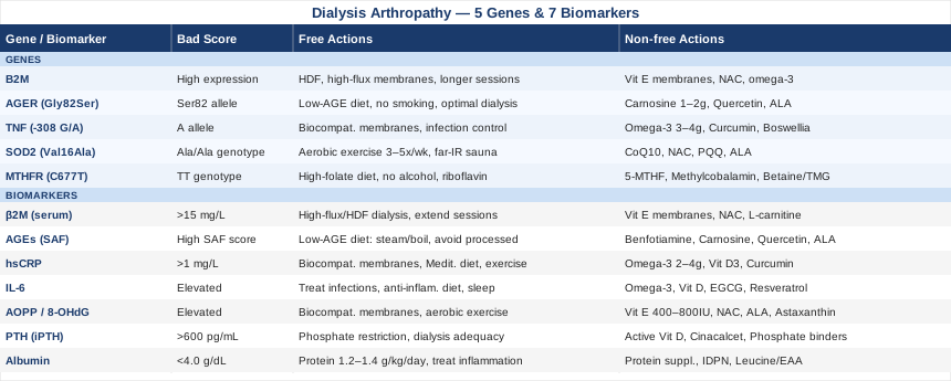
7 Biomarkers That Reveal What Is Happening in Dialysis Arthropathy
Biomarkers do not just confirm what you already suspect. When chosen carefully, they reveal which specific biological pathway is most active — and point toward targeted interventions rather than generic management. For dialysis arthropathy, the following seven markers cover the most clinically meaningful terrain: amyloid load, glycation burden, systemic inflammation, oxidative damage, bone metabolism, and nutritional status.
1. Serum Beta-2 Microglobulin (β2M)
Why it matters and what it reveals: Beta-2 microglobulin is the central pathological molecule in dialysis-related amyloidosis. Normally maintained below 2 mg/L by healthy kidneys, it climbs dramatically in dialysis patients — frequently reaching 15 to 50 mg/L or higher with conventional hemodialysis. At sustained elevated concentrations, β2M misfolds into amyloid fibrils that deposit in joints, carpal tunnels, and vertebral discs, driving pain, stiffness, and structural joint destruction. Serum β2M level is not a perfect proxy for the total tissue amyloid burden already deposited, but it remains the most directly relevant and accessible biomarker for tracking ongoing exposure and dialysis clearance efficiency.
How to measure it
A standard serum β2M assay is ordered through most nephrology and hospital labs. Cost in the US typically ranges from $20 to $80, and it is often included in routine dialysis monitoring panels in Europe. For maximum information, measure it both before and after a dialysis session: the pre-session value reflects accumulation between sessions, while the post-session value reveals clearance efficiency. Frequency: at minimum every three months; monthly when adjusting dialysis modality.
If β2M is elevated: plan without supplements
The most impactful non-pharmacological lever is dialysis modality. High-flux dialysis membranes remove substantially more β2M per session than low-flux membranes. Online hemodiafiltration (HDF) with high substitution volumes — above 23 liters per session — has been shown in the CONTRAST and Turkish OL-HDF trials to reduce pre-dialysis β2M by 30–50% compared to conventional hemodialysis. Increasing dialysis frequency (short daily dialysis or nocturnal dialysis) further reduces β2M exposure time between sessions. Extending session duration beyond four hours per session improves clearance. If your center does not yet offer high-volume HDF, requesting a transition to high-flux membranes is the most accessible first step.
If β2M is elevated: plan with supplements or equipment
Vitamin E-coated dialysis membranes (such as Excebrane) have been studied for their capacity to reduce oxidative modification of β2M during the dialysis process — a modification that makes β2M dramatically more amyloidogenic. These specialized membranes are available in some centers and represent a meaningful equipment-level upgrade. N-acetylcysteine (NAC) at 600 mg twice daily may reduce oxidative β2M modification by replenishing glutathione; evidence in DRA specifically remains early but the safety profile in dialysis patients is established. L-carnitine, administered intravenously at dialysis session end (typically 10–20 mg/kg, three times per week), is used in some centers to address carnitine depletion and improve dialysis tolerance, with secondary oxidative benefits. Reassess β2M every three months when adjusting dialysis modality or beginning new supplements. Side effects: NAC is generally well tolerated; rare nausea at higher doses.
2. Advanced Glycation End Products (AGEs)
Why it matters and what it reveals: AGEs are proteins or lipids that become abnormally modified through non-enzymatic glycation under conditions of metabolic and oxidative stress — both of which are chronically extreme in dialysis patients. Their specific relevance to DRA is critical: AGE-modified β2M is far more amyloidogenic than unmodified β2M. It aggregates faster, forms more insoluble fibrils, and triggers a more intense inflammatory response through its receptor, RAGE. Measuring AGE burden therefore captures a dimension of risk that serum β2M alone misses — two patients with the same β2M level may have very different amyloid progression rates depending on their AGE burden.
How to measure it
Two practical options exist. Skin autofluorescence (SAF), measured with a device such as the AGE Reader (DiagnOptics), offers non-invasive and reproducible tissue AGE quantification that correlates well with systemic AGE accumulation and cardiovascular risk. Cost ranges from free at academic centers to $50–150 per measurement at specialized clinics. Serum methylglyoxal or fluorescent AGE assays are available from specialty labs ($50–150) but are less clinically standardized. Recheck every 6 months when dietary or supplement interventions are in place.
If AGEs are elevated: plan without supplements
Diet is the most direct lever. The AGE-reduced diet prioritizes avoiding dry heat cooking (grilling, frying, broiling, roasting) in favor of moist, lower-temperature methods: steaming, boiling, poaching, slow-cooking in liquid. High-AGE foods include well-done meats, fried foods, and processed snacks. Reducing dietary phosphate and avoiding processed foods simultaneously reduces the substrate for endogenous AGE formation. Staying within dialysis-appropriate fluid limits supports AGE clearance pathways. Structured low-intensity exercise improves metabolic AGE production through better insulin sensitivity.
If AGEs are elevated: plan with supplements or equipment
Benfotiamine (fat-soluble vitamin B1) at 150–300 mg/day diverts toxic glycolytic metabolites through the transketolase pathway, reducing AGE formation at a biochemical level. Carnosine (beta-alanyl-L-histidine) at 1–2 g/day acts as a natural anti-glycation dipeptide that traps reactive carbonyl intermediates before they modify proteins. Quercetin (500–1000 mg/day with food) reduces downstream RAGE activation. Alpha-lipoic acid (300–600 mg/day) reduces both AGE formation and oxidative stress simultaneously. Cycling: cycle benfotiamine for 8–12 weeks with a 4-week washout; carnosine can be taken continuously. Side effects: carnosine may occasionally cause histamine-related reactions in sensitive individuals; benfotiamine is generally well tolerated. Always review with the nephrologist before adding supplements, as dialysis alters medication metabolism.
3. High-Sensitivity C-Reactive Protein (hsCRP)
Why it matters and what it reveals: hsCRP is one of the most robust predictors of dialysis-related complications and a core measure of systemic inflammation. Elevated hsCRP in dialysis patients correlates with cardiovascular risk, malnutrition, and — critically for DRA — accelerated amyloid deposition. Chronic inflammation simultaneously promotes β2M production (β2M is itself an acute-phase reactant) and impairs the mechanisms that limit amyloid accumulation. In Outlive (2023), Peter Attia names hsCRP as one of the foundational inflammatory markers to monitor in any individual aiming to understand and modify their long-term disease trajectory — an argument that applies directly here.
How to measure it
hsCRP is one of the most affordable and widely available biomarkers in medicine: $10–40 in the US, frequently included in dialysis monitoring panels in Europe. Measure on a clinically stable day — acute illness, injury, or recent surgery will transiently spike the value and confound interpretation. Confirm the high-sensitivity version is ordered, as standard CRP lacks precision at the lower ranges where early chronic inflammation lives. Target for meaningful disease risk reduction: below 1 mg/L. Recheck every 3 months when interventions are active.
If hsCRP is elevated: plan without supplements
Begin with dialysis optimization: biocompatible membranes (polysulfone, PMMA) generate significantly less contact-related inflammatory activation than older cellulosic membranes. Endotoxin contamination of dialysate — a common and underappreciated driver of persistent elevated CRP — is addressed by ensuring your center uses ultrapure dialysate. Mediterranean-style dietary pattern — high in vegetables, olive oil, oily fish, nuts, and legumes — has the strongest anti-inflammatory dietary evidence base. Sleep quality independently raises CRP when fragmented: addressing restless legs, pain, and nocturnal symptoms supports a lower inflammatory baseline. Even 20–30 minutes of daily walking reduces hsCRP over weeks to months.
If hsCRP is elevated: plan with supplements or equipment
Omega-3 fatty acids (combined EPA+DHA at 2–4 g/day) have strong and replicated evidence for reducing hsCRP in dialysis populations — multiple randomized controlled trials in this specific population have confirmed clinically meaningful reductions. Vitamin D3 (supplementing to a serum 25-OH vitamin D of 40–60 ng/mL, typically 2,000–5,000 IU/day based on baseline level) reduces hsCRP and IL-6 through immunomodulatory pathways. Curcumin phytosome (formulations with enhanced bioavailability such as Meriva or Longvida, 500–1,000 mg/day) has been shown in multiple small trials to reduce hsCRP. Cycle curcumin for 8–12 weeks with reassessment; continue omega-3s long-term. Avoid high-dose curcumin alongside anticoagulant medications without medical oversight; monitor for any bleeding tendency.
4. Interleukin-6 (IL-6)
Why it matters and what it reveals: IL-6 is a central pro-inflammatory cytokine that adds important precision beyond hsCRP alone. Its relevance to DRA is specific: IL-6 promotes synovial inflammation in affected joints, directly upregulates β2M production (amplifying the primary driver of amyloid accumulation), and activates osteoclasts — contributing to the subchondral bone destruction seen in advanced disease. Elevated IL-6 also suppresses albumin synthesis, creating a mechanistic bridge between the inflammatory and nutritional biomarkers discussed here. Tracking IL-6 separately from hsCRP captures a dimension of immune activation that the two markers together reveal more completely than either alone.
How to measure it
Serum IL-6 is available through most hospital labs and reference laboratories at a cost of $30–100. It has a shorter half-life than CRP and is more biologically variable, so it should ideally be measured on a clinically stable day, away from active infections or recent procedures. It is not routinely included in standard dialysis monitoring at most centers but is increasingly accessible through specialty panels. Recheck every 3 months when trialing interventions.
If IL-6 is elevated: plan without supplements
Treating the upstream cause is critical. Dental infections, vascular access infections, and subclinical bacteremia are the most common drivers of persistent IL-6 elevation in dialysis patients — these must be clinically ruled out before assuming the elevation is a chronic baseline. After infection sources are excluded, the same lifestyle levers that reduce hsCRP apply: anti-inflammatory diet, structured exercise, and sleep quality improvement. Requesting a switch to higher biocompatibility dialysis membranes reduces the membrane-contact inflammatory activation that drives a portion of IL-6 elevation in this population.
If IL-6 is elevated: plan with supplements or equipment
Omega-3 fatty acids (EPA+DHA, 3–4 g/day) reduce IL-6 in addition to hsCRP through overlapping pathways, with evidence specific to dialysis patients. Vitamin D supplementation targeting 40–60 ng/mL reduces IL-6 through multiple immunomodulatory mechanisms. Green tea extract (EGCG) at 400–800 mg/day has been studied in dialysis patients for anti-inflammatory effects including IL-6 reduction, though trials remain small. Resveratrol (250–500 mg/day in a bioavailable formulation) may modulate IL-6 through SIRT1 pathways — early evidence is promising but not yet definitive. Cycling: review IL-6 every 3 months when trialing any new intervention; adjust based on response. Monitor for potential interactions with existing medications when using green tea extract at higher doses.
5. Advanced Oxidation Protein Products (AOPP)
Why it matters and what it reveals: AOPP quantifies the oxidative modification of plasma proteins — a direct readout of oxidative stress burden. Dialysis patients have chronically elevated AOPP driven by the procedure itself generating reactive oxygen species, severely impaired antioxidant defenses in uremia, and the pro-oxidant uremic environment. AOPP is particularly specific to DRA because oxidative modification of β2M converts it into a substantially more amyloidogenic form: modified β2M aggregates faster and more aggressively than its unmodified counterpart. Reducing AOPP is therefore not just about general health — it is a mechanistically direct lever for slowing amyloid deposition.
How to measure it
AOPP is measured from serum or plasma using a spectrophotometric assay, available primarily through research and specialized clinical labs at $50–150 when offered. It is not yet universally part of routine clinical panels but is increasingly included in nephrology research protocols. An accessible alternative is urinary 8-OHdG (8-hydroxy-2'-deoxyguanosine) — an oxidative DNA damage marker available through specialty labs such as Genova Diagnostics or Doctor's Data ($80–200) that captures a related dimension of oxidative stress.
If AOPP is elevated: plan without supplements
Biocompatible dialysis membranes generate substantially less reactive oxygen species per session than older materials. The physical dialysis process creates oxidative stress through blood-air interface exposure and dialysate contaminant interactions — requesting online-prepared, ultrapure dialysate reduces this meaningfully. Moderate aerobic exercise (3–5 sessions per week, 20–30 minutes each at 50–70% maximum heart rate, ideally on non-dialysis days) upregulates endogenous antioxidant enzymes including superoxide dismutase and catalase through Nrf2 pathway activation. Over weeks to months, this measurably reduces baseline AOPP. Minimizing dietary AGEs — as described in the previous section — simultaneously reduces oxidative protein modification.
If AOPP is elevated: plan with supplements or equipment
Vitamin E (natural alpha-tocopherol, 400–800 IU/day) has been studied in controlled trials in dialysis patients, showing consistent reductions in oxidative markers including AOPP. NAC at 600 mg twice daily replenishes intracellular glutathione and has an established safety profile specifically in dialysis populations. Alpha-lipoic acid (300–600 mg/day) is uniquely effective due to its solubility in both water and fat, allowing it to work across cellular compartments. Astaxanthin (4–12 mg/day) is a potent carotenoid antioxidant with preliminary evidence in dialysis-related oxidative stress. Cycling: reassess AOPP every 3–4 months; cycle high-dose antioxidants every 3 months with 4-week washouts. Do not combine high-dose vitamin E with anticoagulant medications without physician oversight. Verify renal clearance considerations for all supplements with your nephrologist.
6. Parathyroid Hormone (PTH)
Why it matters and what it reveals: Secondary hyperparathyroidism is nearly universal in advanced CKD and dialysis patients, and it contributes meaningfully to the bone and joint complications that characterize dialysis arthropathy. Chronically elevated PTH drives osteoclast-mediated bone resorption, creating the subchondral bone cysts that are a radiographic hallmark of advanced DRA and that mechanically weaken joint surfaces. PTH also promotes periarticular soft tissue calcification, further compromising joint function. Tracking PTH provides essential context for the bone component of DRA — context that serum β2M alone cannot supply.
How to measure it
Intact PTH (iPTH) is a standard dialysis lab test, typically costing $20–60 and already part of routine monitoring at most dialysis centers. Targets in dialysis patients differ from the general population: KDIGO guidelines suggest maintaining iPTH between two and nine times the upper limit of normal, roughly 150–600 pg/mL in most labs, though individual nephrologists may use more specific targets based on bone biopsy or clinical presentation. Measure monthly during active PTH management, quarterly when stable.
If PTH is elevated: plan without supplements
Dietary phosphate restriction is the most direct lever: reducing processed foods, cola beverages (which contain highly absorbable phosphate additives), and excess dairy reduces the phosphate burden that continuously stimulates PTH secretion. Ensuring adequate dialysis time and frequency improves phosphate removal per session. Optimizing sun exposure — or using appropriate light therapy — supports endogenous vitamin D status, which is already severely impaired in dialysis patients; however, this is a supportive measure rather than sufficient alone for managing elevated PTH in this context.
If PTH is elevated: plan with supplements or equipment
Active vitamin D analogs — calcitriol, alfacalcidol, or paricalcitol — are the established clinical approach to suppressing PTH in dialysis and should be managed by the nephrologist based on concurrent calcium and phosphate levels. Calcimimetics (cinacalcet) sensitize the parathyroid gland to calcium, reducing PTH without raising serum calcium or phosphate — an important advantage when both are already dysregulated. Phosphate binders (sevelamer, lanthanum carbonate, or calcium-based binders depending on individual calcium levels) reduce dietary phosphate absorption and indirectly reduce PTH stimulus. These are prescription interventions; the practical role of tracking PTH as a biomarker is to ensure it is monitored consistently so adjustments can be made proactively rather than reactively.
7. Serum Albumin
Why it matters and what it reveals: Serum albumin simultaneously functions as a nutritional marker, an inflammatory readout, and a biochemical buffer. In dialysis patients, low albumin reflects both inadequate protein-caloric intake and suppression of hepatic albumin synthesis by chronic inflammation — particularly IL-6. The dual origin of hypoalbuminemia is diagnostically important: if CRP is simultaneously elevated, inflammation is a primary driver and dietary intervention alone will not restore albumin without also addressing the inflammatory burden. Additionally, albumin functions as a natural antioxidant and AGE carrier in circulating blood, meaning low albumin independently increases oxidative and glycative stress — directly worsening the biological environment for DRA.
How to measure it
Serum albumin is among the most basic and affordable labs available: $10–30 and typically included in standard dialysis monitoring. Pre-dialysis values are most informative, as post-dialysis values are affected by hemoconcentration. Target for dialysis patients: ≥4.0 g/dL. Values below 3.5 g/dL signal significant risk. Measure monthly during any nutritional or inflammatory intervention; quarterly when stable.
If albumin is low: plan without supplements
Protein intake optimization is the primary dietary action: dialysis patients generally require 1.2–1.4 g of protein per kilogram of body weight per day — substantially more than the general population recommendation — because dialysis removes amino acids directly and induces a catabolic state. Distributing high-quality protein sources (eggs, fish, poultry, plant proteins within potassium and phosphate constraints) across each meal maximizes muscle protein synthesis throughout the day. If CRP is simultaneously elevated, treating inflammation is equally urgent: biocompatible membranes, infection management, and anti-inflammatory dietary patterns all support albumin recovery by reducing its suppression.
If albumin is low: plan with supplements or equipment
Kidney-appropriate oral protein supplements (such as Renilon or dialysis-specific meal replacement formulas designed for controlled potassium and phosphate) can bridge nutritional gaps that diet alone cannot close. Intradialytic parenteral nutrition (IDPN) — nutrients infused intravenously during each dialysis session — is reserved for patients with severe malnutrition who cannot meet needs orally; small trials have shown meaningful improvements in albumin, body composition, and quality of life. Leucine or essential amino acid supplementation (5–10 g with meals, two to three times daily) directly stimulates muscle protein synthesis and may support albumin recovery over time. Frequency: reassess albumin monthly during active interventions; discuss IDPN eligibility formally with the nephrology dietitian and nephrologist. Side effects of oral supplements: digestive tolerance varies; introduce gradually.
Building on these biomarker insights, the next layer of precision comes from understanding the genetic factors that predispose certain individuals to faster amyloid accumulation or more amplified inflammation — regardless of dialysis practice.
What Recent Genetics Research Suggests About Dialysis Arthropathy
Not all dialysis patients develop severe arthropathy. After ten or fifteen years on dialysis, some patients have significant amyloid joint disease while others maintain relatively preserved joints despite similar β2M exposure histories. Genetic variation explains part of this divergence. Understanding which gene variants may be working against you — and which interventions partially compensate — has become increasingly practical with accessible genetic testing platforms. The five genes below have meaningful evidence linking them to β2M accumulation, amyloid formation, inflammatory amplification, or antioxidant defense.
B2M — The Beta-2 Microglobulin Gene Itself
What it affects: The B2M gene on chromosome 15 encodes the β2-microglobulin protein directly. While rare coding mutations are not the primary clinical concern, variability in B2M gene expression — influenced by epigenetic regulation, promoter polymorphisms, and cytokine-driven transcriptional upregulation — affects how much β2M is produced per unit of inflammatory stimulus. Since β2M is itself an acute-phase reactant, patients with more reactive B2M gene expression may produce substantially more protein per dialysis-related inflammatory event, compounding the clearance deficit inherent to kidney failure. Research on epigenetic modifications to B2M expression in CKD is still emerging, but the gene-level logic is directly applicable to clinical management.
If the gene expression is unfavorable: plan without supplements
Since the gene influences production rate and the amyloid propensity of the resulting protein, the most effective countermeasure is maximizing clearance through high-volume online hemodiafiltration (HDF). Advocating with your nephrologist for HDF over conventional hemodialysis is the most structurally impactful change available. Extending session length, increasing session frequency, and avoiding dialysis conditions that promote oxidative β2M modification (poor dialysate quality, low-biocompatibility membranes) all reduce net amyloid deposition.
If the gene is unfavorable: plan with supplements or equipment
Vitamin E-coated membranes reduce oxidative modification of β2M during the dialysis process — a meaningful equipment upgrade when β2M production is high. NAC (600 mg twice daily) reduces oxidative conversion of β2M to its more aggregation-prone modified forms. No supplement directly suppresses B2M gene transcription, but maintaining a consistently low-inflammation environment — through omega-3 fatty acids (3–4 g/day) and anti-inflammatory dietary patterns — reduces the cytokine-driven upregulation of β2M synthesis that compounds genetic susceptibility. Reassess serum β2M every 3 months when implementing changes.
AGER — The RAGE Gene (Gly82Ser Polymorphism)
What it affects: The AGER gene encodes the receptor for advanced glycation end products (RAGE). The Gly82Ser polymorphism (rs2070600) is the most clinically studied variant: the Ser82 allele confers higher binding affinity for AGE ligands, meaning carriers mount a stronger inflammatory cascade when AGE-modified β2M binds to RAGE. In practical terms, this variant amplifies the inflammation produced per unit of AGE burden — so a Ser82 carrier sustains more synovial damage for the same level of glycation compared to a Gly82 homozygote. Gary Brecka and practitioners in functional genomics have specifically highlighted the RAGE axis as a critical amplifier of chronic inflammatory disease in genetically susceptible individuals.
If the gene is unfavorable (Ser82 allele): plan without supplements
Since the variant amplifies the response to AGE ligands rather than increasing RAGE expression per se, the most direct strategy is reducing the ligand load. Strict AGE-reduced dietary practices — avoiding dry-heat cooking, processed foods, and fried items — reduce the volume of AGE molecules available to activate RAGE. Optimal dialysis clearance of β2M reduces the protein available for AGE modification. Eliminating smoking (which generates massive and rapid AGE formation) is non-negotiable for Ser82 carriers.
If the gene is unfavorable: plan with supplements or equipment
Carnosine (1–2 g/day) operates on two relevant levels simultaneously: it competes with AGE ligands at the RAGE receptor and quenches reactive carbonyl intermediates before they form AGEs in the first place. Quercetin (500–1000 mg/day) directly inhibits the downstream NF-κB and MAPK signaling pathways activated by RAGE. Alpha-lipoic acid (600 mg/day) reduces AGE formation upstream of receptor binding. Cycling: carnosine for 12 weeks, then 4-week washout; quercetin can be continued longer. Side effects: carnosine breaks down to histidine and may occasionally cause mild histamine responses in sensitive individuals; alpha-lipoic acid may interact with thyroid medications in some cases — review with physician.
TNF — Tumor Necrosis Factor Alpha (-308 G/A Polymorphism)
What it affects: The TNF gene promoter polymorphism rs1800629 (-308 G/A) — specifically the A allele — is associated with significantly higher TNF-alpha production in response to inflammatory stimuli. In dialysis patients, where the procedure itself generates repeated inflammatory provocation at each session, carrying the A allele may result in chronically elevated TNF-alpha that directly promotes synovial inflammation, cartilage degradation, and osteoclast-mediated bone erosion. Ali Torkamani's population-scale genomic work has highlighted TNF pathway variants as among the most consequential for inflammatory disease risk stratification across populations.
If the gene is unfavorable (A allele): plan without supplements
Minimizing inflammatory provocation at each dialysis session is the primary strategy. Biocompatible polysulfone or PMMA membranes generate less TNF-alpha per session than older cellulosic membranes. Vigilant management of infections, dental disease, and vascular access infections prevents the repeated strong TNF-stimulating events that compound the genetically elevated baseline. The Mediterranean dietary pattern has specific evidence for reducing TNF-alpha levels — the mechanism involves polyphenols modulating NF-κB activity. Moderate aerobic exercise reduces TNF-alpha through IL-10-mediated counter-regulatory pathways.
If the gene is unfavorable: plan with supplements or equipment
Omega-3 fatty acids (EPA+DHA, 3–4 g/day) reduce TNF-alpha production at the gene expression level — this is one of the most replicated findings in omega-3 research. Curcumin phytosome (500–1000 mg/day) blocks NF-κB transcriptional activity, the pathway through which the -308 A allele amplifies much of its effect. Boswellia serrata extract (5-LOXIN or Shallaki, 100–250 mg AKBA-standardized) inhibits 5-lipoxygenase, reducing leukotriene-mediated inflammation downstream of TNF. Cycling: curcumin for 8–12 weeks with 4-week washout; omega-3s can be continued long-term. Important: avoid high-dose curcumin simultaneously with blood-thinning medications without physician monitoring; reassess inflammatory markers every 3 months during supplementation.
SOD2 — Superoxide Dismutase 2 (Val16Ala Polymorphism)
What it affects: SOD2 encodes manganese superoxide dismutase, the primary mitochondrial antioxidant enzyme. The Val16Ala variant (rs4880) affects the mitochondrial targeting sequence: the Ala allele reduces efficient transport of MnSOD into mitochondria, leading to lower intramitochondrial antioxidant activity. In dialysis patients — where oxidative stress is already dramatically elevated from multiple sources — a genetically reduced MnSOD capacity creates less protection against reactive oxygen species, faster oxidative modification of β2M into amyloidogenic forms, and greater cumulative cellular damage. Gary Brecka has specifically noted that SOD2 Ala/Ala carriers often display higher inflammatory and oxidative markers unless genetically compensated.
If the gene is unfavorable (Ala/Ala genotype): plan without supplements
Regular moderate aerobic exercise is the most validated natural inducer of SOD2 expression: exercise upregulates both SOD2 and catalase through Nrf2 and PGC-1α transcriptional pathways. Three to five sessions weekly of moderate aerobic activity — walking, stationary cycling, aqua exercise — are realistic for most dialysis patients and have been shown to measurably reduce oxidative stress biomarkers. Heat exposure (far-infrared sauna) upregulates heat shock proteins that work synergistically with SOD2 in managing oxidative burden, though sauna use in dialysis patients requires explicit medical clearance due to fluid and electrolyte considerations.
If the gene is unfavorable: plan with supplements or equipment
CoQ10 (100–300 mg/day, ubiquinol form for optimal absorption) supports mitochondrial electron transport chain efficiency and reduces the superoxide burden that MnSOD would otherwise neutralize. NAC (600 mg twice daily) replenishes glutathione — the downstream antioxidant dependent on SOD2 activity — and is well tolerated in dialysis populations. PQQ (pyrroloquinoline quinone, 10–20 mg/day) stimulates mitochondrial biogenesis and upregulates antioxidant enzyme expression including SOD2 itself. Alpha-lipoic acid (300–600 mg/day) both regenerates vitamins C and E and activates Nrf2-mediated antioxidant gene induction. Cycling: reassess AOPP every 3–4 months; cycle PQQ every 3 months with reassessment; continue CoQ10 and NAC with regular lab monitoring. Discuss all supplements with your nephrologist given altered drug clearance in dialysis.
MTHFR — Methylenetetrahydrofolate Reductase (C677T Polymorphism)
What it affects: The MTHFR C677T polymorphism (rs1801133) — particularly the TT homozygous genotype — reduces enzyme activity by 30–70%, impairing conversion of folate to its active form (5-methylTHF) and thereby disrupting the methylation cycle, including the conversion of homocysteine to methionine. In dialysis patients, homocysteine is already substantially elevated due to impaired renal clearance; the MTHFR TT genotype compounds this further. Elevated homocysteine drives endothelial damage, systemic inflammation, and — relevant to DRA — appears to promote oxidative protein modification including of β2M, amplifying its amyloidogenic behavior. Additionally, the impaired methylation from MTHFR dysfunction affects gene expression regulation broadly, potentially influencing inflammatory gene activity.
If the gene is unfavorable (TT genotype): plan without supplements
A high natural folate dietary pattern provides 5-methylTHF in its natural form, which does not require MTHFR for utilization: spinach, arugula, asparagus, avocado, lentils, and broccoli (within dialysis-appropriate potassium constraints) are rich sources. Eliminating alcohol is essential — alcohol directly depletes folate stores. Adequate dietary riboflavin (vitamin B2 from eggs, dairy, lean meats) matters because riboflavin is a cofactor for MTHFR itself and dietary adequacy supports whatever residual enzyme activity remains. Broad B-vitamin sufficiency across the diet supports the methylation network holistically.
If the gene is unfavorable: plan with supplements or equipment
5-MTHF (methylfolate, 400–800 mcg/day) — the active, pre-converted form of folate — bypasses the blocked MTHFR enzyme entirely and is strongly preferred over standard folic acid for TT carriers. Methylcobalamin (B12, 500–1000 mcg/day) works in synergy with methylfolate in the methylation cycle. Betaine/TMG (trimethylglycine, 500–1500 mg/day) provides an alternative methyl donor route that is entirely independent of MTHFR. Riboflavin (B2, 25–50 mg/day) may partially restore residual MTHFR enzyme activity in TT carriers — a finding from human intervention studies that is frequently overlooked in clinical practice. Frequency: daily; reassess homocysteine every 3–6 months to titrate dosing. Monitor B-vitamin status with the nephrologist, as dialysis alters B-vitamin handling and certain B-vitamin excesses carry their own risks in renal populations.
The genetic and biomarker evidence creates a detailed biological picture. The next section draws on longevity medicine — a field increasingly relevant to dialysis patients — for a broader framework that integrates these findings.
Ten Insights From Longevity Medicine That Change How You Think About Dialysis Arthropathy
Outlive: The Science and Art of Longevity by Peter Attia (2023) is written primarily for a general audience seeking to extend healthy lifespan, but its framework for understanding inflammation, protein aggregation, metabolic health, and biomarker-driven medicine applies with unusual directness to dialysis arthropathy. The book draws on hundreds of peer-reviewed studies to argue that most chronic disease shares a manageable set of root drivers. For dialysis patients navigating accelerated biological aging and amyloid accumulation, the following ten insights from this framework stand out as most impactful.
1. Inflammaging Is Cumulative and Measurable
Attia distinguishes between short-term acute inflammation and the chronic, low-grade inflammatory state — "inflammaging" — that drives progressive tissue damage over years. In dialysis patients, this state is dramatically elevated at baseline from multiple converging sources. The key insight: even modest reductions in chronic inflammatory markers — not eliminating inflammation entirely — meaningfully reduce cumulative joint and amyloid damage over time. Tracking hsCRP and IL-6 regularly, not only when something seems acutely wrong, enables early intervention before the inflammatory load becomes structurally irreversible.
2. Protein Aggregation Is a Cross-Disease Biological Thread
Attia writes extensively about amyloid diseases — primarily Alzheimer's and certain cardiomyopathies — noting that misfolded protein aggregation is a convergent mechanism across multiple conditions. The same environmental factors that accelerate neuronal amyloid deposition — oxidative stress, AGEs, elevated inflammatory cytokines, poor metabolic health — accelerate β2M amyloid in dialysis joints. Strategies developed in the neurodegeneration prevention space (lifestyle, antioxidant management, glycemic control) therefore have direct mechanistic relevance for DRA patients.
3. Measuring the Right Things Transforms Behavior
One of Attia's central arguments is that tracking specific biomarkers regularly enables better clinical and personal decisions — not because data is inherently powerful, but because it converts vague risk into concrete, actionable numbers. For dialysis patients, this argues directly for advocating with your care team for β2M, IL-6, AGE markers, and AOPP measurements even when they are not standard at a given center. You cannot optimize what you cannot measure.
4. Exercise Is the Highest-Return Medical Intervention Available
Attia places consistent, structured exercise above all supplements and most medications in terms of healthspan impact per unit of effort. For dialysis patients, intradialytic cycling — pedaling a stationary bike during dialysis sessions — has been studied in randomized controlled trials and shown to reduce inflammatory markers, improve physical function, reduce fatigue, and improve cardiovascular markers. The anti-inflammatory mechanisms are partly mediated by IL-10 upregulation, myokine secretion, and Nrf2 antioxidant pathway activation.
5. Metabolic Health Directly Drives AGE Formation
Poor metabolic health — insulin resistance, elevated fasting glucose, dyslipidemia — dramatically accelerates AGE production and RAGE activation. Attia argues that glycemic control (tracked through fasting glucose, HbA1c if applicable, and fasting insulin) is foundational to inflammation management. In dialysis patients, even those without clinical diabetes often have significant underlying insulin resistance that drives AGE burden. Improving metabolic health through diet, exercise, and sleep is a lever that reduces DRA progression through the AGE pathway.
6. Sleep Quality Is an Undervalued Inflammatory Driver
Attia devotes significant attention to sleep as a fundamental determinant of inflammation and cellular repair. Sleep deprivation acutely raises IL-6, TNF-alpha, and CRP — precisely the inflammatory profile that worsens DRA. Dialysis patients frequently experience disrupted sleep due to restless legs syndrome, chronic pain, nocturnal symptoms, and the physical and psychological burden of treatment. Addressing sleep disruption through treatment of restless legs where present, sleep hygiene, and evidence-appropriate interventions (melatonin, in some cases) has measurable downstream effects on inflammatory biomarkers.
7. Protein Adequacy Is Systematically Underestimated
Attia argues that most people — particularly older adults — chronically under-consume high-quality protein, creating a deficit that progressively erodes muscle mass, immune function, and tissue repair capacity. For dialysis patients, this problem is structurally amplified: dialysis removes amino acids per session, chronic inflammation suppresses anabolism, and the anorexia of uremia reduces appetite. Consistently meeting protein targets (1.2–1.4 g/kg/day) is not optional — it is foundational to all other interventions.
8. Zone 2 Cardio Is the Most Accessible Antioxidant Available
Sustained aerobic exercise at conversational pace — approximately 65–75% of maximum heart rate — specifically trains mitochondrial oxidative efficiency and upregulates endogenous antioxidant enzymes through PGC-1α activation. Attia recommends 150–200 minutes per week for general healthspan. For dialysis patients, even 60–90 minutes weekly divided across three to four sessions has shown measurable benefits in published trials. Intradialytic stationary cycling achieves Zone 2 training without requiring additional time beyond what dialysis already occupies.
9. Standard Monitoring Panels Arrive Too Late
Attia's consistent argument across multiple contexts is that waiting for standard clinical abnormalities to appear means waiting until disease is already established. He advocates for earlier, more frequent, and broader testing — including markers like hsCRP, oxidative stress assays, and AGE measurements that are not part of many standard clinical panels. For DRA patients, this directly argues for requesting β2M trending, AGE assessment, and IL-6 monitoring even if none are standard at your center.
10. Engaged Patients Have Measurably Better Outcomes
Attia's overarching conclusion — relevant to every complex chronic condition — is that informed, engaged patients who understand their own biology consistently do better over time. Understanding your biomarkers, knowing which genetic factors may be working against you, asking specific questions of your care team, and making consistent habit changes compounds in effect over years and decades. For dialysis arthropathy, treating its management as an active, ongoing project rather than a passive consequence is itself among the highest-value interventions available.
Complementary Approaches With Clinical Evidence for Dialysis Joint Health
The following modalities are selected for having at least some human clinical evidence applicable to dialysis-related joint pain, systemic inflammation, or functional disability. They are not substitutes for conventional medical management — but used alongside standard dialysis care, several have shown meaningful improvements in pain, function, and inflammatory markers.
Tai Chi
Tai chi is a slow, deliberate movement practice combining balance, coordination, mindful breathing, and postural control. For dialysis patients with joint pain, reduced mobility, and elevated fall risk — all consequences of advanced DRA — tai chi is particularly well-suited because it is low-impact, joint-friendly, and directly addresses the balance deficits that make conventional exercise programs difficult to sustain.
A randomized controlled trial in hemodialysis patients (published in Clinical Rehabilitation) found that a 12-week tai chi program — three 45-minute sessions per week — significantly improved physical function scores, self-reported pain, and quality of life compared to a sedentary control group. Biologically, tai chi reduces cortisol and modestly lowers IL-6 over the course of a program, providing an anti-inflammatory mechanism relevant to DRA.
In practice, begin with a beginner-level class or a video-adapted program designed for older or medically limited adults. Twenty to thirty minutes, three times weekly on non-dialysis days, is a practical and achievable starting target. Inform the instructor about your dialysis status and any specific joint limitations from existing DRA so modifications can be made for affected joints. Discuss with your nephrologist before beginning if you have significant bone disease or fall history.
Mindfulness Meditation / MBSR
Mindfulness-Based Stress Reduction (MBSR) is an eight-week structured program developed by Jon Kabat-Zinn, involving weekly group sessions and daily home practice of meditation, body scan, and mindful movement. Among mind-body interventions for chronic pain, MBSR has one of the most robust human evidence bases — with applicability to the chronic joint pain that characterizes dialysis arthropathy.
A randomized trial in dialysis patients showed that MBSR significantly reduced pain intensity, fatigue, and depression scores at eight weeks compared to usual care. The mechanism partly involves HPA axis regulation — MBSR reduces cortisol reactivity and, over sustained practice, modestly lowers IL-6 — providing both a subjective pain relief effect and a measurable anti-inflammatory one.
Standard MBSR programs are available in-person at many hospital systems and online through validated platforms. Committing to the full eight weeks — including the recommended 20–30 minutes of daily home practice — yields the strongest results. Post-program, maintaining a daily 15–20 minute practice sustains benefits. There are no significant physiological side effects; occasional mild emotional discomfort during early practice is normal and typically resolves within the first two weeks.
Low-Level Laser Therapy / Photobiomodulation
Low-level laser therapy (LLLT) uses specific wavelengths of light (600–1,000 nm) at low intensities to stimulate mitochondrial energy production, reduce synovial inflammation, and accelerate tissue repair. It has accumulated substantial clinical evidence for musculoskeletal pain over two decades of research.
A systematic review by Brosseau et al., evaluating LLLT for knee osteoarthritis — the condition with the most evidence overlap with DRA joint symptoms — found significant reductions in pain and improvements in joint function at wavelengths of 780–860 nm and 904 nm. In the context of DRA, photobiomodulation's mechanisms are particularly mechanistically apt: it reduces pro-inflammatory cytokine production, promotes mitochondrial activity (directly complementing the SOD2 vulnerability discussed in the genetics section), and reduces synovial swelling.
For practical access, search for a physiotherapy clinic offering LLLT or photobiomodulation. Sessions are typically 10–15 minutes per affected joint area, two to three times weekly for four to eight weeks. Clinical-grade devices under trained supervision are preferable to consumer home devices, which vary considerably in intensity and quality. Side effects are minimal when protocols are followed correctly; avoid direct eye exposure and treatment over actively progressing cancers.
Massage Therapy
Therapeutic massage — particularly connective tissue massage and Swedish techniques applied to affected regions — reduces musculoskeletal pain, improves local circulation, and reduces both anxiety and pain perception through central nervous system mechanisms. For dialysis arthropathy, carpal tunnel syndrome, shoulder periarthritis, and diffuse joint stiffness are the most common targets where massage provides direct symptom benefit.
A systematic review in Pain Medicine found that therapeutic massage consistently reduced chronic musculoskeletal pain intensity by 25–35% compared to control conditions, with effects sustained for up to six months with ongoing sessions. For dialysis patients specifically, upper extremity massage — targeting the joints most commonly affected by DRA (wrists, shoulders, small hand joints) — is most clinically applicable and can be safely provided by a trained therapist.
In practice, sessions of 45–60 minutes once to twice weekly for the initial month, then once every one to two weeks for maintenance, is a reasonable schedule. Always inform the therapist about your dialysis access site — arteriovenous fistulas and grafts must be avoided during massage to prevent damage or thrombosis. Choose a therapist with experience working with medically complex patients; a brief orientation about dialysis-specific precautions is worth doing before the first session. Side effects are generally limited to mild post-session muscle soreness.
Microbiome-Directed Therapies
The gut-kidney axis — the bidirectional relationship between gut microbiome composition and renal and inflammatory status — has emerged as a significant area in nephrology research. Dialysis patients consistently show severely disrupted microbiome composition: reduced diversity, overgrowth of proteolytic bacteria, reduced butyrate-producing species, and elevated production of uremic toxins including indoxyl sulfate and p-cresol sulfate. These gut-derived toxins directly promote systemic inflammation and oxidative stress — both of which accelerate dialysis arthropathy through the mechanisms described throughout this article.
A randomized controlled trial by Rossi et al. published in Nephrology Dialysis Transplantation found that prebiotic supplementation in dialysis patients significantly reduced serum indoxyl sulfate and improved inflammatory markers over twelve weeks. Probiotic trials in dialysis patients have shown mixed results, but multi-strain formulations including Lactobacillus and Bifidobacterium species have in several trials demonstrated reductions in uremic toxin levels and hsCRP.
In practice, a three-pronged approach is reasonable: (1) prebiotic fiber from appropriate foods — chicory, asparagus, garlic, and onion within dialysis dietary restrictions for potassium and phosphate; (2) a high-quality multi-strain probiotic (10–50 billion CFU/day with Lactobacillus and Bifidobacterium species); (3) minimizing unnecessary microbiome-disruptive medications, particularly proton pump inhibitors which are commonly over-prescribed in dialysis patients and are profoundly dysbiotic. Work with the nephrology dietitian to ensure prebiotic food choices are compatible with your specific dietary restrictions.
Conclusion
Dialysis arthropathy responds poorly to passive management. Its biology — β2M accumulation, AGE-driven amyloid formation, genetic amplifiers of inflammation and oxidative damage — is specific enough that targeted, informed action creates a meaningfully different trajectory than generic advice. The seven biomarkers covered here give you a measurable window into which biological pathway is most active in your specific case. The five genetic factors help explain why two patients with identical dialysis histories can have such different outcomes. And the complementary approaches offer a non-pharmacological layer of support that is consistently underutilized in dialysis care despite meaningful human evidence.
The clearest next step is to review which of these biomarkers are already being monitored in your current care and which are not. Bring this list to your next nephrology or dialysis appointment and ask specifically about β2M trending, hsCRP, and IL-6. If genetic testing is accessible, understanding your TNF, SOD2, AGER, and MTHFR variants adds precision that can guide which dietary, lifestyle, and supplementation strategies offer the most leverage for your biology. Better data, consistent habits, and an engaged partnership with your care team are the three things that compound most reliably over time.
Musculoskeletal: Bone Conditions Joint Conditions
Autoimmune: Inflammatory Conditions Connective Tissue Conditions
Urological: Kidney Conditions