This article contains AI generated content.
· UpdatedDiffuse-Type Giant Cell Tumor of Tendon Sheath: 5 Genes and 6 Biomarkers to Track
Introduction
If you or someone close to you has been diagnosed with diffuse-type giant cell tumor of the tendon sheath — also known as tenosynovial giant cell tumor, diffuse type, or historically as pigmented villonodular synovitis — you are dealing with one of the rarest soft-tissue conditions in orthopedic oncology. Most physicians will encounter it only a handful of times in their careers. That rarity has real consequences: patients often leave appointments with a surgical plan but little understanding of what is actually driving the tumor, why it tends to recur, or what can be tracked to stay ahead of it.
Generic advice does not apply here. DTGCT-TS is not an inflammatory arthritis or a lifestyle-driven degenerative condition. It is a molecularly specific neoplasm driven by the overproduction of a signaling protein that floods the joint tissue with a macrophage-attracting signal. The tumor's behavior — its local aggression, its recurrence pattern, its response to targeted therapy — is shaped by a specific set of genes and the inflammatory environment they create. Understanding that mechanism changes what you should be measuring, what you ask your specialist, and which adjunctive decisions might actually matter.
What the standard conversation around this condition often misses is how much actionable biological information sits in blood panels, synovial fluid analysis, and genetic profiling. These data points do not replace surgery or targeted therapy. But they can tell you whether the CSF1 signaling pathway is active, whether systemic inflammation is rising, and whether the tissue environment is tilted toward regrowth. That kind of information is worth having.
This article takes that deeper approach. The first section covers six biomarkers that can be tracked at different cost levels to monitor disease activity and guide clinical conversations. The genetics section then explains the five most important genes in DTGCT-TS — including what happens when each is altered and what the evidence says about compensating for that. You will also find applied insights from Peter Attia's cancer surveillance framework, plus three complementary approaches with meaningful clinical evidence for managing pain and inflammation in this context. Better information, here, genuinely can lead to better decisions.
Summary
Diffuse-type giant cell tumor of the tendon sheath is driven by a specific genetic translocation that floods the joint with a macrophage-recruiting signal called CSF1. Six key biomarkers — serum M-CSF, CRP/ESR, MMP-3, absolute monocyte count, VEGF, and synovial fluid IL-6 — can reveal whether the molecular environment is active or calm, inform decisions about targeted therapy like pexidartinib, and detect early recurrence after surgery. Five core genes — CSF1, CSF1R, COL6A3, TP53, and RANKL — determine how aggressively the tumor behaves and whether it is likely to respond to CSF1R inhibition. For each biomarker and gene, the article details a concrete action plan both with and without supplements, including dosing, cycling, and side effects. Beyond the molecular picture, Peter Attia's metabolic cancer framework highlights how insulin resistance, visceral fat, and poor sleep amplify the inflammatory signals that feed this tumor type. Three complementary approaches — photobiomodulation, MBSR, and massage therapy — add practical, evidence-backed tools for managing the daily burden of this condition. If you want to move from diagnosis to an informed, trackable plan, this article is the starting point.
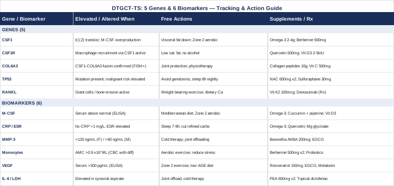
6 Biomarkers Worth Tracking in Diffuse-Type Giant Cell Tumor of the Tendon Sheath
DTGCT-TS is not a tumor you monitor passively. Its recurrence rate after surgery alone runs as high as 50% in the diffuse form, and the biological activity that predicts regrowth often shows up in measurable signals before imaging does. The six biomarkers below are selected for their mechanistic relevance to the CSF1-driven biology of this condition, their clinical trackability, and their practical value in guiding treatment decisions — from timing surgical review to evaluating eligibility for CSF1R inhibitor therapy.
Serum M-CSF (Macrophage Colony-Stimulating Factor / CSF1 Protein)
M-CSF, the protein product of the CSF1 gene, is the central molecular driver of DTGCT-TS. A subset of tumor cells carries a translocation that fuses the CSF1 gene to COL6A3, causing sustained, high-level secretion of M-CSF into the surrounding tissue. This signal recruits and activates CSF1R-positive macrophages, which form the bulk of what is seen as the "tumor mass." Elevated serum M-CSF is not merely a bystander finding; it reflects active CSF1 pathway signaling and correlates with disease burden. Tracking it gives you a real-time proxy for whether the molecular driver of the condition is currently active.
How to Measure It
Serum M-CSF is measured via ELISA-based assay through specialty or academic medical center labs. It is not a standard panel item at most commercial labs, so it may require a physician order specifying the assay. Cost ranges from approximately $80 to $250 depending on the laboratory. It is most useful as a serial measurement — a baseline before surgery followed by measurements at 3, 6, and 12 months post-intervention provides meaningful trend data. Some academic centers specializing in sarcoma and bone tumors track this routinely in DTGCT-TS patients.
If M-CSF Is Elevated — Plan Without Supplements
The most evidence-backed non-pharmacological approach to elevated M-CSF in this context is reducing systemic inflammatory burden. A Mediterranean-pattern diet — rich in oily fish, olive oil, vegetables, and low in ultra-processed food — reduces the upstream cytokine environment that amplifies CSF1 pathway signaling. Visceral adipose tissue is a significant source of M-CSF and IL-6; reducing abdominal adiposity through sustained caloric modesty and regular Zone 2 aerobic exercise (30–45 minutes, four to five days per week at a pace where you can hold a conversation) is the most impactful non-supplemental lever. Reducing alcohol consumption also directly lowers inflammatory cytokine output. The clinical priority when M-CSF is persistently elevated is a discussion with your oncologist about CSF1R inhibitor therapy — pexidartinib (Turalio) is the only FDA-approved agent for this indication and showed a 38% overall response rate in the ENLIVEN phase 3 randomized trial.
If M-CSF Is Elevated — Plan With Supplements or Equipment
Omega-3 fatty acids (EPA/DHA): 2–4g combined EPA/DHA daily with a fatty meal. No cycling required; continuous use is standard. Side effects include a mild blood-thinning effect (discuss with surgeon before procedures), fishy breath, and occasional GI upset. These are important to discuss with your oncologist if you are on pexidartinib.
Curcumin with piperine: 500–1000mg curcumin plus 5–10mg piperine (black pepper extract), twice daily with food. Cycling 8 weeks on, 2 weeks off is a reasonable protocol. Side effects include GI discomfort at high doses; curcumin can inhibit CYP3A4, which metabolizes many oncology drugs — this interaction must be reviewed with your treating physician.
Vitamin D3/K2: If serum 25-OH-D is below 50 ng/mL, supplementing 2000–4000 IU D3 daily with 100–200mcg MK-7 K2 is reasonable. Monitor 25-OH-D levels every 3 months. No cycling required; adjust based on test results.
C-Reactive Protein (CRP) and Erythrocyte Sedimentation Rate (ESR)
CRP and ESR are non-specific markers of systemic inflammation, but in DTGCT-TS they carry specific meaning. The macrophage-rich tumor microenvironment produces large amounts of inflammatory cytokines — including TNF-alpha, IL-1, and IL-6 — that spill into systemic circulation and elevate both markers. Persistently elevated CRP and ESR after surgery suggest residual disease activity or early recurrence. High-sensitivity CRP (hs-CRP) is more useful than standard CRP for tracking low-grade chronic inflammation, and a target of below 1 mg/L is associated with the lowest inflammatory burden in chronic disease contexts.
How to Measure It
hs-CRP is available at virtually every commercial laboratory and ranges from $10 to $40. ESR is similarly ubiquitous and inexpensive ($10–$30). Both can be ordered by any physician. For DTGCT-TS monitoring, measuring every 3–6 months alongside imaging follow-up provides useful trend data. Ferritin can be added as a third inflammatory marker at minimal cost and provides additional context.
If CRP/ESR Is Elevated — Plan Without Supplements
Sleep is the most underappreciated lever here. Sleeping fewer than 7 hours per night raises CRP significantly, independent of other factors. Prioritizing 7–9 hours of high-quality sleep — with consistent sleep and wake times, a dark and cool room, and no bright screens in the 60 minutes before bed — produces measurable reductions in hs-CRP within 4–6 weeks. Eliminating refined carbohydrates and seed oils reduces systemic prostaglandin production. Moderate aerobic exercise (not extreme exertion, which transiently spikes CRP) lowers CRP chronically. Stress management through structured relaxation also reduces cortisol-mediated inflammatory signaling.
If CRP/ESR Is Elevated — Plan With Supplements or Equipment
Omega-3 EPA/DHA: As above. This is the most robustly supported anti-inflammatory supplement with relevant human trial data. Quercetin: 500–1000mg daily with vitamin C (to enhance absorption). Cycle 6 weeks on, 2 weeks off. Side effects are minimal; potential interaction with some antibiotics. Magnesium glycinate: 300–400mg at night. Continuous use is appropriate. Well-tolerated; loose stools at higher doses. Magnesium deficiency independently elevates CRP and is common in Western diets. Infrared sauna: 3–4 sessions per week at 60°C for 20–30 minutes has emerging evidence for lowering CRP and improving inflammatory load. Avoid if hemodynamically unstable or within 4 weeks of surgery.
Matrix Metalloproteinase-3 (MMP-3 / Stromelysin-1)
MMP-3 is a protease that degrades extracellular matrix components including collagen and proteoglycans. In DTGCT-TS, the macrophage-rich inflammatory environment drives significant MMP-3 upregulation within the synovium, contributing to the local tissue destruction and invasive behavior characteristic of the diffuse form. MMP-3 can be measured in both serum and synovial fluid, with synovial fluid levels being considerably higher and more locally reflective. Elevated serum MMP-3 is a sensitive marker for synovial inflammation and is more commonly used in rheumatoid arthritis monitoring, but its relevance extends to any condition involving aggressive synovial macrophage activity, including DTGCT-TS. Multiple studies have examined MMP-3 as a synovial biomarker, and the pattern seen in DTGCT-TS mirrors what occurs in aggressive inflammatory synovitis.
How to Measure It
Serum MMP-3 is available through specialty labs and some academic hospital labs; cost ranges from $60–$150. It is not a standard metabolic panel item but can be ordered by a rheumatologist or orthopedic oncologist. Reference ranges vary by lab and sex; values above 120 ng/mL in females and above 60 ng/mL in males are typically flagged as elevated. Tracking it pre- and post-surgery and during any targeted therapy gives useful signal about synovial activity.
If MMP-3 Is Elevated — Plan Without Supplements
Joint protection strategies are foundational. Avoiding high-impact activity that loads the affected joint reduces mechanical stimulation of synovial macrophage activity. Physical therapy focused on strengthening the muscles surrounding the affected joint — without direct joint compression — can reduce inflammatory load at the synovial level. Cold therapy (10–15 minutes of ice application to the affected area, 2–3 times daily during active inflammation) transiently reduces local MMP activity. Weight management, as above, reduces systemic MMP-3 elevation through adipokine pathways.
If MMP-3 Is Elevated — Plan With Supplements or Equipment
Boswellia serrata (AKBA extract): 100–200mg AKBA standardized extract, twice daily with food. The boswellic acid AKBA directly inhibits MMP-3 and other matrix metalloproteinases through 5-LOX inhibition. Cycle 12 weeks on, 2 weeks off. Side effects are generally mild (GI upset); no significant drug interactions identified, but review with oncologist. Green tea extract (EGCG): 400–600mg standardized EGCG daily with food. Cycle 4–6 weeks on, 2 weeks off. Side effects include possible caffeine sensitivity and, at doses above 800mg/day, rare hepatotoxicity — stay within the lower range and monitor liver enzymes if using other potentially hepatotoxic agents (pexidartinib itself carries a liver safety profile that requires monitoring). Collagen peptides: 10–15g daily, supporting extracellular matrix integrity and potentially reducing MMP-3 demand at the synovial interface. Continuous use; minimal side effects.
Absolute Monocyte Count
This is one of the most practical and inexpensive biomarkers available for DTGCT-TS, and it is almost always already included in a standard complete blood count with differential. Monocytes are the circulating precursors to the macrophages that form the bulk of the DTGCT-TS tumor mass. They express CSF1R on their surface and are actively recruited by the elevated M-CSF produced by the translocation-bearing tumor cells. An elevated monocyte count — above 0.9 × 10⁹/L — suggests heightened CSF1R pathway activity and active macrophage recruitment. Tracking monocyte count over time is a low-cost proxy for the systemic activity of the CSF1 signaling axis.
How to Measure It
A complete blood count with differential is available at any clinical lab for $15–$50 and is typically already part of routine oncology follow-up. Request the absolute monocyte count (AMC) specifically from the differential. Serial measurement every 3 months in the first 2 years post-surgery is a reasonable protocol. An upward trend in AMC, even within normal range, is worth noting and correlating with imaging and clinical symptoms.
If Monocyte Count Is Elevated — Plan Without Supplements
Elevated AMC in this context is most meaningfully addressed by reducing CSF1 pathway stimulation at the systemic level. This again points to reducing visceral adiposity (a major source of CSF1), improving sleep quality, and avoiding chronic psychological stress (which elevates monocyte output through glucocorticoid resistance mechanisms). Structured exercise — particularly moderate aerobic exercise — reduces circulating monocyte counts over time by improving immune regulation. If AMC is persistently elevated alongside clinical symptoms, this is a relevant data point to bring to your oncologist for discussion of CSF1R inhibitor therapy.
If Monocyte Count Is Elevated — Plan With Supplements or Equipment
Berberine: 500mg twice daily with meals. Berberine modulates macrophage polarization and has evidence for reducing monocyte-derived inflammatory output. Cycle 8 weeks on, 2 weeks off. Side effects: GI upset (especially in first week), potential interaction with metformin and some drug-metabolizing enzymes. Review with your physician. Probiotics (Lactobacillus rhamnosus, Bifidobacterium species): A daily multi-strain probiotic reduces gut-derived inflammatory signaling that contributes to monocyte activation. Continuous use is appropriate. Side effects are minimal; transient bloating in the first 2 weeks.
Vascular Endothelial Growth Factor (VEGF)
VEGF drives the formation of new blood vessels — a process that supplies nutrients and oxygen to tumor tissue and is required for sustained tumor growth. In DTGCT-TS, macrophages within the tumor mass produce significant quantities of VEGF, and elevated serum VEGF correlates with more aggressive local behavior. Tracking VEGF also has emerging relevance in the context of anti-VEGF adjunctive strategies that are being explored in CSF1R inhibitor-refractory cases. More practically, VEGF level serves as an indicator of how metabolically active the tumor microenvironment currently is, making it useful between imaging studies. Research on VEGF in TGCT macrophage biology supports its relevance as a monitoring tool.
How to Measure It
Serum VEGF is measured via ELISA through specialty labs; cost ranges from $80–$180. Normal serum VEGF is typically below 500 pg/mL; values above that warrant attention in the context of a known or previously treated DTGCT-TS. Measure at baseline and 6-month intervals. Plasma VEGF is less stable and should be specified as serum when ordering.
If VEGF Is Elevated — Plan Without Supplements
Regular moderate-intensity aerobic exercise has a paradoxically normalizing effect on VEGF over time: while acute intense exercise transiently spikes it, chronic moderate exercise downregulates pathological VEGF overexpression through improved metabolic efficiency. Reducing dietary advanced glycation end products (AGEs) — found primarily in high-temperature processed foods — lowers pro-angiogenic signaling. Reducing simple sugar intake and improving insulin sensitivity through dietary changes reduces the glucose-driven Warburg-effect metabolism that upregulates VEGF in tumor-adjacent cells.
If VEGF Is Elevated — Plan With Supplements or Equipment
Resveratrol: 150–500mg daily, with a fat-containing meal (improves absorption). Resveratrol inhibits HIF-1α, the primary transcription factor for VEGF upregulation. Cycle 6 weeks on, 2 weeks off. Side effects: mild GI upset; potential interaction with blood thinners. EGCG (green tea extract): As above (see MMP-3 section). EGCG independently inhibits VEGF receptor signaling. Melatonin: 1–5mg at bedtime. Beyond its sleep benefit, melatonin has demonstrated anti-angiogenic effects in human cell and animal models by suppressing VEGF expression. Continuous use at low dose is appropriate; at higher doses, cycling every 2–3 months is prudent. Minimal side effects at 1–3mg; grogginess if taken too late.
Synovial Fluid Interleukin-6 (IL-6) and Lactate Dehydrogenase (LDH)
When synovial fluid is accessible — whether through clinical aspiration or joint drainage — measuring IL-6 and LDH provides a direct window into the local tumor microenvironment rather than a systemic proxy. IL-6 is produced in large quantities by activated macrophages within DTGCT-TS tissue, and local levels can be orders of magnitude higher than serum IL-6. Elevated synovial IL-6 reflects active macrophage-mediated inflammation and is a marker of local disease burden. LDH in synovial fluid reflects cellular turnover and metabolic activity within the joint; elevated levels suggest high tissue remodeling activity consistent with active tumor.
How to Measure It
Synovial fluid IL-6 and LDH are measured from joint aspirate, typically at the time of clinical aspiration. LDH is a standard chemistry assay (low cost, $10–$30). Synovial IL-6 requires an ELISA-based assay ($60–$150). These biomarkers are most useful at the time of symptomatic flare, recurrence evaluation, or pre-surgical planning. Not all centers routinely measure synovial IL-6; request it specifically when aspiration is being performed.
If Synovial IL-6 or LDH Is Elevated — Plan Without Supplements
Elevated synovial IL-6 is most directly addressed through disease management rather than lifestyle alone. If levels are high in the context of a known or suspected recurrence, this is a data point to bring to your orthopedic oncologist urgently. Non-pharmacologically, joint offloading (reducing weight-bearing on the affected joint through crutches, a cane, or a brace), cold therapy to reduce local cytokine production, and elimination of dietary pro-inflammatory triggers are the available levers while definitive management is arranged.
If Synovial IL-6 or LDH Is Elevated — Plan With Supplements or Equipment
Palmitoylethanolamide (PEA): 600mg twice daily with food. PEA is an endocannabinoid-like molecule with strong evidence for reducing local macrophage-driven inflammation without the side-effect profile of NSAIDs. Continuous use is appropriate. Side effects are very mild; no known significant drug interactions. Topical diclofenac gel (prescription or OTC depending on region): Applied directly over the affected joint 2–3 times daily. Reduces local prostaglandin production with minimal systemic absorption. Cycle as needed with flare activity. Low-level laser therapy device (home or clinical): 660–904nm wavelength applied to the joint for 10–15 minutes per session, 3–4 times per week. See the complementary approaches section for more detail on this modality.
The Genetic Architecture of DTGCT-TS: 5 Key Genes
The biomarkers above tell you what is happening right now in the tissue. The genetics below explain why it is happening — and, increasingly, what can be done about it at a molecular level. DTGCT-TS is one of the few benign tumors with a well-characterized single genetic driver, making the genetic picture clearer here than in most other musculoskeletal conditions.
CSF1 — The Master Switch
CSF1 (Colony Stimulating Factor 1) is the gene that encodes M-CSF, the macrophage-recruiting signal protein described in the biomarker section. The critical finding in DTGCT-TS, established through landmark research on the CSF1 "landscape effect", is that a chromosomal translocation fusing CSF1 to the COL6A3 promoter causes the affected cells to produce CSF1 at dramatically elevated levels. Only a minority of cells in the tumor (2–16%) carry this translocation, but their output of M-CSF is high enough to drive the recruitment of large numbers of normal macrophages — which become the bulk of the visible tumor mass. This is not a conventional tumor where every cell is malignant; it is a molecularly orchestrated recruitment event.
If CSF1 Is Overexpressed — Plan Without Supplements
Since CSF1 overexpression in this context is caused by a fixed chromosomal translocation rather than an epigenetic state that responds readily to lifestyle modification, the most direct intervention is medical: surgical resection removes the source cells, and CSF1R inhibitors block the downstream receptor. Non-pharmacologically, reducing systemic M-CSF through the lifestyle strategies outlined under the M-CSF biomarker section — visceral fat reduction, anti-inflammatory diet, Zone 2 aerobic exercise — lowers the broader CSF1 pool in circulation and can reduce the amplitude of macrophage recruitment even when the translocation-bearing cells remain active.
If CSF1 Is Overexpressed — Plan With Supplements or Equipment
No supplement directly blocks the CSF1-COL6A3 fusion product. The adjunctive approach is to reduce the systemic inflammatory conditions that amplify the response to elevated CSF1. Omega-3 EPA/DHA (2–4g/day, continuous): reduces macrophage activation thresholds systemically. Berberine (500mg twice daily, 8 weeks on/2 weeks off): modulates macrophage polarization, shifting tissue macrophages toward a less inflammatory phenotype. These are supportive measures; the primary therapeutic discussion for high CSF1 activity should always be with your oncologist.
CSF1R — The Receptor and the Therapeutic Target
CSF1R is the cell-surface receptor that receives the M-CSF signal and drives macrophage survival, proliferation, and activation. All recruited macrophages in DTGCT-TS express CSF1R, making it the most important therapeutic target in the condition. Pexidartinib (Turalio), the only FDA-approved drug for DTGCT-TS, is a small-molecule inhibitor of CSF1R that blocks this receptor and induces tumor regression in a meaningful proportion of patients. In the ENLIVEN randomized phase 3 trial, pexidartinib achieved a 38% overall response rate versus 0% for placebo, with significant improvements in range of motion and patient-reported outcomes. CSF1R inhibition does not address the source mutation (the translocation), but it breaks the downstream signaling chain that drives macrophage recruitment.
If CSF1R Pathway Is Overactive — Plan Without Supplements
Discussion of pexidartinib eligibility with your oncologist is the most important step when CSF1R pathway activity is confirmed to be high — through elevated serum M-CSF, elevated monocyte count, or histopathological confirmation of CSF1R overexpression in surgical tissue. Non-pharmacologically, reducing CSF1R ligand availability (by lowering M-CSF as described) is the only lifestyle lever available. Avoiding substances that upregulate macrophage CSF1R expression — including high dietary saturated fat, alcohol, and high-fructose corn syrup — reduces the tonic activation of this pathway in systemic tissue.
If CSF1R Pathway Is Overactive — Plan With Supplements or Equipment
Quercetin: 500–1000mg daily with vitamin C. Quercetin has been shown in preclinical models to partially inhibit CSF1R signaling. It is not a replacement for pexidartinib but may be useful as an adjunct in patients who are not candidates for targeted therapy. Cycle 6 weeks on, 2 weeks off. Vitamin D3: Ensure serum 25-OH-D is in the 50–80 ng/mL range. Vitamin D receptors regulate macrophage differentiation downstream of CSF1R; deficiency amplifies the macrophage response to CSF1 signaling. 2000–5000 IU daily, no cycling required, monitor every 3–6 months.
COL6A3 — The Translocation Partner
COL6A3 is a collagen gene whose promoter region is the fusion partner in the t(1;2)(p13;q37) translocation that defines most DTGCT-TS cases. The promoter is highly active in synovial fibroblasts, which is why — when it becomes fused to the CSF1 coding sequence — CSF1 expression is dramatically upregulated specifically in the synovial environment. COL6A3 itself is also a structural component of the extracellular matrix; its disruption by translocation may contribute to matrix remodeling abnormalities that facilitate tumor expansion. Detecting the CSF1-COL6A3 fusion through fluorescence in situ hybridization (FISH) on surgical tissue is the most definitive diagnostic confirmation of DTGCT-TS and distinguishes it from other synovial proliferations.
If COL6A3 Fusion Is Confirmed — Plan Without Supplements
Confirmation of the CSF1-COL6A3 fusion by FISH is both a diagnostic certainty and a signal that this tumor has the classical molecular driver that responds to CSF1R inhibition. The direct management implication is surgical resection (aiming for complete synovectomy) with discussion of pexidartinib for diffuse disease. Supporting extracellular matrix integrity through joint protection, physiotherapy, and avoidance of high-impact loading reduces the structural instability that the COL6A3 disruption contributes to. Resistance training of periarticular muscles — keeping loads modest and avoiding the affected joint's pain threshold — preserves structural support and reduces macrophage-activating mechanical stress at the synovial interface.
If COL6A3 Fusion Is Confirmed — Plan With Supplements or Equipment
Collagen peptides: 10–15g daily. The COL6A3 translocation disrupts one copy of the collagen gene; supplementing collagen peptides may support extracellular matrix repair in adjacent tissue. Continuous use; minimal side effects. Vitamin C: 500–1000mg daily. Required cofactor for collagen hydroxylation; deficiency impairs matrix repair. Continuous use; water-soluble and excreted in excess, so side effects at this dose are minimal.
TP53 — Tumor Suppressor and Malignant Transformation Risk
TP53 encodes p53, the most important tumor suppressor protein in human biology. In conventional DTGCT-TS, TP53 is usually intact, which is why the condition remains benign. However, in the rare cases of malignant transformation — malignant TGCT — TP53 mutations are frequently identified alongside other genomic instability markers. Altered TP53 function allows cells to escape apoptosis, accumulate further mutations, and potentially progress toward malignancy. Monitoring for p53 pathway dysfunction (through TP53 sequencing in surgical tissue) is most relevant in cases with unusually aggressive local behavior, rapid recurrence, or atypical histological features.
If TP53 Shows Alterations — Plan Without Supplements
TP53 alterations in this context signal a need for more intensive oncological monitoring — closer imaging intervals, consideration of additional molecular profiling (whole exome sequencing), and expert sarcoma center review. Lifestyle factors known to reduce the rate of somatic TP53 mutation accumulation include avoiding genotoxic exposures (tobacco, alcohol, high-dose ionizing radiation), maintaining high antioxidant intake from food, and prioritizing sleep (during which DNA repair mechanisms are most active). Aerobic exercise improves DNA repair capacity through AMPK-pathway activation.
If TP53 Shows Alterations — Plan With Supplements or Equipment
N-acetylcysteine (NAC): 600mg twice daily. NAC is the direct precursor to glutathione, the cell's primary antioxidant and DNA-protective molecule. Cycle 8 weeks on, 2 weeks off. Side effects: GI upset at high doses; rare bronchospasm in asthma. Sulforaphane (from broccoli sprout extract): 30–60mg standardized sulforaphane equivalent daily. Sulforaphane activates Nrf2, which upregulates the entire endogenous antioxidant and DNA repair network. Cycle 6 weeks on, 2 weeks off. Side effects: mild GI symptoms; avoid in thyroid conditions without physician guidance. Discuss both agents with your oncologist before use — the interaction profile with chemotherapy or radiation matters in higher-risk cases.
RANKL (TNFSF11) — Bone Remodeling and Giant Cell Formation
RANKL (Receptor Activator of Nuclear Factor Kappa-B Ligand, encoded by the TNFSF11 gene) is the primary signal that drives osteoclast differentiation and bone resorption. In DTGCT-TS, the macrophage-rich environment produces RANKL, which drives the formation of the multinucleated giant cells characteristic of the tumor on pathological examination. These giant cells are osteoclast-like in behavior and contribute to the bony erosion sometimes seen in aggressive DTGCT-TS affecting the knee or hip. Denosumab, a monoclonal antibody against RANKL, has been explored in DTGCT-TS — particularly in CSF1R inhibitor-refractory cases — and can reduce giant cell burden and erosive disease. Elevated serum RANKL and a low OPG/RANKL ratio indicate active giant cell formation.
If RANKL Pathway Is Elevated — Plan Without Supplements
Weight-bearing exercise in a protected and pain-free range normalizes the RANKL/OPG ratio through mechanical loading of bone — a well-established effect. Dietary calcium (1000–1200mg/day from food sources) and vitamin D sufficiency are foundational to maintaining appropriate RANKL regulation. Avoiding proton pump inhibitors (PPIs) if not medically necessary is relevant, as chronic PPI use impairs calcium absorption and indirectly elevates RANKL activity. If joint erosion is documented on imaging alongside elevated RANKL activity, the conversation with your oncologist should include denosumab as an adjunct option.
If RANKL Pathway Is Elevated — Plan With Supplements or Equipment
Vitamin K2 (MK-7): 100–200mcg daily. K2 activates osteocalcin and matrix Gla protein, both of which modulate RANKL/OPG balance and reduce osteoclast-mediated bone loss. Continuous use; very low side-effect profile. Avoid if on warfarin without physician guidance. Calcium from food + D3: As above. Strontium ranelate: Available in some European markets; this compound inhibits RANKL-mediated osteoclast differentiation. Only use under physician supervision; it is not widely available in the US without compounding.
What Peter Attia's Cancer Framework Reveals About Monitoring and Metabolism in DTGCT-TS
The molecular and biomarker picture of DTGCT-TS exists within a broader biological context: the systemic metabolic and immune environment either supports or suppresses the tumor's activity. Peter Attia's Outlive: The Science and Art of Longevity — while not specific to DTGCT-TS — provides one of the most practical frameworks for understanding how metabolic health, cancer surveillance, and lifestyle interact. Ten of its most impactful points are directly applicable here.
Early Detection Surveillance Beats Reactive Imaging
Attia's central argument about cancer is that the medicine of the future catches tumors and their biological signatures years before they become clinically apparent. For DTGCT-TS, this means not waiting for pain or limited range of motion to prompt imaging — it means tracking serum biomarkers like M-CSF and monocyte count on a regular schedule so that recurrence is caught at the molecular signal stage, not the symptomatic stage.
Insulin Resistance Creates a Pro-Tumor Microenvironment
High insulin levels drive IGF-1 signaling, mTOR activation, and glucose availability — all of which feed the macrophage-rich metabolic activity in DTGCT-TS tissue. Attia argues that maintaining a fasting insulin level below 6 μIU/mL is one of the most protective metabolic interventions available. Reducing refined carbohydrate intake, improving sleep, and exercising regularly are the primary tools.
Zone 2 Cardio Improves Mitochondrial Function and Reduces Systemic Inflammation
Attia prescribes 3–4 hours per week of Zone 2 aerobic exercise — an intensity where you can hold a conversation but feel challenged. This level of exercise improves mitochondrial efficiency, reduces circulating inflammatory cytokines (including IL-6 and TNF-alpha), and lowers fasting insulin. For DTGCT-TS patients, it also improves joint-supporting muscle mass without the impact stress that aggravates the affected joint.
Continuous Glucose Monitoring Reveals Hidden Metabolic Dysfunction
A CGM worn for 2–4 weeks can reveal post-meal glucose spikes that a fasting glucose or HbA1c misses entirely. Glycemic variability — large swings in blood sugar — drives oxidative stress and inflammatory signaling at the cellular level. Identifying and correcting these patterns through dietary adjustment is one of the fastest ways to reduce the pro-inflammatory environment that amplifies CSF1-pathway macrophage activity.
Sleep Is an Immune Surveillance Mechanism
During slow-wave sleep, the brain clears inflammatory metabolites and the immune system performs surveillance of peripheral tissues. Attia cites data showing that even one week of sleeping less than 7 hours per night measurably impairs natural killer cell activity and elevates inflammatory markers. For someone managing a recurrent neoplastic condition, chronic sleep restriction is a concrete immune suppression strategy — in the wrong direction.
Visceral Adiposity Is the Most Potent Modifiable Inflammatory Signal
Visceral fat — fat stored around the abdominal organs — is hormonally active and produces significant quantities of M-CSF, IL-6, and TNF-alpha. Attia argues that reducing visceral adiposity, tracked through DEXA scan body composition or waist-to-height ratio, produces broader anti-inflammatory effects than almost any supplement protocol. For DTGCT-TS patients with elevated M-CSF, this is not a minor point — it is a central one.
Strength Training Preserves the Metabolic Sink
Muscle tissue is the primary site of glucose disposal. Maintaining or increasing muscle mass through resistance training 2–3 times per week keeps insulin sensitivity high and glucose clearance efficient — directly reducing the metabolic substrate for tumor-adjacent cell activity. Modified resistance training that avoids loading the affected joint while training the surrounding musculature is achievable under physiotherapy guidance.
ApoB and Lipid Particles Influence the Inflammatory Environment
Attia's work on cardiovascular risk biomarkers — particularly ApoB as a more accurate marker of atherogenic particle burden than standard LDL — extends to general inflammatory health. High ApoB and oxidized LDL particles promote systemic endothelial inflammation that amplifies macrophage activation. Lowering ApoB through diet, exercise, and if needed medical intervention reduces the overall inflammatory load in which DTGCT-TS macrophages operate.
Liquid Biopsy and Circulating Tumor DNA Are the Next Frontier
Attia discusses emerging liquid biopsy technologies that can detect circulating tumor DNA (ctDNA) in blood — potentially before imaging shows recurrence. While no validated ctDNA test for DTGCT-TS currently exists commercially, research in this area is progressing. Staying informed about these technologies through an academic sarcoma center may give early access to monitoring tools that are far more sensitive than standard imaging.
Biomarker Surveillance Is a Mindset, Not Just a Test
The underlying principle of Attia's framework is that you cannot manage what you do not measure, and that proactive measurement — before symptoms appear — is fundamentally different from reactive testing. For DTGCT-TS, building a personal biomarker tracking practice (a simple spreadsheet of M-CSF, CRP, monocyte count, and VEGF plotted over time) transforms follow-up appointments from passive check-ins into data-driven conversations with your care team.
Complementary Approaches with Clinical Evidence
The molecular and metabolic strategies above address the biology of DTGCT-TS directly. The following three modalities have meaningful human evidence for managing the pain, inflammation, and quality-of-life burden that accompany this condition day to day.
Low-Level Laser Therapy and Photobiomodulation
Low-level laser therapy (LLLT), also called photobiomodulation, uses specific wavelengths of red and near-infrared light (630–1064nm) to penetrate tissue and stimulate mitochondrial cytochrome c oxidase, reducing oxidative stress and local inflammatory cytokine production. In the context of DTGCT-TS, where macrophage-driven inflammation at the joint causes significant pain and tissue damage between surgical interventions, LLLT offers a non-invasive tool to reduce local inflammatory activity. It is particularly relevant for managing synovial pain and soft tissue inflammation without the GI side effects of chronic NSAID use.
The evidence base for LLLT in musculoskeletal pain is substantial. A comprehensive review of RCTs and meta-analyses, catalogued on PubMed, consistently shows clinically significant reductions in pain scores and inflammatory markers for conditions involving synovial tissue and periarticular inflammation. Specific parameters matter: doses in the 4–8 joules/cm² range using 810–904nm diodes show the strongest outcomes in joint-adjacent soft tissue applications.
For DTGCT-TS, a practical protocol involves 3–4 clinical sessions per week for an initial 8-week course, followed by maintenance sessions as needed. Home devices using 660nm + 850nm wavelengths are available for $200–$600 and can supplement clinical treatment for daily symptom management. Avoid applying directly over known tumor tissue during active disease; periarticular application targeting the inflamed synovium is the goal. LLLT has an excellent safety profile; the main precaution is avoiding direct application over eyes and not using it directly over sites of active infection.
Mindfulness-Based Stress Reduction (MBSR)
Chronic pain from DTGCT-TS — whether from active disease, post-surgical recovery, or anticipatory anxiety around recurrence — significantly impairs quality of life and can itself drive inflammatory signaling through cortisol-mediated neuroimmune pathways. MBSR, the 8-week structured program developed by Jon Kabat-Zinn at the University of Massachusetts Medical School, uses body scanning, sitting meditation, and mindful movement to train sustained present-moment awareness and reduce the pain catastrophization cycle that amplifies chronic pain perception.
The evidence for MBSR in chronic musculoskeletal pain is well-established. A landmark meta-analysis of MBSR in chronic pain conditions demonstrated significant reductions in pain severity, pain-related disability, and depression compared to active control conditions. The effect sizes are modest but durable — participants maintain benefit at 12-month follow-up, which is clinically meaningful for a condition like DTGCT-TS where the disease course is measured in years.
The standard protocol is an 8-week program with weekly 2.5-hour group sessions and 45 minutes of daily home practice. Online, instructor-led programs are now widely available for $200–$500. The practice is particularly useful during the uncertainty of monitoring periods between imaging studies, when anxiety tends to spike and pain thresholds tend to drop. It requires genuine daily commitment to be effective; approaches expecting quick results typically do not sustain the neuroplastic changes that drive benefit.
Massage Therapy
Massage therapy addresses the soft tissue tightness, lymphatic congestion, and periarticular muscle guarding that develop around an affected joint in DTGCT-TS — both as a consequence of the disease itself and as a residual effect of surgical intervention. Regular massage by a therapist trained in oncological massage (a subspecialty that accounts for the presence of tumor tissue and surgical history) can reduce pain, improve range of motion, and support lymphatic drainage in the post-surgical joint environment.
Human clinical evidence for massage in musculoskeletal pain and post-surgical recovery includes multiple RCTs and a growing body of trial data showing significant reductions in pain scores, anxiety, and inflammatory biomarkers including CRP. While no studies are specific to DTGCT-TS (the rarity of the condition precludes this), the biological mechanisms by which massage reduces local inflammation and periarticular muscle tension apply directly to the joint environment seen in this tumor type.
The recommended protocol is weekly sessions of 45–60 minutes with a certified oncological massage therapist. Crucially, the therapist must be informed of the diagnosis, the surgical history, and the location of any known or suspected tumor tissue — direct pressure over tumor sites or fresh post-surgical areas must be avoided. Gentle lymphatic drainage technique over the affected joint is the most relevant modality in the acute post-surgical phase; deeper trigger-point and myofascial work becomes appropriate 6–12 weeks post-surgery once incision integrity is confirmed.
Conclusion
Diffuse-type giant cell tumor of the tendon sheath is driven by a specific, trackable molecular mechanism — one that has, for the first time, led to an FDA-approved targeted therapy and a clearer understanding of what can be monitored over time. The six biomarkers covered here — M-CSF, CRP/ESR, MMP-3, absolute monocyte count, VEGF, and synovial IL-6 — give you a practical surveillance toolkit that goes beyond waiting for symptoms. The five genes — CSF1, CSF1R, COL6A3, TP53, and RANKL — explain the architecture of the condition and point toward both medical and adjunctive strategies for each abnormality. Metabolic health, sleep, visceral fat, and exercise are not peripheral considerations for this tumor; they directly modulate the inflammatory environment in which the CSF1 pathway operates.
The next smart step is to bring a prioritized biomarker request to your next specialist appointment — starting with hs-CRP, absolute monocyte count, and serum M-CSF if accessible. Build a simple log. Track the trends. And if targeted therapy has not yet been discussed, the ENLIVEN trial data makes that a conversation worth initiating. Better data leads to better decisions, and this is a condition where that principle has real clinical weight.
Musculoskeletal: Bone Conditions Joint Conditions Tendon & Ligament Conditions
Autoimmune: Inflammatory Conditions