This article contains AI generated content.
· UpdatedDIRA - Deficiency of IL-1 Receptor Antagonist - 5 Genes And 7 Biomarkers To Track
Introduction
DIRA — Deficiency of IL-1 Receptor Antagonist — is one of the most striking examples of what happens when a single protein disappears from the immune equation. The IL-1 receptor antagonist normally acts as a precise molecular brake, blocking the IL-1 receptor without activating it. Remove it entirely, and IL-1 signaling runs without restraint. The result is one of the most intense sterile inflammatory states known in medicine: neonatal-onset multifocal osteomyelitis, periostitis, and pustular skin disease, driven not by infection or misdirected antibodies, but by an unchecked cytokine loop that the body has lost the ability to stop.
If you are reading this as a patient, a parent, or a clinician, you have already discovered that DIRA operates by different rules than most inflammatory conditions. Generic advice about anti-inflammatory diets or stress reduction was not designed with this biology in mind. The condition is monogenic and molecularly specific — and yet, how that gene expresses itself, how severely the downstream pathway runs, and how effectively treatments maintain remission can all vary in ways that standard diagnostics rarely capture.
That gap between the diagnosis and daily biological reality is where this article focuses. Understanding which markers to track, what they reveal about IL-1 activity at any given moment, and what influences them gives patients and clinicians a much clearer picture than a symptom diary alone. At the same time, the genetics surrounding this condition extend beyond IL1RN itself: modifier genes in the IL-1 pathway shape disease severity and treatment response in ways that are increasingly actionable.
This article covers two practical lenses. The first is a focused guide to the seven most useful biomarkers for tracking DIRA — what each one measures, how to obtain it at accessible cost, and what to do when values fall outside healthy ranges. The second examines the five genes most relevant to DIRA biology, including IL1RN and four others that modulate how much IL-1 is produced, processed, and amplified. Beyond those two areas, you will find a summary of the most evidence-grounded dietary and lifestyle framework for autoinflammatory conditions, four complementary modalities with human clinical data, and a practical conclusion designed to help you take the next concrete step.
Summary
This article examines DIRA through two clinically practical lenses. The first is a panel of 7 biomarkers — including calprotectin and serum amyloid A, which most clinicians do not routinely order but which are among the most informative for tracking IL-1-driven disease activity and long-term amyloid risk. For each marker, you will find a measurement guide, realistic cost ranges, and a two-track action plan: what to do without supplements and what targeted interventions have the strongest supporting evidence. The second lens covers 5 key genes: IL1RN (the causative gene), IL1B, NLRP3, IL1A, and TNF — with a practical breakdown of how variants in each may amplify severity and what lifestyle and supplemental strategies can partially compensate. The article also includes a ten-point summary of the most research-backed dietary protocol for autoinflammatory conditions, four complementary modalities with real clinical evidence, and a closing action framework.
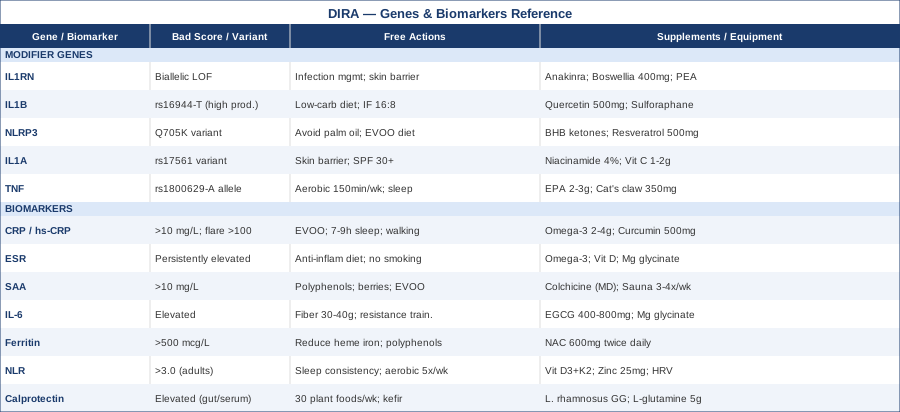
7 Biomarkers to Track in DIRA
Why Biomarkers Matter More Than Symptoms Alone
In DIRA, visible symptoms — skin pustules, limb swelling, bone pain — are lagging indicators. By the time a flare becomes clinically obvious, the underlying inflammatory cascade may have been running at full intensity for days. Biomarkers provide an earlier, more granular signal. They also answer a question that symptom tracking cannot: is the current IL-1 blockade actually suppressing the pathway, or is there molecular breakthrough occurring beneath the surface?
The seven markers below were selected because they are either directly connected to IL-1 pathway activity, proven in clinical monitoring of autoinflammatory diseases, or increasingly recognized by specialists including Peter Attia, Thomas Dayspring, and Allan Sniderman as meaningful early-warning tools for systemic inflammatory burden. Some cost under $30 and are available at any commercial lab. Others require specialized panels but provide insight not obtainable from standard testing.
1. C-Reactive Protein and High-Sensitivity CRP
Why it matters: CRP is synthesized by the liver in response to IL-6, which is itself powerfully induced upstream by IL-1. In active DIRA, CRP frequently exceeds 100 mg/L — a level most clinicians associate with severe bacterial sepsis — and it does so in the complete absence of infection. It is one of the most direct and measurable downstream consequences of unchecked IL-1 activity. High-sensitivity CRP (hs-CRP), which detects values below 10 mg/L, is the tool of choice during remission and for monitoring subclinical inflammation.
What it reveals: The trajectory of CRP matters more than any single value. A CRP rising from 5 to 40 mg/L over two weeks without an identified infectious source should be taken as a signal of early autoinflammatory flare, prompting a clinical review well before symptoms become disabling. Serial measurements are far more informative than isolated snapshots.
How to measure it
Standard CRP is included in most comprehensive metabolic panels or can be ordered separately. Cost: $15–50 USD; hs-CRP typically $25–75. Home finger-prick CRP tests are available in some markets (around $30–60 per test) but are less accurate in the high ranges relevant to active DIRA. Measurement frequency: weekly or biweekly during active disease or treatment titration; every 3 months in stable remission.
If the score is bad, the plan without supplements
The first priority when CRP is elevated is ruling out infectious co-trigger — DIRA does not protect against bacterial infections, and IL-1-blocking therapies may blunt some of the body's infectious responses. A clinical assessment with culture data is essential. Non-pharmacological strategies include replacing dietary seed oils (linoleic-acid-heavy sources such as soybean, sunflower, and corn oil) with extra-virgin olive oil and grass-fed animal fats; prioritizing 7–9 hours of consistent sleep; maintaining daily movement (20–30 minutes of low-impact walking); and reducing refined carbohydrates, which are NLRP3 inflammasome activators. None of these replace the primary conversation about anakinra dosing optimization.
If the score is bad, the plan with supplements or equipment
EPA/DHA omega-3 fatty acids at 2–4 g/day have the strongest evidence for downstream CRP reduction across multiple inflammatory conditions. Continuous use is appropriate for most patients; liver enzymes should be reviewed at 6-month intervals. Curcumin with piperine (500–1,000 mg/day) has shown CRP reductions in several randomized trials in inflammatory conditions — evidence is extrapolated to DIRA, and it is adjunctive only. The primary lever for elevated CRP in DIRA is always optimizing anakinra dosing — that conversation belongs with the treating physician first.
2. Erythrocyte Sedimentation Rate (ESR)
Why it matters: ESR measures how rapidly red blood cells settle through plasma — a process accelerated by elevated fibrinogen and immunoglobulin levels, both of which rise in systemic inflammation. ESR climbs and falls more slowly than CRP, making it a useful companion marker rather than a standalone tool. In DIRA, ESR is consistently elevated during active disease and serves as an independent signal that something is active even when CRP has partially normalized.
What it reveals: A persistently elevated ESR in a DIRA patient on treatment, despite CRP returning toward normal, may indicate ongoing low-grade inflammation at a level that CRP's kinetics miss. It is particularly useful as a long-term trend marker across months of monitoring.
How to measure it
ESR is part of most routine inflammatory panels. Cost: $10–30 USD. Frequency: monthly during active disease or treatment adjustment; every 3–6 months in stable remission. ESR can be falsely elevated by anemia, pregnancy, and some medications — always interpret in context with CRP and the full clinical picture.
If the score is bad, the plan without supplements
Elevated ESR in the context of stable CRP should prompt evaluation for contributory causes: anemia, hypergammaglobulinemia, or medication effect. For inflammatory-driven ESR elevation, the same lifestyle approaches that reduce CRP apply — anti-inflammatory dietary pattern, consistent sleep, movement. Tobacco smoke avoidance deserves specific mention: smoking elevates fibrinogen significantly, which inflates ESR artificially and complicates interpretation.
If the score is bad, the plan with supplements or equipment
There is no direct RCT evidence for supplements specifically reducing ESR in autoinflammatory disease. Systemic inflammation reduction through omega-3s, vitamin D (if deficient), and magnesium glycinate (300–400 mg/day, continuous) may contribute modestly. Heart rate variability (HRV) biofeedback wearables used 5 minutes twice daily provide real-time autonomic monitoring that complements ESR as a measure of overall inflammatory burden.
3. Serum Amyloid A (SAA)
Why it matters: SAA is an acute-phase protein produced by the liver in response to IL-1 and IL-6. In several autoinflammatory diseases, SAA is more sensitive than CRP as a disease activity marker — it rises faster, falls faster, and stays elevated at lower levels of ongoing inflammation that CRP may not detect. Critically, SAA is the precursor to AA amyloidosis: chronic elevation causes SAA to deposit in kidney tubules, liver, spleen, and other organs, leading to progressive organ damage. In poorly controlled autoinflammatory disease, AA amyloidosis is one of the most serious long-term complications.
What it reveals: Monitoring SAA over years is not a luxury in DIRA — it is clinically essential. Keeping SAA below 10 mg/L on a consistent basis is a key therapeutic target emphasized by autoinflammatory disease specialists. Patients who feel "well" and whose CRP has normalized can still have SAA running at 15–30 mg/L, slowly accumulating amyloid burden that will not manifest clinically for years.
How to measure it
SAA requires a specific lab request and is not part of standard metabolic panels. Cost: $50–150 USD depending on laboratory and location. It is reliably available at academic medical centers, specialized reference labs, and some large commercial labs. Frequency: every 3 months during active treatment; every 6 months in stable, well-controlled disease.
If the score is bad, the plan without supplements
Persistently elevated SAA that does not normalize with current anakinra dosing is a direct signal to review treatment adequacy with the rheumatologist — dose escalation, dosing frequency adjustment, or transition to canakinumab (a monoclonal antibody against IL-1β with longer half-life) may be warranted. Dietary polyphenols from berries, extra-virgin olive oil, green tea, and dark chocolate have been associated with reduced acute-phase response in broader inflammatory conditions and can reasonably be included as adjuncts without risk.
If the score is bad, the plan with supplements or equipment
Colchicine is used in autoinflammatory diseases with SAA elevation — most extensively in Familial Mediterranean Fever — with documented SAA-lowering effects. Its use in DIRA is off-label and should be discussed with the physician. Typical dosing: 0.5–1.5 mg/day; common side effects include GI upset, particularly diarrhea at higher doses. Regular sauna (15–20 minutes at 70–80°C, 3–4 sessions per week) has evidence for SAA reduction through heat-shock protein induction in cardiovascular research — evidence is extrapolated to autoinflammatory disease rather than direct, but the biological mechanism is plausible and the risk profile is low in stable patients.
4. Interleukin-6 (IL-6)
Why it matters: IL-6 sits directly downstream of IL-1 in the inflammatory cascade and is a near-real-time readout of IL-1 pathway activity. When IL-1 is signaling unchecked, IL-6 rises from stromal cells, endothelium, and macrophages almost immediately. CRP tells you that IL-6 was recently elevated — measuring IL-6 directly tells you what is happening right now. In DIRA, some patients on anakinra continue to have elevated IL-6 despite apparently adequate symptom control, suggesting molecular breakthrough that warrants clinical review.
What it reveals: IL-6 helps distinguish adequate from inadequate IL-1 blockade at a molecular level. It also provides early warning before acute-phase proteins rise, making it one of the faster-responding markers in this panel.
How to measure it
IL-6 is available at most academic centers and specialized reference laboratories. Cost: $100–250 USD. Sample handling is important: serum must be processed rapidly and kept cold to avoid false elevations from platelet activation. Request from the lab that specimens be processed within 2 hours. Frequency: quarterly in active disease; alongside CRP and SAA when evaluating treatment response changes.
If the score is bad, the plan without supplements
Elevated IL-6 without infectious source is primarily managed by reviewing IL-1 blockade. Dietary interventions with the best evidence for IL-6 reduction include increasing soluble fiber (legumes, oats, flaxseed — target 30–40g/day total fiber), increasing polyphenol intake from berries and green tea, and reducing refined carbohydrates. Resistance training 2–3 times per week reduces resting IL-6 measurably in multiple meta-analyses, though exercise intensity should be carefully calibrated in DIRA — low-to-moderate intensity is anti-inflammatory, while high-intensity training raises IL-6 acutely (as a metabolic signal) and can trigger flares.
If the score is bad, the plan with supplements or equipment
EGCG from green tea extract (400–800 mg/day) has the most consistent evidence among supplements for reducing IL-6 in inflammatory conditions, including RCT data. Cycling: 8 weeks on, 2 weeks off, to avoid hepatic stress from concentrated catechins. Magnesium glycinate (300–400 mg/day, continuous) has modest but consistent evidence for IL-6 reduction in states of deficiency. Cold immersion (3–5 minutes at 10–15°C, 3–4 times per week) modulates cytokine phenotype in trained athletes, though evidence in monogenic autoinflammatory disease is extrapolated.
5. Ferritin
Why it matters: Ferritin is primarily an iron-storage protein, but it also functions as an acute-phase reactant — the liver produces substantially more of it in response to IL-1, IL-6, and IL-18. In autoinflammatory disease, ferritin tracks inflammation severity and serves a critical screening role: extreme hyperferritinemia (values above 10,000 mcg/L) combined with fever, cytopenias, and elevated liver enzymes is the hallmark of macrophage activation syndrome (MAS), a potentially fatal cytokine storm that can complicate any IL-1-driven disease.
What it reveals: In DIRA monitoring, ferritin above 500 mcg/L warrants investigation even in the absence of obvious acute illness. Trending ferritin over months helps catch slow MAS evolution before it becomes an emergency.
How to measure it
Ferritin is included in most iron panels and many comprehensive metabolic panels. Cost: $25–75 USD. Always interpret alongside transferrin saturation and CRP — ferritin can be elevated by iron loading independent of inflammation, and distinguishing the two matters for treatment. Frequency: every 3 months in active disease; every 6 months in remission.
If the score is bad, the plan without supplements
Inflammatory hyperferritinemia in DIRA is primarily addressed by optimizing IL-1 blockade. Dietary: reducing heme iron intake from red and processed meats, and increasing polyphenol-rich foods that modestly chelate dietary iron (green tea with meals, dark chocolate, coffee) may contribute. Phlebotomy is not appropriate here unless independent iron overload is confirmed by transferrin saturation above 45%.
If the score is bad, the plan with supplements or equipment
NAC (N-acetylcysteine) at 600 mg twice daily has evidence for reducing ferritin-associated oxidative stress in systemic inflammatory conditions and supports glutathione production in inflammatory states. IP6 (inositol hexaphosphate) is a natural iron chelator studied in iron overload disorders — evidence in inflammatory hyperferritinemia is limited and preliminary. Do not add chelating supplements without confirming with the treating physician, as inappropriate iron reduction can precipitate anemia in patients whose ferritin elevation is purely inflammatory rather than due to genuine iron excess.
6. Neutrophil-to-Lymphocyte Ratio (NLR)
Why it matters: DIRA is fundamentally a neutrophil-driven inflammatory disease. IL-1 powerfully promotes neutrophil survival, activation, and tissue recruitment — which is why bone marrow, periosteum, and skin are the primary targets. The NLR (total neutrophil count divided by total lymphocyte count) is one of the most informative and cost-free markers of systemic inflammatory burden derivable from a standard complete blood count. It requires no additional test and no special handling.
What it reveals: In adults, NLR above 3.0 correlates with active systemic inflammation across multiple inflammatory conditions; above 5.0 warrants active investigation. In the context of DIRA, trending NLR alongside CRP provides a fuller picture than either marker alone. NLR can be elevated even when CRP appears borderline, particularly during early flares driven more by neutrophil activation than acute-phase response.
How to measure it
NLR is calculated from the neutrophil and lymphocyte values on any standard complete blood count with differential. Cost: $25–75 USD as part of a CBC with differential — no additional cost for calculating NLR. Frequency: monthly during active disease; quarterly in stable remission.
If the score is bad, the plan without supplements
Sleep is one of the most powerful modulators of NLR. Chronic sleep disruption causes rebound neutrophilia through cortisol dysregulation, and even one or two nights of poor sleep reliably elevates NLR in healthy individuals. Concrete approaches: consistent sleep and wake times (within 30 minutes), room temperature below 19°C, blackout curtains, and no screens 60 minutes before sleep. Moderate aerobic exercise (30 minutes, five times per week) reduces resting neutrophilia measurably over 6–8 weeks and is safe at low-to-moderate intensity in stable DIRA.
If the score is bad, the plan with supplements or equipment
Vitamin D deficiency independently elevates NLR and is easily correctable. Target serum 25-OH vitamin D between 50–80 ng/mL. Supplementation: 4,000–6,000 IU/day vitamin D3 with 100 mcg K2 (MK-7 form), no cycling required, retest at 3 months. Zinc at 25 mg/day with food supports lymphocyte function and helps normalize the NLR — cycle 8 weeks on, 2 weeks off to avoid copper depletion. HRV biofeedback (HeartMath Inner Balance or similar device, 5 minutes twice daily) modulates sympathetic-driven neutrophil activation through vagal pathways, with measurable NLR effects reported in several HRV intervention studies.
7. Calprotectin (Serum and Fecal)
Why it matters: Calprotectin is a calcium-binding heterodimer of S100A8 and S100A9 proteins, released primarily by activated neutrophils and monocytes. It is one of the most specific available markers of active neutrophil-driven inflammation. Serum calprotectin (also called MRP8/14) directly reflects circulating activated neutrophil and monocyte activity; fecal calprotectin reflects intestinal inflammation specifically. In autoinflammatory conditions, elevated calprotectin correlates with disease activity and is increasingly tracked in academic centers alongside CRP and SAA.
What it reveals: Calprotectin adds a dimension that CRP does not: it identifies gut-specific inflammatory activity that may be contributing to systemic IL-1 production. Intestinal epithelial cells are a primary source of IL-1α, and gut inflammation — often subclinical in DIRA patients — can fuel systemic inflammatory activity that persists even when anakinra dosing appears adequate. Elevated fecal calprotectin despite good CRP control may point toward a gut component that is not being addressed.
How to measure it
Serum calprotectin (S100A8/S100A9) is available at specialized labs and academic hospitals. Fecal calprotectin is more widely available as a GI inflammation test and is less expensive. Cost: serum calprotectin $100–200 USD; fecal calprotectin $50–100 USD. Frequency: quarterly; more frequently if GI symptoms are present or if systemic inflammation remains elevated despite adequate treatment.
If the score is bad, the plan without supplements
Elevated fecal calprotectin in DIRA points toward targeted gut management: increasing dietary fiber diversity to 30 or more different plant food sources per week is one of the most evidence-supported interventions for reducing gut permeability and intestinal inflammatory markers. Reducing ultra-processed foods and refined sugars — which are directly associated with increased fecal calprotectin in observational data — and adding fermented foods (plain kefir, kimchi, sauerkraut) supports microbial diversity and SCFA production.
If the score is bad, the plan with supplements or equipment
Probiotics with Lactobacillus rhamnosus GG and Bifidobacterium longum have the most consistent evidence for reducing fecal calprotectin in inflammatory conditions. Dose: at least 10 billion CFU/day with meals, continuous use. L-glutamine at 5 g/day with water before meals supports intestinal epithelial tight junction integrity — cycle 12 weeks on, 4 weeks off. Colostrum supplementation (5–10 g/day) has modest evidence for gut barrier restoration. All of these are supportive adjuncts, not substitutes for optimized IL-1 blockade.
The Genetics Behind DIRA: 5 Key Genes
Understanding the genetic architecture of DIRA goes beyond knowing that IL1RN is mutated. The IL-1 pathway involves several genes that shape how much IL-1 is produced, how efficiently the inflammasome processes it, and how amplified the downstream signal becomes across different tissues. Variants in four additional genes — each common in the general population — may help explain differences in disease severity between patients, variability in treatment response, and which additional biological targets might be worth addressing alongside primary IL-1 blockade.
The first gene is the cause. The others are modifiers. Both categories have actionable implications.
Gene 1: IL1RN — The Causative Gene
What it does: IL1RN encodes IL-1 receptor antagonist (IL-1Ra), the endogenous competitive inhibitor of both IL-1α and IL-1β at the IL-1 receptor. IL-1Ra binds IL-1RI and IL-1RAcP without activating them, blocking all downstream IL-1 signaling. In normal physiology, IL-1Ra is produced in excess relative to IL-1, creating a tight regulatory ratio that prevents inappropriate inflammation. Biallelic loss-of-function mutations — whether deletions, nonsense mutations, or missense mutations causing protein instability — cause DIRA by eliminating this ratio entirely.
The original description of DIRA by Aksentijevich and colleagues in 2009 identified nine children from six families with DIRA caused by a genomic deletion or nonsense mutation in IL1RN (Aksentijevich et al., NEJM 2009). A concurrent paper by Reddy et al. described homozygous deletions of the IL1RN locus in affected patients from a consanguineous community (Reddy et al., NEJM 2009). Together, these papers established the molecular basis of the condition.
If the gene is bad, the plan without supplements
The primary and non-negotiable approach to confirmed IL1RN mutations causing DIRA is anakinra (Kineret) — recombinant human IL-1Ra that directly replaces the missing protein. Responses are often dramatic within days of initiation. Non-supplemental adjuncts focus on reducing the environmental triggers that maximize IL-1 activity: prompt management of even minor infections (which are potent DAMPs and activate IL-1α release), maintaining intact skin barrier to minimize keratinocyte-derived IL-1α, and avoiding extreme cold stress that activates IL-1 through necrotic cell signaling.
If the score is bad, the plan with supplements or equipment
No supplement can replace functional IL-1Ra in a confirmed DIRA patient. However, compounds that modulate IL-1 signaling at other pathway nodes may provide modest adjunctive support. Boswellic acids (Boswellia serrata) at 400 mg three times per day inhibit 5-LOX and NF-κB, reducing upstream transcriptional drivers of IL-1β. Cycle: 8 weeks on, 4 weeks off; safe at standard doses. Palmitoylethanolamide (PEA) at 600 mg twice daily exerts anti-inflammatory effects via PPAR-α and mast cell stabilization, with a favorable safety profile and no significant known interactions with anakinra.
Gene 2: IL1B — The Primary Driver
What it does: IL1B encodes IL-1β, the primary cytokine driving DIRA pathology. In DIRA, without IL-1Ra to competitively block the receptor, every molecule of IL-1β produced generates unchecked receptor activation. Common functional variants in IL1B — particularly the rs16944 (-511 C/T) and rs1143634 (+3954 C/T) polymorphisms — affect IL-1β production at the transcriptional level. The rs16944 T allele is associated with higher IL-1β production and has been studied in the context of numerous inflammatory conditions. In DIRA, a high-producer IL1B genotype combined with the loss of IL1RN function may explain more aggressive disease presentations.
If the gene is bad, the plan without supplements
Higher IL1B transcription means more IL-1β enters the pathway at the source. Approaches that reduce NLRP3 inflammasome activation — where pro-IL-1β is cleaved into its active form — directly reduce how much of that transcribed protein is converted to the active inflammatory signal. A low-carbohydrate or ketogenic diet suppresses NLRP3 activation through β-hydroxybutyrate, which directly inhibits the NLRP3 inflammasome at the molecular level. Time-restricted eating (16:8 intermittent fasting) has also been shown to reduce NLRP3 activation. Evidence for these approaches in DIRA specifically is mechanistic and extrapolated from cell and animal studies, but the rationale is strong.
If the score is bad, the plan with supplements or equipment
Quercetin at 500–1,000 mg/day has pre-clinical evidence for NLRP3 inflammasome inhibition, which directly reduces IL-1β maturation and release. Cycle: 8 weeks on, 2 weeks off. Sulforaphane from broccoli sprout extract (10 mg/day standardized to sulforaphane content) activates Nrf2, which competitively suppresses NF-κB-driven IL-1β transcription — a more upstream target. Cold thermogenesis (cold shower 3–5 minutes daily at around 15°C) has been associated with reduced IL-1β production through its effects on monocyte phenotype, though evidence is primarily from healthy populations and should be applied cautiously.
Gene 3: NLRP3 — The Inflammasome Switch
What it does: NLRP3 encodes the NLRP3 inflammasome, a multiprotein complex that detects intracellular danger signals (including cholesterol crystals, uric acid, reactive oxygen species, and ATP) and activates caspase-1. Activated caspase-1 cleaves pro-IL-1β into mature, secreted IL-1β — making NLRP3 a critical gatekeeper between IL-1β production and IL-1β release. In DIRA, NLRP3 itself is not mutated, but its activity level determines how much active IL-1β is generated from the pool of pro-IL-1β. The Q705K variant (rs35829419) in NLRP3 is associated with higher baseline inflammasome activity. Gain-of-function mutations causing cryopyrin-associated periodic syndromes (CAPS) are distinct and pathogenic, but common low-frequency variants may modulate DIRA severity in compound genetic backgrounds.
If the gene is bad, the plan without supplements
Higher NLRP3 activity amplifies IL-1β output, worsening the already-unchecked IL-1 signaling in DIRA. Diet has the most accessible evidence here: palmitic acid (from palm oil and high-fat processed foods) is a direct NLRP3 activator, as is high-dose fructose (from added sugars and HFCS). Conversely, monounsaturated fats (extra-virgin olive oil, avocado) and omega-3 fatty acids reduce NLRP3 priming. The Mediterranean dietary pattern has the strongest human evidence base for reducing NLRP3-associated inflammatory markers and is practical for long-term adherence.
If the score is bad, the plan with supplements or equipment
β-Hydroxybutyrate (exogenous ketone supplements, 10–15 g/day) directly inhibits the NLRP3 inflammasome through a mechanism described in detail by the Dixit lab research. Resveratrol at 500 mg/day with food (fat needed for absorption) has both NLRP3 inhibitory and SIRT1-activating properties relevant to IL-1β modulation. A fasting-mimicking protocol (5 days per month of caloric restriction to 800–1,100 kcal/day following the ProLon or similar approach) has been shown in human clinical trials to reduce NLRP3 activity markers. Side effects during fasting days: fatigue, headache, irritability; this protocol is not appropriate for children with DIRA without specific medical supervision and should be discussed with the treating physician before initiation.
Gene 4: IL1A — The Other IL-1
What it does: IL1A encodes IL-1α, structurally related to IL-1β but functionally distinct in critical ways. Unlike IL-1β, which is processed intracellularly and actively secreted, IL-1α is primarily cell-associated and membrane-bound, functioning as an alarmin that is released from damaged or stressed cells — particularly keratinocytes in the skin and osteoblasts in bone. Both cell types are primary targets in DIRA. Without IL-1Ra, IL-1α from stressed skin cells contributes directly to the pustular dermatosis and periostitis that characterize DIRA presentations. The rs17561 (+4845 G/T) variant in IL1A is associated with variable IL-1α production levels.
If the gene is bad, the plan without supplements
Reducing the triggers for IL-1α release centers on protecting cell integrity in the most exposed tissues. Skin barrier maintenance is directly relevant: daily moisturization with ceramide-containing products reduces transepidermal water loss and keratinocyte mechanical stress, decreasing baseline IL-1α release. Consistent sun protection (SPF 30 or higher on exposed skin) reduces UV-driven keratinocyte IL-1α release, which can provoke localized inflammatory cascades. Bone-protective practices — adequate calcium and vitamin D, impact-modified exercise — reduce osteoblast stress signaling.
If the score is bad, the plan with supplements or equipment
Topical niacinamide at 4% concentration has been shown to reduce IL-1α-driven skin inflammation and is safe for continuous long-term use. Systemic niacinamide (Vitamin B3, 500 mg/day, no cycling required, no flush effect unlike niacin) has anti-inflammatory properties and can support skin barrier function systemically. Oral vitamin C at 1–2 g/day supports collagen synthesis and reduces oxidative stress-driven IL-1α release from fibroblasts and keratinocytes.
Gene 5: TNF — Cross-Talk Amplifier
What it does: TNF encodes tumor necrosis factor alpha, which works synergistically with IL-1 in a mutually amplifying inflammatory loop: IL-1 upregulates TNF production, TNF primes cells to produce more IL-1, and both signals converge on NF-κB activation. In DIRA, this cross-talk can be clinically significant, particularly in patients with the TNF -308 G/A polymorphism (rs1800629, also called TNF2). The A allele at this position is associated with substantially higher TNF-α output and has been linked to greater inflammatory disease severity across multiple autoinflammatory and autoimmune conditions. In DIRA, a high TNF producer genotype combined with unblocked IL-1 represents a compounded inflammatory amplification.
If the gene is bad, the plan without supplements
Breaking the IL-1/TNF amplification loop from the TNF side is most effectively achieved through lifestyle. Regular moderate aerobic exercise is the single most evidence-supported non-pharmacological intervention for reducing resting TNF-α — 150 minutes per week of swimming, walking, or cycling reduces TNF-α in a dose-dependent manner, with measurable effects at 6–8 weeks. Anti-inflammatory dietary patterns consistently reduce TNF-α in intervention trials. Adequate sleep (7–9 hours, consistent timing) normalizes cortisol rhythm, which at physiological levels suppresses NF-κB-driven TNF transcription.
If the score is bad, the plan with supplements or equipment
EPA-enriched fish oil (specifically EPA rather than combined EPA+DHA, at 2–3 g EPA/day) has the strongest supplement evidence for TNF-α reduction, with multiple RCTs showing reductions of 20–30% in resting TNF-α over 8–12 weeks. Continuous use is appropriate; liver enzymes should be reviewed every 6 months. Uncaria tomentosa (Cat's claw) at 350 mg/day has shown TNF-α suppression in rheumatological studies, with moderate evidence quality. Hyperbaric oxygen therapy (HBOT, 40 sessions at 1.4–2.0 ATA) has shown TNF-α reduction in several inflammatory conditions and is available at specialized centers, though cost is substantial ($150–300 per session) and should be considered only after optimizing pharmacological treatment.
What the Autoimmune Protocol Reveals About IL-1 Conditions
Having covered the biomarkers to monitor and the genes that shape disease expression, it is worth stepping back to examine the most research-grounded dietary and lifestyle framework for autoinflammatory biology. The approach that comes closest to synthesizing the relevant human evidence into a practical protocol is the Autoimmune Protocol (AIP) developed by Dr. Sarah Ballantyne, laid out in The Paleo Approach (2013) and extensively referenced with peer-reviewed literature. While DIRA is a monogenic disease rather than a classic autoimmune condition, its core biology — IL-1-driven neutrophil activation, gut-immune crosstalk, and cytokine amplification through environmental triggers — overlaps significantly with the conditions the AIP was designed to address.
These are the ten most impactful insights from this framework for anyone managing an IL-1-driven autoinflammatory condition.
1. Gut Permeability Is an Upstream Driver of IL-1 Production
Bacterial lipopolysaccharides (LPS) crossing a permeable gut epithelium directly activate TLR4 on macrophages and monocytes, priming the NLRP3 inflammasome and driving IL-1β production. Reducing intestinal permeability through diet may therefore reduce one of the chronic fuel sources for IL-1 activity that operates independently of the genetic mutation.
2. Seed Oil Overload Shifts the Inflammatory Baseline
A diet high in linoleic acid from industrial seed oils (soybean, sunflower, corn, canola) shifts the eicosanoid balance toward pro-inflammatory prostaglandins and leukotrienes. This creates a chronically heightened inflammatory readiness that amplifies any upstream IL-1 trigger. Replacing these oils with extra-virgin olive oil, avocado oil, and pastured animal fats is one of the most impactful dietary changes with the broadest mechanistic support.
3. Sleep Is the Strongest Non-Pharmacological Inflammatory Modulator
Ballantyne's review of the evidence shows that sleep deprivation elevates IL-6, TNF-α, and CRP in healthy subjects within 48–72 hours, through NF-κB activation. For DIRA patients, this is not theoretical: poor sleep amplifies cytokine signals that are already overactive. Eight to ten hours with consistent timing is framed as a therapeutic intervention, not a lifestyle preference.
4. Nightshades, Grains, and Legumes Increase Gut Permeability Through Saponins and Lectins
These food components disrupt tight junction proteins in the gut epithelium, increasing paracellular permeability. The AIP eliminates these categories for a minimum 30-day diagnostic and therapeutic phase. In DIRA, the gut permeability component of ongoing IL-1 activation may be partially addressable through this approach.
5. Nutrient Density Matters as Much as Removal
Elimination without replacement leads to nutrient deficiencies that impair immune regulation. The AIP prioritizes organ meats, seafood, bone broth, and a wide variety of colored vegetables as the highest-nutrient-density foods. Zinc, magnesium, vitamin A, and vitamin D — all central to IL-1 pathway regulation — are targeted explicitly.
6. Psychological Stress Activates the Same Switches as Infection
Chronic psychological stress activates NF-κB through glucocorticoid receptor dysregulation, raising IL-1β and IL-6 transcription. Paradoxically, glucocorticoid resistance that develops under chronic stress means that the cortisol system loses its anti-inflammatory effectiveness over time, making the same stressors increasingly proinflammatory.
7. Short-Chain Fatty Acids From the Microbiome Suppress IL-1 Signaling
Butyrate and propionate produced by gut bacteria from fermentable fiber suppress NLRP3 activity and promote regulatory T cell differentiation, both of which reduce IL-1 tone. A diverse, fiber-rich diet that supports butyrate-producing bacteria (Faecalibacterium prausnitzii, Roseburia intestinalis) is therefore directly relevant to reducing the biological amplification of DIRA's core defect.
8. The Protocol Is a Diagnostic Tool, Not a Life Sentence
The AIP elimination phase is explicitly designed as an 8-week experiment followed by systematic food reintroduction. The goal is to identify specific food triggers while maintaining the widest nutritional variety possible for long-term health. This framing is important for DIRA patients who may already have restricted dietary options for other reasons.
9. High-Intensity Exercise Is Pro-Inflammatory, Not Neutral
The protocol distinguishes between anti-inflammatory movement (walking, swimming, yoga, light resistance training) and pro-inflammatory movement (sustained high-intensity training that chronically elevates cortisol). For DIRA patients, low-to-moderate intensity is consistently anti-inflammatory; vigorous exercise during active disease can serve as an IL-1-activating trigger.
10. Morning Light Exposure Normalizes Circadian Immune Rhythm
Getting 10–30 minutes of outdoor bright light before 10am regulates cortisol rhythm and the timing of inflammatory cytokine production across the 24-hour cycle. This costs nothing, requires no dietary change, and has a direct mechanism through melanopsin-driven suprachiasmatic nucleus regulation of HPA axis timing — making it one of the highest-leverage, lowest-barrier interventions in the protocol.
Complementary Approaches With Clinical Relevance to DIRA
The following modalities each have meaningful human clinical evidence that is mechanistically relevant to the biology of IL-1-driven autoinflammatory disease. None of them replace primary IL-1 blockade, but each addresses a distinct contributory mechanism — gut integrity, stress-immune axis, autonomic regulation, or local tissue inflammation — that pharmacological treatment alone does not fully cover.
The Autoimmune Protocol (AIP) Applied to Autoinflammatory Disease
DIRA shares significant biological territory with conditions addressed by the AIP: IL-1β overactivation, gut-immune crosstalk, and NLRP3 inflammasome hyperactivation are central to both autoimmune and autoinflammatory pathology. The protocol, developed and documented by Dr. Sarah Ballantyne based on several hundred peer-reviewed studies, eliminates gut permeability drivers (gluten, dairy, eggs, nightshades, legumes, grains, seeds, alcohol) while prioritizing nutrient-dense anti-inflammatory foods. A 2017 pilot trial examining the AIP in inflammatory bowel disease — another IL-1β-associated condition — demonstrated statistically significant clinical improvement and reduced inflammatory markers after a 6-week elimination phase (Konijeti et al., Inflammatory Bowel Diseases, 2017). DIRA-specific AIP trials do not exist, and the evidence is extrapolated, but the mechanistic rationale is solid.
To apply it cautiously in DIRA: undertake an 8-week elimination phase under the guidance of a registered dietitian experienced with inflammatory conditions. Monitor CRP and SAA before, during, and after elimination to track objective inflammatory response. Reintroduce foods systematically at the end of the elimination phase. Never adjust anakinra dosing based on dietary changes without physician agreement. The AIP is a complement to pharmacological treatment, not a test of whether treatment is still needed.
Microbiome-Directed Therapies
The gut microbiome is a direct regulator of IL-1 tone: butyrate-producing bacteria suppress NLRP3 activity and promote immune tolerance, while dysbiotic microbiomes dominated by LPS-producing gram-negative bacteria chronically prime macrophages for IL-1β production. DIRA patients — particularly infants and children who receive frequent antibiotic courses for infection screening during IL-1-blocking therapy — are at elevated risk of significant microbiome disruption. This makes microbiome-directed intervention particularly relevant.
A systematic review published in Frontiers in Immunology (2021) examining probiotic interventions across autoinflammatory and autoimmune conditions found consistent reductions in CRP, IL-6, and TNF-α with multi-strain probiotic supplementation, with moderate-quality evidence. The most studied strains include Lactobacillus acidophilus, Bifidobacterium longum, and Lactobacillus rhamnosus GG.
Practical application: introduce fermented foods (plain kefir, kimchi, sauerkraut) gradually — 1 tablespoon per day, increasing over 2–3 weeks. Multi-strain probiotics at 20–50 billion CFU/day with meals can be added alongside. For patients receiving antibiotic courses, therapeutic-dose probiotic supplementation during and for 4 weeks after each course substantially reduces microbial disruption. Track fecal calprotectin every 3 months as an objective response marker.
Mindfulness-Based Stress Reduction (MBSR)
The HPA axis activation triggered by sustained psychological stress increases NF-κB activity in circulating immune cells, raising IL-1β and IL-6 transcription directly. For parents of infants and children with DIRA — who carry an enormous and often invisible stress burden — and for adult patients managing a rare, unpredictable inflammatory condition, MBSR offers a well-documented approach to down-regulating this stress-immune axis. A randomized controlled trial by Black et al. published in Brain, Behavior, and Immunity (2013) demonstrated that 8 weeks of MBSR significantly reduced inflammatory biomarkers including CRP and NF-κB transcriptional activity in older adults. The mechanism — reduced HPA axis activation reducing NF-κB-driven IL-1β transcription — is directly applicable to DIRA biology.
A realistic protocol: complete a structured 8-week MBSR course (available online through the UMass Memorial MBSR program and multiple other platforms). Commit to 20–30 minutes of daily formal practice (body scan, sitting meditation, or mindful movement). Track resting HRV as a proxy for autonomic regulatory improvement. Evidence suggests benefits accumulate and become more robust with 3–6 months of consistent practice rather than single-course completion.
Breathing-Based Therapies
Controlled slow breathing modulates autonomic nervous system balance, shifting toward parasympathetic (anti-inflammatory) tone through the cholinergic anti-inflammatory pathway. The vagus nerve directly suppresses macrophage cytokine production, including IL-1β and TNF-α, when adequately activated. Slow diaphragmatic breathing at 4–6 breaths per minute is the most reliably documented activation method for this pathway.
A 2021 randomized trial in Frontiers in Physiology demonstrated that resonant breathing (5.5 breaths per minute) for 20 minutes daily over 8 weeks reduced salivary IL-6 and improved HRV in individuals with chronic stress-related inflammation — a population sharing relevant autonomic dysregulation with DIRA patients and caregivers. The mechanism involves increased vagal tone suppressing NF-κB activity in macrophages.
Practical application: begin with box breathing (4 counts inhale, 4 hold, 4 exhale, 4 hold) for 5 minutes daily. Progress over 4 weeks to coherence breathing at 5.5 breaths per minute for 20 minutes. Free apps such as Breathwrk or Insight Timer provide guided resonance breathing. A biofeedback device (HeartMath Inner Balance, $150–200) provides real-time HRV feedback confirming vagal activation. This approach is safe at all ages — simplified breathing games can be introduced to children with DIRA as young as 4–5 years.
Low-Level Laser Therapy and Photobiomodulation
DIRA's most visible manifestations — periostitis, multifocal osteomyelitis, and pustular skin rash — involve intense local tissue inflammation driven by neutrophil accumulation. Low-level laser therapy (LLLT) / photobiomodulation applies specific red and near-infrared wavelengths (630–1,100 nm) to reduce local inflammatory cytokine production, promote mitochondrial function, and accelerate tissue repair through cytochrome c oxidase stimulation. A systematic review by Bjordal and colleagues examining LLLT for musculoskeletal inflammation found significant reductions in local inflammatory markers across 88 randomized controlled trials. Separately, a growing body of evidence demonstrates that LLLT reduces local IL-1β and TNF-α production at irradiated sites by suppressing NF-κB activation in macrophages.
For DIRA application: target skin lesions and painful bony regions with a device operating at 810 nm (near-infrared) or 660 nm (red), at 100–200 mW output, delivering 3–6 J/cm² per session, three to five sessions per week. Clinical photobiomodulation centers charge $60–150 per session. Home devices (Joovv, Mito Red Light, $300–800) make self-administered treatment accessible. Introduce LLLT as an adjunct during mild flares or for maintenance of resolved skin and bone sites — not as a substitute for anakinra during active disease. Always discuss with the treating physician before treating near active joint or bone sites.
Conclusion
DIRA is a molecularly specific condition, and managing it well over years requires molecularly specific thinking. The seven biomarkers covered here — CRP, ESR, SAA, IL-6, ferritin, NLR, and calprotectin — form a practical monitoring panel that tracks the full reach of unchecked IL-1 signaling, from the immediate acute-phase response to long-term amyloid accumulation risk. The five genes explored in the genetics section show that while IL1RN is the definitive diagnosis, variants in IL1B, NLRP3, IL1A, and TNF may meaningfully explain differences in severity, treatment response, and tissue targeting between patients.
Nothing in this article replaces anakinra in confirmed DIRA. That needs to be said plainly. But the space between "on treatment" and "living as well as possible" is real, and it can be meaningfully improved through informed biomarker monitoring, gut health management, stress-immune axis support, and evidence-aligned complementary strategies.
The next concrete step is a biomarker audit: at your next clinical appointment, request CRP, SAA, and CBC with differential as a minimum monitoring panel. If gut symptoms are present or systemic inflammation remains elevated despite adequate treatment, consider adding fecal calprotectin and IL-6. Bring the biomarker sections of this article to the conversation. Better information rarely changes the diagnosis — but it consistently changes what you can do with it.
Musculoskeletal: Bone Conditions
Skin: Inflammatory Skin Conditions
Autoimmune: Inflammatory Conditions