This article contains AI generated content.
· UpdatedDistal Femur Fracture Genes Biomarkers - 5 Genes And 7 Biomarkers To Track
Introduction
A distal femur fracture is not simply a broken bone you wait to heal. It is a complex biological event shaped by your metabolic environment, your hormonal status, your nutritional state, and — in ways that are only now becoming clinically actionable — your genetic makeup. Two people with the same fracture, the same surgical fixation, and the same rehabilitation protocol can follow dramatically different healing trajectories. Most people never find out why.
The standard conversation after a distal femur fracture focuses on surgery, immobilization, and physiotherapy. That conversation is necessary. But it leaves out a body of evidence that has accumulated over the past two decades — evidence showing that blood biomarkers and genetic variants can identify why healing slows, why bones were fragile in the first place, and what personalized interventions can meaningfully improve the outcome. Generic advice to "take calcium and vitamin D" treats every patient as identical. They are not.
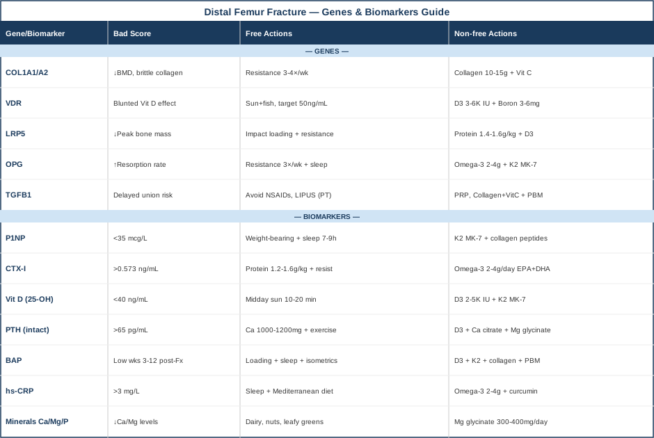
This article takes a more precise approach. It presents two tools: a set of seven blood biomarkers that reveal what is happening at the molecular level during fracture healing and bone remodeling, and five genetic variants with the strongest replicated evidence for influencing bone quality and healing capacity. For each, the goal is not just identification but action — what to do differently depending on what you find.
Better data does not guarantee better outcomes, but it makes better decisions possible. The biomarker section is designed to give you a practical monitoring protocol you can begin immediately. The genetics section is designed to help you understand whether standard protocols need to be adjusted for your biology. Both sections offer concrete, graduated plans — with and without supplementation — grounded in the current evidence base.
7 Biomarkers to Track for Distal Femur Fracture Recovery and Risk
Bone is a living tissue undergoing constant remodeling, and fracture healing is a precisely orchestrated biological process that moves through distinct phases: acute inflammation, soft callus formation, hard callus mineralization, and remodeling. Each phase leaves measurable chemical traces in the blood. Tracking these biomarkers provides a window into the repair site that X-rays cannot — imaging confirms structure, but blood markers reveal activity. Here are the seven most clinically useful.
Biomarker 1: P1NP — The Gold-Standard Bone Formation Signal
Why it matters
P1NP (Procollagen Type 1 N-terminal Propeptide) is the most sensitive and specific bone formation marker currently available. When osteoblasts build new collagen scaffolding, they cleave a fragment called P1NP from the collagen precursor — and that fragment enters the bloodstream in proportion to osteoblastic activity. The International Osteoporosis Foundation and the International Society for Clinical Densitometry jointly designated P1NP as the reference bone formation marker for clinical studies. In fracture healing, a meaningful rise in P1NP during the first 4 to 8 weeks is an early biological sign that repair is underway.
A persistently low P1NP weeks after a distal femur fracture is a red flag. It may reflect nutritional deficiency, hormonal insufficiency, suppression from previously prescribed bisphosphonates, or an underlying metabolic bone condition. It is a number that changes the clinical conversation.
How to measure it
P1NP is drawn from a fasting morning blood sample. Cost: $50–$150 depending on the lab and insurance coverage. It is available through Quest Diagnostics, LabCorp, and most academic medical center labs. Optimal adult levels: above 35 mcg/L. Post-fracture, expect measurable elevation within the first two months if healing is progressing normally.
If the score is low — the plan without supplements
Mechanical loading is the most powerful non-pharmacological stimulus for osteoblast activity. Even limited weight-bearing, cleared by your surgeon, sends piezoelectric signals through bone that upregulate bone-forming cells. Upper-body resistance training and isometric quadriceps contractions during lower-limb immobilization maintain systemic anabolic signals. Sleep quality matters considerably here: growth hormone — a primary driver of bone formation — is secreted in pulsatile bursts during slow-wave sleep. Aiming for 7–9 hours of uninterrupted sleep is not generic advice; it is targeted bone biology. Frequency: loading activities every 2–3 days; sleep hygiene every night.
If the score is low — the plan with supplements or equipment
Vitamin K2 in the MK-7 form (100–200 mcg/day) activates osteocalcin, the protein that anchors calcium into bone matrix and supports mature osteoblast function. Collagen peptides (10–15 g/day taken 30–60 minutes before a loading session with vitamin C) have shown promise in small RCTs for improving bone turnover markers when combined with exercise. Silicon as orthosilicic acid (10–25 mg/day) supports collagen cross-linking at the matrix level. These can be taken continuously; no specific cycling is required, but a 3-month recheck of P1NP levels is a reasonable checkpoint.
Biomarker 2: CTX-I — Measuring How Fast Bone Is Being Broken Down
Why it matters
CTX-I (C-terminal Telopeptide of Type I Collagen) is the complementary reference marker to P1NP — it reflects osteoclastic activity, the bone resorption side of the remodeling equation. Some resorption is biologically necessary and expected; the problem arises when it significantly outpaces formation, which is the hallmark of osteoporosis and a contributor to both fragility fracture risk and impaired healing.
Elevated CTX-I in someone presenting with a distal femur fracture after a low-energy mechanism (a fall from standing height, for example) is a clinically significant finding. It suggests that bone resorption has been chronically exceeding formation — the very metabolic environment that renders the femur susceptible to a fracture that younger, healthier bone would have withstood. Tracking CTX-I during recovery also helps determine whether resorption is appropriately balanced with the formation signal seen in P1NP.
How to measure it
CTX-I must be drawn fasting in the early morning — it exhibits strong diurnal variation, with levels highest before eating and declining substantially after a meal. Cost: $50–$150. In postmenopausal women, values above 0.573 ng/mL are generally considered elevated. A mild elevation immediately post-fracture is expected and benign; persistently high CTX-I at 6–8 weeks without a corresponding P1NP rise warrants evaluation.
If the score is high — the plan without supplements
Resistance exercise is the most evidence-backed lifestyle intervention for reducing excessive bone resorption. It activates Wnt signaling and modulates the RANKL/OPG ratio toward lower osteoclast activity. Equally important: adequate protein intake. Despite conventional concern about protein "acidifying" bone, the research consistently shows that protein restriction in older adults increases bone resorption. Target 1.2–1.6 g of protein per kilogram of body weight per day during recovery. Alcohol and smoking both independently elevate CTX-I; eliminating these is among the highest-leverage lifestyle changes available.
If the score is high — the plan with supplements or equipment
Omega-3 fatty acids (2–4 g/day of combined EPA+DHA from fish oil or algae) have shown consistent resorption-reducing effects in multiple randomized trials. Calcium from dietary sources (1,000–1,200 mg total daily from all sources) helps suppress the PTH-driven resorption signal. If CTX-I remains elevated despite 3–6 months of lifestyle optimization, a referral to an endocrinologist or metabolic bone disease specialist is appropriate before considering pharmacotherapy (bisphosphonates, denosumab). These medications are effective but have direct implications for fracture healing timing — a critical consideration during active recovery.
Biomarker 3: 25-OH Vitamin D — The Nutrient Most Fracture Patients Are Deficient In
Why it matters
Vitamin D deficiency is among the most modifiable and underappreciated contributors to both fracture risk and healing impairment. Calcidiol (25-hydroxyvitamin D) — the storage form measured in blood — controls calcium absorption in the gut, supports the mineralization of callus during fracture healing, modulates the immune microenvironment at the repair site, and influences the expression of multiple genes involved in bone remodeling. Levels below 20 ng/mL are associated with significantly impaired fracture healing and increased nonunion risk in multiple cohort studies.
Peter Attia, whose longevity-medicine approach has popularized rigorous biomarker monitoring, targets 40–60 ng/mL for optimal bone and systemic health — a range that has progressively replaced the older, more conservative threshold of 30 ng/mL in the metabolic bone medicine community.
How to measure it
A standard blood test, widely covered by insurance; cost under $50 out-of-pocket. Specify 25-OH vitamin D (calcidiol), not 1,25-OH vitamin D (calcitriol), which measures the active form and is not a reliable index of stores. Recheck at 8–12 weeks after initiating supplementation; aim for the 40–60 ng/mL range.
If the score is low — the plan without supplements
Midday sun exposure (10–20 minutes on arms and legs, without sunscreen) stimulates cutaneous vitamin D synthesis, though efficacy depends heavily on latitude, season, and skin pigmentation. Dietary sources — oily fish, egg yolks, fortified dairy — contribute modestly. For most people with frank deficiency (below 30 ng/mL), lifestyle alone is insufficient to reach optimal levels in a reasonable timeframe.
If the score is low — the plan with supplements or equipment
Vitamin D3 (cholecalciferol) at 2,000–5,000 IU/day, taken with the largest fat-containing meal, is appropriate for most adults with confirmed deficiency. Pair with vitamin K2 (MK-7, 100–200 mcg/day) to direct calcium appropriately into bone rather than soft tissue — an important safety consideration at higher D3 doses. For severe deficiency (below 10 ng/mL), a physician-supervised loading protocol (e.g., 50,000 IU D3 weekly for 8 weeks) may be appropriate. Recheck at 3 months. Toxicity becomes a concern above sustained 10,000 IU/day; monitoring blood levels removes the guesswork.
Biomarker 4: Intact PTH — The Hidden Driver of Bone Loss
Why it matters
Parathyroid hormone (PTH) is the body's primary calcium regulator. When serum calcium falls — whether from low dietary intake, poor absorption, or vitamin D deficiency — the parathyroid glands release PTH to restore it. In the short term, this is adaptive; PTH mobilizes calcium from bone. Chronically elevated PTH (secondary hyperparathyroidism) drives persistent bone resorption and is a major, often unrecognized, contributor to bone fragility. A substantial number of patients presenting with distal femur fractures have PTH elevations that were never previously identified or treated.
In a healing context, elevated PTH signals that the systemic environment is actively working against consolidation — the body is still in a calcium-deficit mode, leaching the very mineral needed to mineralize the callus.
How to measure it
Intact PTH from a morning fasting blood draw. Cost: $50–$100. Normal range approximately 15–65 pg/mL; optimal bone health is generally associated with levels in the lower half of this range. Always interpret together with serum calcium and 25-OH vitamin D — the combination tells the full story.
If the score is elevated — the plan without supplements
Adequate dietary calcium is the most direct lever: 1,000–1,200 mg/day from food sources (dairy, fortified plant milks, leafy greens, canned fish with bones) reliably suppresses excess PTH secretion. Vitamin D sufficiency is a prerequisite — the gut cannot absorb calcium adequately without it. Weight-bearing physical activity has mild, consistent PTH-suppressing effects in clinical studies. If PTH remains elevated despite correcting these foundational deficiencies, primary hyperparathyroidism (a parathyroid gland adenoma) should be excluded with an endocrinology referral.
If the score is elevated — the plan with supplements or equipment
Vitamin D3 correction, as described above, is the primary pharmacological intervention. Calcium supplementation using calcium citrate (500 mg with meals, not exceeding 1,200 mg total daily from all sources) can support this if dietary intake is genuinely insufficient. Magnesium glycinate (300–400 mg/day) deserves particular attention: magnesium is required for PTH synthesis and release, and hypomagnesemia blunts the response to both vitamin D supplementation and calcium repletion — meaning that correcting the other deficiencies without addressing magnesium can leave PTH elevated despite apparent compliance with other interventions. Recheck PTH alongside vitamin D at 3 months.
Biomarker 5: Bone-Specific Alkaline Phosphatase (BAP) — The Healing Activity Marker
Why it matters
Total alkaline phosphatase (ALP) appears on most standard liver function panels, but it conflates liver and bone-derived ALP — two enzymes with entirely different clinical meanings. Bone-specific alkaline phosphatase (BAP or BSAP) isolates the osteoblast-derived fraction, making it a precise, liver-independent indicator of bone formation activity.
During active fracture healing — particularly the hard callus phase between weeks 3 and 12 — BAP rises substantially and naturally. This rise is expected and is a positive sign. A failure of BAP to rise during this window, or a premature return to baseline, may indicate inadequate osteoblast recruitment or premature suppression of the formation response. In clinical settings, BAP is particularly useful when P1NP is unavailable or when elevated liver enzymes confound total ALP interpretation.
How to measure it
BAP is available through specialty and reference labs, including quest and LabCorp specialty panels. Cost: $75–$200. It is less commonly covered by insurance for this indication but can be ordered directly by a physician or through direct-to-consumer lab services. Consistent fasting morning timing is recommended.
If the score is low during healing — the plan without supplements
Mechanical loading is the most potent osteoblast activator available without a prescription. Isometric quadriceps contractions, upper-body resistance training, aquatic therapy, and even electrical muscle stimulation during the immobilized period maintain systemic anabolic signals that reach bone. Sleep optimization (7–9 hours, dark and cool environment) sustains growth hormone pulses that drive osteoblast activity overnight. These are not peripheral considerations — they are the first-line interventions.
If the score is low during healing — the plan with supplements or equipment
Vitamin D3 and K2 at the doses described earlier support osteoblast differentiation and maturation. Collagen peptides with vitamin C continue to be relevant here. Photobiomodulation (low-level laser therapy applied locally) has shown early evidence of stimulating osteoblast activity and BAP expression in both animal models and human pilot trials — a modality discussed further in the complementary section. For cases of severely impaired healing (delayed union at 3+ months), a specialist conversation about teriparatide (synthetic PTH as an anabolic agent) is warranted — it is one of the most powerful bone formation stimulators available and has shown accelerated callus formation in published case series.
Biomarker 6: hs-CRP and IL-6 — The Inflammatory Backdrop to Healing
Why it matters
Inflammation is not the enemy of fracture healing — it is the initiator of it. The acute inflammatory phase, marked by a local spike in cytokines including IL-6 and TNF-alpha, recruits the stem cells and macrophages that begin the repair cascade. The problem is chronic, systemic, low-grade inflammation: elevated hs-CRP and IL-6 in the background reflect a metabolic environment that interferes with the transition from the inflammatory phase to the proliferative phase, predisposing to delayed union, impaired callus quality, and slower rehabilitation.
Patients with obesity, metabolic syndrome, type 2 diabetes, or chronic psychological stress typically carry elevated baseline inflammatory markers — and research in these populations consistently shows worse orthopedic outcomes. Identifying and addressing this inflammatory backdrop is one of the most underutilized tools in fracture management.
How to measure it
hs-CRP: $20–$60, widely available, frequently covered by insurance. IL-6: $50–$150, less commonly ordered but available through most reference labs. Optimal hs-CRP: below 1 mg/L. Values above 3 mg/L signal high systemic inflammatory burden; values above 10 mg/L suggest acute illness or injury (expected immediately post-fracture, but should normalize substantially within 2–4 weeks). IL-6 optimal: below 2 pg/mL.
If the score is high — the plan without supplements
Sleep quality is the highest-leverage non-pharmacological anti-inflammatory intervention known. Even three nights of short sleep (less than 6 hours) meaningfully raises IL-6 and hs-CRP; restoring consistent, high-quality sleep can reduce these markers within weeks. A Mediterranean-style dietary pattern — rich in vegetables, olive oil, fatty fish, legumes, and low in ultra-processed foods — is the best-replicated dietary approach to reducing hs-CRP. Moderate aerobic activity, when medically cleared, also reduces systemic inflammatory markers; enforced immobility during fracture recovery often raises them, making cleared movement during healing doubly important.
If the score is high — the plan with supplements or equipment
Omega-3 fatty acids (2–4 g/day EPA+DHA) reduce both hs-CRP and IL-6 across multiple meta-analyses with robust effect sizes. Curcumin in a bioavailable form (BCM-95 or Meriva formulation, 500–1,000 mg/day) has demonstrated modest but consistent anti-inflammatory effects; cycle 8 weeks on, 2–4 weeks off to maintain receptor sensitivity and avoid habituation effects. Magnesium glycinate (300–400 mg/day) reduces hs-CRP in individuals with deficiency. One important caution: avoid high-dose NSAIDs during active fracture healing without physician guidance. While anti-inflammatory, NSAIDs inhibit prostaglandin synthesis — a pathway that is pro-resorptive locally but is also required for the anabolic phase of callus formation, and NSAID use during fracture repair is associated with impaired healing in several clinical studies.
Biomarker 7: Serum Calcium, Magnesium, and Phosphorus Panel — The Mineralization Substrates
Why it matters
Hydroxyapatite — the mineral crystal that gives bone its rigidity — is composed primarily of calcium and phosphorus. Magnesium occupies roughly 60% of its body stores in bone and is required for PTH secretion, vitamin D activation, and osteoblast enzyme function. Deficiency in any one of these three minerals impairs the final mineralization step of fracture healing, even when every other variable is optimized.
Isolated hypomagnesemia is particularly common and particularly underdiagnosed. It is often asymptomatic, not captured on standard metabolic panels unless specifically looked for, and creates a cascade that undermines bone health — impairing PTH regulation, reducing vitamin D conversion to its active form, and blunting osteoblast function. This is one reason some patients do not respond to calcium and vitamin D supplementation as expected: the magnesium deficiency is never identified.
How to measure it
Standard metabolic panel or dedicated bone/mineral panel. Cost: $30–$80. Optimal targets: serum calcium 9.0–10.0 mg/dL; serum magnesium 2.0–2.5 mg/dL (though serum magnesium is a poor proxy for total body stores — red blood cell magnesium is more accurate and available through specialty labs at $50–$100); phosphorus 3.0–4.5 mg/dL. Request the panel after fasting; some labs allow direct-to-consumer ordering.
If the scores are off — the plan without supplements
Dietary calcium from dairy, fortified plant milks, leafy greens, and canned fish with bones. Magnesium from nuts, seeds (especially pumpkin seeds), legumes, dark chocolate, and whole grains. Phosphorus is almost universally adequate in adults consuming adequate protein; deficiency arises primarily in malabsorption syndromes or with chronic antacid overuse (antacids bind phosphate). Reducing alcohol, caffeine, and refined carbohydrates decreases urinary mineral losses across all three.
If the scores are off — the plan with supplements or equipment
Magnesium glycinate or malate (300–400 mg elemental magnesium/day with food, starting at a lower dose and titrating to avoid GI effects) is the preferred form for tolerability and absorption. Calcium citrate (500 mg with meals, not exceeding 1,200 mg total from all sources) if genuinely deficient and dietary sources are insufficient. Avoid calcium carbonate, which requires gastric acid for absorption and is poorly bioavailable in individuals on acid-reducing medications. Recheck the mineral panel every 3–6 months and adjust doses accordingly.
Understanding your biomarkers gives you a map. But the terrain of bone biology also includes a layer that biomarkers alone cannot fully capture — the genetic scaffolding that shapes how your body responds to loading, nutrition, and healing signals in the first place.
5 Genes That Shape Fracture Risk and Healing Capacity
Genetics does not predetermine outcomes — but it does define the range of physiological response and the effort required to achieve it. The five gene variants below have the strongest, best-replicated human evidence linking them to bone quality, fracture susceptibility, and healing biology. Each section includes what the variant means in practice and what you can do about it.
Gene 1: COL1A1 and COL1A2 — The Collagen Blueprint
What these genes do
COL1A1 and COL1A2 encode the two alpha chains of type I collagen — the primary structural protein of bone, constituting approximately 90% of the organic matrix. Collagen scaffolding gives bone its tensile flexibility; without it, calcium crystals are brittle and susceptible to fracture under load. Severe mutations cause osteogenesis imperfecta (brittle bone disease). Common polymorphisms — particularly the COL1A1 Sp1 binding site variant (rs1800012) — are more subtle but significantly associated with lower bone mineral density and increased fragility fracture risk across multiple meta-analyses involving tens of thousands of participants.
Individuals carrying the less-favorable alleles have collagen scaffolding that is quantitatively or qualitatively inferior, making the bone matrix less resilient to impact and increasing the likelihood that a given force will result in fracture rather than bruising.
If the gene variant is present — the plan without supplements
Progressive resistance training is the most potent physiological stimulus for upregulating type I collagen synthesis in bone. Mechanical stress activates fibroblasts and osteoblasts to increase COL1A1 gene expression — the variant shifts the baseline, but loading raises expression above it. Compound movements (squats, deadlifts, loaded carries) with progressive overload generate the kind of axial forces that specifically stimulate femoral collagen production. Frequency: 3–4 sessions per week consistently over months; adaptation is cumulative, not acute. The collagen response to exercise is time-dependent, peaking 60–90 minutes post-session, which has implications for supplement timing.
If the gene variant is present — the plan with supplements or equipment
Vitamin C (500–1,000 mg/day from food and supplements, split across two doses) is a cofactor for prolyl and lysyl hydroxylase — the enzymes responsible for collagen cross-linking. Without adequate vitamin C, collagen fibers form but cannot cross-link properly, regardless of how much collagen is synthesized. Hydrolyzed collagen peptides (10–15 g/day taken 30–60 minutes before a loading session, with 50 mg vitamin C co-administered) have shown in RCTs to measurably improve collagen synthesis markers in connective tissue. Glycine (3–5 g/day) — the most abundant amino acid in collagen — supports the substrate pool. These can be taken continuously with no meaningful side effects; recheck bone biomarkers (P1NP) every 3 months as a functional readout.
Gene 2: VDR — How Well You Actually Use Vitamin D
What this gene does
The VDR gene encodes the receptor through which vitamin D exerts its biological effects in virtually every tissue: bone cells, gut epithelium, immune cells, and muscle. Without an adequate receptor, even high circulating vitamin D levels produce a blunted downstream response — less calcium absorption, less osteoblast activation, less immune modulation at the fracture site.
Several polymorphisms in VDR are clinically relevant: FokI (rs2228570), BsmI (rs1544410), TaqI (rs731236), and ApaI (rs7975232). The FokI variant is particularly important because it directly affects the length and transcriptional activity of the VDR protein. Meta-analyses across large European and Asian cohorts have found significant associations between VDR genotypes and fracture risk, BMD at the hip and spine, and vitamin D supplementation response — meaning that at the same blood vitamin D level, VDR variant carriers may have meaningfully lower biological activity.
If the gene variant is present — the plan without supplements
The compensatory strategy is to raise the upstream vitamin D signal high enough that even a less efficient receptor maintains adequate signaling. This means more disciplined attention to sun exposure (earlier in the morning, longer duration when latitude allows), increased dietary vitamin D from oily fish and eggs, and — critically — tighter blood level monitoring. Rather than settling for 30 ng/mL, VDR variant carriers should aim for the upper optimal range of 50–60 ng/mL, checked every 6 months.
If the gene variant is present — the plan with supplements or equipment
Vitamin D3 at 3,000–6,000 IU/day is a reasonable starting point for VDR variant carriers who are documented as deficient — always titrated to blood levels. Boron (3–6 mg/day from boron glycinate or food sources) has shown the ability to raise levels of the active form of vitamin D (1,25-dihydroxyvitamin D) and to extend its half-life, partially compensating for receptor inefficiency. Magnesium (300–400 mg/day) is a required cofactor for hepatic and renal vitamin D hydroxylation steps; VDR variants create greater functional reliance on having an optimal magnesium status to maximize the conversion of whatever D3 is ingested. Recheck blood vitamin D every 6 months; adjust dose based on results, not by fixed protocol.
Gene 3: LRP5 — The Wnt Pathway Gateway to Bone Mass
What this gene does
LRP5 encodes a co-receptor in the Wnt signaling pathway — the most critical molecular pathway for bone mass accrual and maintenance across the lifespan. Activating mutations cause unusually high bone density; loss-of-function mutations cause the rare osteoporosis-pseudoglioma syndrome. Common polymorphisms, including A1330V (rs3736228), are associated with lower peak bone mass, reduced adult BMD, and increased fracture risk in genome-wide association studies involving hundreds of thousands of participants.
Crucially, LRP5/Wnt signaling mediates a significant portion of bone's mechanical adaptation response — the anabolic signal that translates loading into bone formation. LRP5 variant carriers may require more loading stimulus to achieve the same bone adaptation as those with higher-functioning LRP5, making consistent, adequate exercise not merely beneficial but biologically compensatory.
If the gene variant is present — the plan without supplements
High-impact loading is the strongest physiological activator of Wnt/LRP5 pathway signaling in bone. Jumping, bounding, and resistance training with axial loading (leg press, squats, deadlifts) generate mechanical signals that, through osteocyte mechanosensing, feed into the LRP5/Wnt cascade. Whole-body vibration platforms (30–50 Hz, 10–20 minutes, 3–5 days/week) have shown modest but consistent BMD benefits in studies including populations with impaired LRP5 function and may be a useful adjunct during immobilization recovery periods. Frequency: daily impact activity when possible; structured resistance training 3–4 times per week.
If the gene variant is present — the plan with supplements or equipment
No over-the-counter supplement directly activates the Wnt/LRP5 pathway. The indirect support strategy is to ensure the systemic anabolic environment is as favorable as possible: adequate protein (1.4–1.6 g/kg/day), vitamin D sufficiency (50–60 ng/mL), and sleep quality. For individuals with confirmed LRP5 variants, low BMD, and high fracture risk, the pharmaceutical class of sclerostin antibodies (romosozumab — activates the same downstream pathway that LRP5 feeds into) represents the most direct pharmacological analog, though it requires a specialist referral and careful risk-benefit assessment. This is a conversation worth initiating with a metabolic bone disease specialist if LRP5 variants are identified in the context of a fragility fracture.
Gene 4: TNFRSF11B (OPG) — The Bone Resorption Brake
What this gene does
TNFRSF11B encodes osteoprotegerin (OPG), a soluble decoy receptor that intercepts RANKL — the primary signal for osteoclast activation — and prevents it from binding to its receptor on osteoclast precursors. The RANKL/OPG ratio is the central molecular switch for bone resorption: high RANKL or low OPG shifts bone biology toward net loss. Several OPG gene polymorphisms are associated with reduced OPG expression, a higher effective RANKL/OPG ratio, and consequently increased fracture risk and lower BMD — findings replicated in both European and Asian cohort studies.
In practical terms: lower OPG function means the bone resorption brake is less effective. The skeleton is more readily broken down in response to hormonal changes, inflammation, disuse, or nutritional deficiency.
If the gene variant is present — the plan without supplements
Exercise is the most direct non-pharmacological OPG upregulator available. Both resistance and endurance training increase serum OPG and reduce RANKL expression in bone tissue — an effect documented in multiple clinical studies. In postmenopausal women (a population with both lower estrogen and potentially lower OPG), this is particularly meaningful because estrogen is a primary driver of OPG expression; the exercise-driven OPG upregulation partially compensates for the post-menopause hormonal reduction. Adequate sleep and stress management reduce cortisol, which otherwise suppresses OPG expression. Frequency: at least 3 resistance sessions per week sustained consistently over months.
If the gene variant is present — the plan with supplements or equipment
Omega-3 fatty acids (2–4 g EPA+DHA/day) reduce RANKL expression and appear to support OPG activity in bone cell studies. Vitamin K2 (MK-7, 100–200 mcg/day) modulates the OPG/RANKL balance and independently protects against vascular calcification — relevant because calcium released from bone under high-resorption conditions can deposit in vessel walls if not properly directed. Denosumab — a prescription monoclonal antibody that directly mimics OPG by blocking RANKL — is the pharmacological direct analog for OPG insufficiency; it is highly effective and often considered for individuals with OPG variants in the context of confirmed osteoporosis or fragility fracture. This is a specialist conversation, not a self-managed intervention.
Gene 5: TGFB1 — The Healing Recruiter
What this gene does
TGFB1 encodes Transforming Growth Factor Beta 1, a multifunctional growth factor with central roles in fracture healing specifically. During the resorption phase of remodeling, TGF-β1 is released from the bone matrix and acts as a chemotactic signal — recruiting mesenchymal stem cells to the fracture site, stimulating periosteal expansion, and coordinating the transition from cartilaginous callus to woven bone. Several functional polymorphisms in TGFB1, including the codon 25 variant (rs1800471) and the codon 10 variant (rs1982073), affect baseline TGF-β1 production levels and have been linked to differences in fracture healing speed and complication rates in clinical studies.
This gene is worth particular attention for anyone with a history of delayed union, nonunion, or a fracture that healed more slowly than expected — a pattern that may have a partly genetic explanation.
If the gene variant is present — the plan without supplements
Platelet-rich plasma (PRP), delivered locally at the fracture site either intraoperatively or as an adjunct procedure, is a concentrated source of TGF-β1 and other growth factors. While not a supplement, it is a clinical intervention that directly compensates for genetically reduced TGF-β1 signaling at the repair site. Discuss this with your orthopedic surgeon if you have known TGFB1 variants and face a complex or high-risk fracture configuration. Avoiding NSAIDs during active healing is especially important in TGFB1 variant carriers, as prostaglandin pathways and TGF-β1 signaling interact during callus formation. Ultrasound therapy (low-intensity pulsed ultrasound, LIPUS) upregulates TGF-β1 expression at the repair site and is available through physical therapy settings.
If the gene variant is present — the plan with supplements or equipment
Collagen peptides and vitamin C support the downstream collagen synthesis that TGF-β1 normally drives, at the doses described in the COL1A1 section. Photobiomodulation (low-level laser therapy, applied locally 3–5 times per week during the healing phase) has been shown to upregulate TGF-β1 expression in both bone and soft tissue healing contexts — making it particularly relevant for TGFB1 variant carriers and discussed further below. Glycine (3–5 g/day), as a key component of TGF-β1-stimulated collagen synthesis, provides substrate-level support. These interventions have good tolerability profiles; none require cycling and can be maintained throughout the healing phase.
With the genetic picture in view, it is worth examining how one of the most comprehensive frameworks in modern longevity medicine approaches the bone health question — and what it says that most orthopedic clinicians are not routinely telling their patients.
What Peter Attia's "Outlive" Gets Right About Bone Health That Most Doctors Miss
Peter Attia's 2023 book Outlive: The Science and Art of Longevity dedicates significant attention to what he calls the "four horsemen" of mortality — but equally important in the book's framework is the "Centenarian Decathlon": the physical and metabolic capacities required to function well in the final decade of life. Within this framework, bone density and fracture prevention emerge as central, underappreciated priorities — not because fractures are immediately lethal, but because a distal femur fracture in a 75-year-old is often the event that precipitates a cascade: immobility, muscle loss, hospital-acquired complications, and cognitive decline.
Here are ten of the most impactful insights from Attia's approach as they apply to distal femur fractures and bone health.
1. Falls Kill More Slowly Than People Realize — and That Makes Them Worse
Attia argues compellingly that the medical system dramatically underestimates the mortality risk of fall-related fractures in aging populations. The hip fracture 1-year mortality figure (roughly 20–30% in older adults) is widely cited; distal femur fractures carry comparable functional consequences. His central point: prevention infrastructure needs to begin decades earlier, not after the first fracture.
2. Bone Density Testing Is Done Too Late and Too Rarely
The standard recommendation (DXA scan at age 65 for women) is, in Attia's framing, decades too late to intervene on peak bone mass — which is largely set by age 30. He advocates for baseline DXA scans in the late 30s or early 40s, with subsequent monitoring tied to trajectory rather than a single threshold. Knowing whether BMD is stable, improving, or declining is far more actionable than knowing whether it has crossed a diagnostic cutoff.
3. The DEXA Scan Alone Is Incomplete — Trabecular Bone Score Matters
Attia emphasizes that bone mineral density (BMD from DXA) does not capture bone quality — the microarchitectural integrity of trabecular bone, which is a major determinant of fracture resistance. The Trabecular Bone Score (TBS), calculated from the same DXA data, adds texture analysis that captures structural quality. Two patients with identical BMD T-scores can have dramatically different fracture risk depending on their TBS. Request TBS alongside standard DXA interpretation.
4. Zone 2 Aerobic Training Matters for Bone — But Only If Combined with Loading
Attia is a strong advocate for Zone 2 aerobic exercise (low-intensity, sustained effort in a conversational heart rate zone) for metabolic health, but he is equally clear that bone health requires mechanical loading above and beyond Zone 2. Impact loading — running, jumping, resistance training — generates the compressive and tensile forces that stimulate osteoblast activity. Walking alone is insufficient to maintain or build bone in most adults.
5. Muscle Mass and Bone Mass Are Biologically Inseparable
Muscle contractions generate forces that act directly on bone via tendon attachment points — the primary mechanical driver of bone adaptation. Sarcopenia (muscle loss with aging) is therefore not only a weakness problem; it is a bone loss problem. Attia frames muscle mass as the single most important modifiable variable for both fracture prevention and post-fracture recovery. Target protein intake of 1.6 g/kg/day minimum, combined with consistent resistance training, to preserve the muscle-bone unit.
6. The Three Key Blood Biomarkers Attia Monitors for Bone Health
Attia specifically advocates tracking P1NP, CTX-I, and 25-OH vitamin D as a minimum bone health biomarker panel — exactly the first three biomarkers in the primary strategy above. He uses P1NP and CTX-I together to understand the direction of bone turnover: are you net-forming or net-resorbing? He describes cases where vitamin D appeared adequate by conventional thresholds (30 ng/mL) but bone turnover markers still indicated net resorption — resolved by pushing vitamin D to 50+ ng/mL.
7. Fall Prevention Is a Trainable Skill, Not Just a Fate
Attia's physical training framework for older adults specifically targets balance, single-leg stability, hip abductor strength, and reaction time — all direct determinants of whether a stumble becomes a fall and whether a fall becomes a fracture. The distal femur specifically takes impact from fall dynamics involving knee and hip position; training proprioception and lateral stability in the months and years before a fall is the most effective intervention available.
8. Hormone Status Is a Key Bone Variable — In Both Sexes
Attia addresses testosterone and estrogen as bone-health variables in both men and women. Low testosterone in men is associated with accelerated bone loss and fracture risk that rivals the estrogen-BMD relationship in women. He advocates monitoring sex hormone levels alongside bone biomarkers — particularly in men over 50 and women approaching perimenopause — and making hormone optimization part of a comprehensive bone health strategy.
9. Grip Strength Is the Simplest Functional Bone-Health Proxy
Among the low-tech assessments Attia values highly, grip strength consistently emerges as a surrogate for musculoskeletal health, fracture risk, and overall longevity. It requires nothing more than a hand dynamometer (under $30) and correlates with total muscle mass, cortical bone density, and fall risk. A grip strength below the 25th percentile for age and sex warrants serious attention to the muscle-bone axis.
10. The Window After a Fracture Is an Opportunity, Not Just a Recovery
Attia's "Medicine 3.0" framing — proactive, personalized, prevention-focused — applies directly to the post-fracture period. A distal femur fracture, in this view, is not just a repair problem. It is a diagnostic event that reveals the underlying bone health status and opens a window for comprehensive evaluation and intervention that was not available before. It is the best opportunity to identify and treat the root causes that made the fracture possible — before the next one.
Beyond blood tests and genetic reports, there are several evidence-backed therapeutic modalities that have demonstrated meaningful effects on bone healing, pain management, and rehabilitation outcomes for distal femur fractures.
Complementary Approaches With Clinical Evidence
Low-Level Laser Therapy (Photobiomodulation)
Photobiomodulation (PBM) uses red and near-infrared light (typically 600–1,000 nm) to stimulate cellular energy production and tissue repair processes at the mitochondrial level. For bone healing, PBM has been shown to upregulate osteoblast activity, stimulate collagen synthesis, increase local TGF-β1 expression, and accelerate callus formation in both animal models and human pilot studies. It is one of the more promising non-invasive adjuncts to fracture repair, particularly for cases where healing is delayed or in patients with compromised healing biology (older adults, diabetics, individuals with poor vascular supply).
A randomized controlled pilot trial published in Photobiomodulation, Photomedicine, and Laser Surgery examined PBM in post-surgical bone healing and found accelerated callus formation and reduced healing time in the treated group compared to sham. The effect is most plausible for superficial sites with adequate light penetration — the distal femur presents some depth challenges, but higher-powered Class IV laser devices (available in sports medicine and physiotherapy clinics) achieve meaningful tissue penetration at relevant wavelengths.
Practically: seek a physiotherapy or sports medicine clinic offering Class IV laser therapy or therapeutic PBM. Typical protocols involve daily or every-other-day sessions of 10–15 minutes over the fracture site for 4–8 weeks during the active healing phase. Costs vary from $30–$80 per session. There are no significant side effects when used by a trained practitioner. At-home devices (red light therapy panels or handhelds) are available but typically deliver lower power; they may be useful as adjuncts but should not replace clinical protocols during active healing.
Mindfulness-Based Stress Reduction (MBSR)
MBSR — the structured 8-week program developed by Jon Kabat-Zinn — addresses the psychological and neurological dimensions of pain and recovery that are consistently underestimated in orthopedic rehabilitation. Chronic pain after fracture and surgical fixation is partly a central sensitization phenomenon: the nervous system's amplification of pain signals that persists beyond the tissue injury itself. Mindfulness training demonstrably alters this amplification by changing the cognitive and emotional appraisal of pain, reducing its intensity without altering the physical stimulus.
A meta-analysis published in JAMA Internal Medicine (2014, PMID 24395196) found that mindfulness meditation programs produced moderate reductions in pain, depression, and anxiety across a range of clinical conditions, with effects sustained at follow-up. For post-fracture pain specifically, the relevance is high: individuals managing significant pain with opioids or high-dose NSAIDs (with their bone-healing implications) may find mindfulness an effective adjunct that reduces analgesic requirements.
Practically: the full MBSR program is available online through certified instructors and platforms such as the University of Massachusetts Center for Mindfulness. The time commitment is approximately 45 minutes per day during the 8-week course. For those unable to commit to the full program, even consistent daily breathing practice (10–15 minutes of diaphragmatic breathing or body scan) shows documented effects on pain perception and inflammatory markers. Apps such as Waking Up, Insight Timer, or Headspace provide accessible starting points. Cost: free to $50/month depending on the platform.
Tai Chi — Rehabilitation and Fall Prevention
Tai chi is a slow, controlled movement practice involving coordinated weight shifting, balance challenges, and proprioceptive training. For the population most commonly affected by distal femur fractures — older adults — tai chi has accumulated one of the strongest evidence bases of any single modality for fall prevention, balance improvement, and functional recovery post-fracture. This matters because the fracture itself is often not the endpoint; the risk of a subsequent fall on the affected or contralateral limb in the months following recovery is substantial and frequently underaddressed.
A landmark meta-analysis of 17 randomized controlled trials involving over 3,800 older adults, published in the Annals of Internal Medicine (2004), found that tai chi practice significantly reduced fall rate and fall risk in older adults. Subsequent studies have confirmed these effects across both healthy older adults and those with prior fracture history, with 12–26 week programs showing the most consistent benefits.
Practically: tai chi should be introduced after the acute healing phase, when the surgeon has cleared gradual weight-bearing and balance activities. Community classes are widely available through senior centers and recreation facilities; YMCA locations in many cities offer adapted programs for post-fracture rehabilitation. A 24-form Yang-style program, practiced 3–5 times per week for at least 12 weeks, is the most studied and most accessible protocol. Cost: typically $10–$20 per class; many community-based programs offer free or sliding-scale pricing. The physical demands should be discussed with both the physiotherapist and the surgeon before beginning.
Biofeedback for Muscle Rehabilitation
After distal femur fracture and surgical fixation, quadriceps atrophy begins within days of immobilization and can be profound — with functional deficits in quad strength persisting for months and sometimes years if not aggressively addressed. Electromyographic (EMG) biofeedback provides real-time visual or auditory feedback of muscle electrical activity, allowing patients to identify and selectively activate muscle fibers that have become neurologically inhibited by pain, swelling, and disuse — a phenomenon known as arthrogenic muscle inhibition (AMI).
AMI is a well-documented phenomenon in knee-area trauma and surgery: the joint effusion and pain signals from the fracture site reflexively suppress quadriceps motor neuron activity, creating a disconnect between voluntary effort and actual muscle activation. Standard physiotherapy exercises may not overcome this if the patient cannot perceive the quality of their muscle activation. EMG biofeedback makes the invisible activation visible, breaking the inhibition cycle.
Clinical studies in post-knee surgery populations have demonstrated that EMG biofeedback-assisted exercise significantly improves quadriceps recruitment and functional outcome compared to exercise alone. A randomized trial in subjects post-total knee arthroplasty — a population with comparable muscle inhibition dynamics — found meaningful advantages in extension strength and functional mobility for the biofeedback group. Practically: request biofeedback-assisted physiotherapy at your rehabilitation center during the intermediate recovery phase (typically 4–12 weeks post-surgery). Sessions typically last 30–45 minutes and are integrated into standard physiotherapy appointments. Cost: covered by many physiotherapy packages or available as an add-on at $20–$60 per session. At-home consumer EMG biofeedback devices are available but less precise than clinical systems.
Conclusion
A distal femur fracture is a biological event that sits at the intersection of structure, metabolism, genetics, and healing biology. The advice most people receive after the fracture is managed — eat calcium, take vitamin D, do your physiotherapy — is a starting point, not a complete picture.
The seven biomarkers covered here (P1NP, CTX-I, 25-OH vitamin D, PTH, BAP, hs-CRP, and the mineral panel) give you a real-time read on how your bone is healing, whether your metabolic environment supports recovery, and whether foundational deficiencies are silently working against you. The five genetic variants (COL1A1, VDR, LRP5, OPG, TGFB1) explain why some people do not respond to standard protocols and what compensatory strategies apply to each. The complementary modalities — photobiomodulation, mindfulness, tai chi, biofeedback — offer evidence-supported adjuncts that go beyond what most rehabilitation programs include by default.
The next smart step is to choose one or two biomarkers from this list — ideally 25-OH vitamin D and P1NP — and get a baseline measurement. Take the results to your treating physician, your endocrinologist, or a metabolic bone disease specialist. Ask the right questions. If your genetics are available through a consumer genomics panel, cross-reference the five variants discussed here and bring that conversation to a clinician who works with bone health at a functional medicine level. Better data, acted upon intelligently, is how outcomes improve — not by waiting for the next fracture to prompt the investigation that should have happened before it.
Musculoskeletal Endocrine & Metabolic
Musculoskeletal: Bone Conditions
Autoimmune: Inflammatory Conditions