This article contains AI generated content.
Distal Iliotibial Band Tear: 5 Genes and 7 Biomarkers to Track
Introduction
A distal iliotibial band tear rarely comes out of nowhere. Most people who search for this term have already tried the standard advice — rest, ice, foam roll, stretch, strengthen the glutes — and either the pain came back, healed slower than expected, or never fully resolved near the lateral knee where the band inserts around Gerdy's tubercle and the distal femur. That pattern, more than any single symptom, is what usually brings someone to look past the generic physiotherapy handout and ask a more specific question: why does my tissue behave this way, and what, biologically, is different about me.
Generic advice exists because it works for the average case, and the average case is not you. Two runners with identical training loads and nearly identical biomechanics can have very different connective tissue resilience, different baseline inflammation, different collagen-building capacity, and different genetic tendencies toward tendon and ligament injury. None of that shows up on an X-ray or in a standard physiotherapy assessment, which is exactly why the same rehab protocol can fix one person's IT band pain in six weeks and barely touch another's after six months.
This article takes a more granular approach. Instead of repeating stretch-and-strengthen advice you have likely already heard, it looks at the underlying biology: the blood biomarkers that reflect how well your body builds and maintains collagen-rich tissue like the IT band, and the genetic variants that research has linked to tendon and ligament injury risk more broadly. Neither of these replaces a proper orthopedic or physiotherapy evaluation of a distal IT band tear — imaging, load management, and biomechanical correction still matter enormously. But they can explain why recovery is stalling and where to direct effort next.
None of this is about miracle fixes. It is about narrowing the list of plausible explanations so that the next decision — a supplement trial, a loading protocol, a conversation with a physician — is based on something more concrete than guesswork. The sections ahead cover the biomarkers worth tracking first, what current genetics research actually shows (and does not yet show), a training framework drawn from tendon biology research that challenges some conventional rehab timelines, and a short review of complementary approaches with real supporting evidence.
Summary
Before going deeper, here is the shape of what follows. The core of this article walks through seven blood biomarkers — vitamin D, hs-CRP, the Omega-3 Index, HbA1c, ferritin, vitamin C, and testosterone/IGF-1 — that influence how well collagen-rich tissue like the iliotibial band is built, repaired, and kept resilient under load. For each one you will find what a poor result actually means for tendon and ligament tissue, how it is measured and what it costs, and two concrete plans: one using only diet, sunlight, sleep, and training adjustments, and one that adds supplements or equipment, complete with dosing, frequency, and side effects worth knowing about before you start.
After the biomarkers, a shorter section looks at five genes — COL5A1, COL1A1, GDF5, MMP3, and TNC — that human studies have repeatedly linked to tendon and ligament injury risk, with an honest account of how strong that evidence actually is. From there, a research-backed training framework built around collagen synthesis timing and tissue-loading windows explains why some rehab protocols underperform, followed by a review of complementary approaches — massage, biofeedback-based gait retraining, and photobiomodulation — with a clear read on where the evidence is solid and where it is still thin.
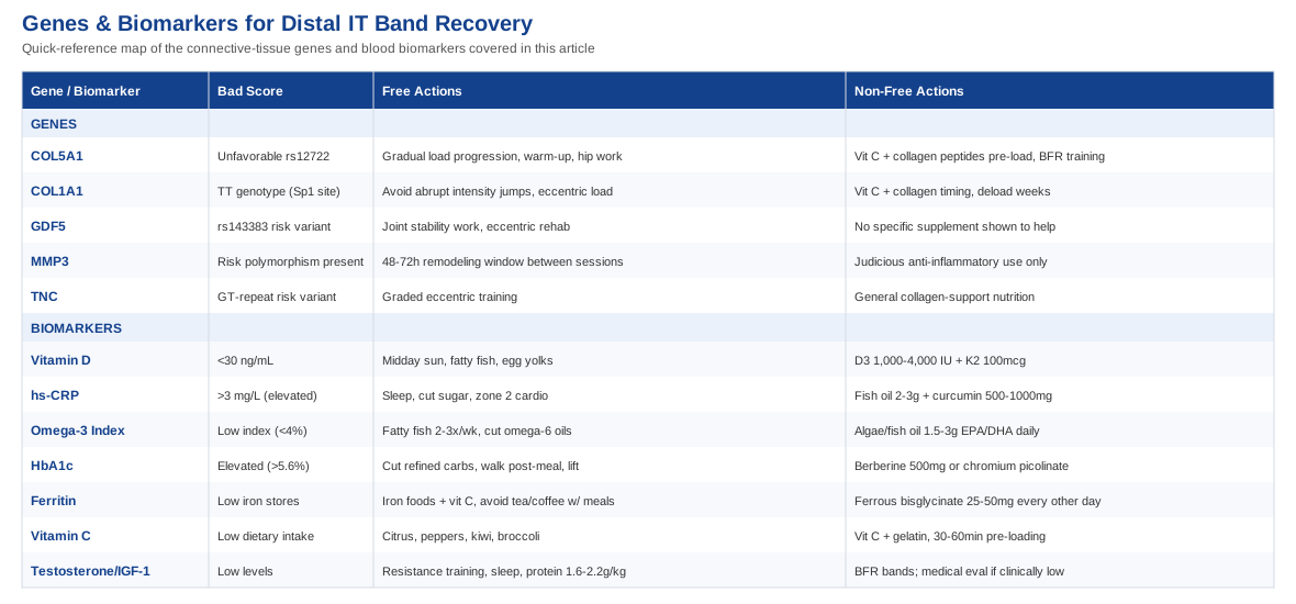
The 7 Biomarkers Worth Tracking When a Distal IT Band Tear Won't Fully Resolve
The iliotibial band is dense, largely collagenous connective tissue with a modest blood supply, which means its capacity to repair itself depends heavily on the raw materials, hormonal signals, and inflammatory environment circulating in your blood. That is precisely what these seven biomarkers reflect. None of them diagnoses a distal IT band tear on their own, but together they build a picture of whether your body is currently set up to rebuild collagen efficiently or working against itself.
Vitamin D (25-hydroxyvitamin D)
Why it matters: Vitamin D receptors are present in tendon and ligament fibroblasts, and vitamin D plays a role in muscle function and neuromuscular control — both of which affect how much compensatory strain gets funneled into the IT band when the hip and glute muscles are not firing efficiently. A comprehensive review on vitamin D and tendon health links deficiency to a higher rate of tendon disorders and slower healing, and low vitamin D has also been associated with a higher risk of other distal soft-tissue injuries in large patient cohorts.
How to measure it: A standard 25-hydroxyvitamin D blood draw, typically $30 to $50 out of pocket or bundled into an annual panel; at-home finger-prick kits run roughly $40 to $70.
If the score is low — the plan without supplements: Aim for 15 to 30 minutes of midday sun exposure on bare skin several times a week, adjusted for skin tone and latitude, plus dietary sources like fatty fish, egg yolks, and fortified dairy. Regular weight-bearing exercise also supports the muscle-function side of the equation.
If the score is low — the plan with supplements: Vitamin D3, typically 1,000 to 4,000 IU daily depending on how deficient the baseline result is, paired with 100 mcg of vitamin K2 to support proper calcium handling. This is taken daily rather than cycled, with a retest at 8 to 12 weeks to adjust the dose. Doses above 10,000 IU/day for extended periods without monitoring risk hypercalcemia, so self-dosing that high is not advisable without lab follow-up.
hs-CRP (high-sensitivity C-reactive protein)
Why it matters: hs-CRP is a general marker of systemic low-grade inflammation. It will not tell you anything specific about the IT band itself, but chronically elevated levels suggest a body that is running an inflammatory load in the background — from poor sleep, excess body fat, or a diet high in refined carbohydrates — which tends to slow soft-tissue healing and make overuse injuries more likely to linger or recur.
How to measure it: A blood test costing $15 to $40, often included automatically in cardiovascular risk panels ordered by primary care physicians.
If the score is high — the plan without supplements: Prioritize sleep consistency, reduce ultra-processed food and added sugar, manage body composition, and add regular zone 2 cardio, which reliably lowers hs-CRP over 8 to 12 weeks in most people.
If the score is high — the plan with supplements: Fish oil providing 2 to 3 g combined EPA/DHA daily, and curcumin with piperine at 500 to 1,000 mg twice daily, cycled 8 to 12 weeks with a 2 to 4 week break. Side effects: fish oil can cause mild GI upset and has a mild blood-thinning effect worth flagging if you take anticoagulants; curcumin can cause GI discomfort and should be avoided by anyone with gallstones.
Omega-3 Index
Why it matters: Rather than a snapshot like hs-CRP, the Omega-3 Index reflects how much EPA and DHA have actually been incorporated into your cell membranes over the preceding months. These fatty acids are precursors to resolvins and protectins, the molecules that actively resolve inflammation rather than just suppressing it — relevant to how efficiently a tearing or overstressed IT band moves through its repair phases. The index itself was first proposed as a risk marker by Harris and von Schacky.
How to measure it: A finger-prick test (commercial kits such as OmegaQuant) for roughly $50 to $90; not yet part of routine panels.
If the score is low — the plan without supplements: Two to three servings per week of fatty fish such as salmon, sardines, or mackerel, while reducing intake of omega-6-heavy vegetable oils that compete for the same enzymatic pathways.
If the score is low — the plan with supplements: Algae-based or fish oil providing 1.5 to 3 g combined EPA/DHA daily, retesting the index at 3 to 4 months since membrane turnover is slow. Side effects: fishy aftertaste, occasional GI upset, and a mild bleeding risk at higher doses — worth pausing before any planned surgery.
HbA1c
Why it matters: Chronically elevated blood glucose accelerates the formation of advanced glycation end-products (AGEs), which create abnormal, non-enzymatic crosslinks within collagen fibers. Research in tenocytes has shown that AGEs increase transglutaminase activity and stiffen tendon tissue, and this glycation process specifically affects the same collagen sites that healthy crosslinking depends on. Stiffer, less pliable collagen is more prone to failure under repetitive strain — exactly the mechanical pattern behind most distal IT band injuries.
How to measure it: A standard HbA1c blood test, $15 to $30; a continuous glucose monitor ($50 to $100/month) gives more granular, real-time feedback if you want to see how specific meals and training sessions affect glucose.
If the score is elevated — the plan without supplements: Reduce refined carbohydrates and added sugar, take a short walk after larger meals, add resistance training (which improves insulin sensitivity independent of weight loss), and protect sleep quality.
If the score is elevated — the plan with supplements: Berberine, 500 mg two to three times daily, cycled 3 months on and 1 month off, or chromium picolinate at 200 to 400 mcg daily. Side effects: berberine commonly causes GI upset and interacts with several medications metabolized through the same liver enzymes, so it should be discussed with a physician if you take other prescriptions.
Ferritin and iron status
Why it matters: Iron is a required cofactor for the prolyl and lysyl hydroxylase enzymes that stabilize the collagen triple helix and form proper crosslinks. Both deficiency and excess iron impair this process — deficiency by starving the enzymes, excess by promoting oxidative stress in the tissue.
How to measure it: A serum ferritin and iron panel, $20 to $50.
If ferritin is low — the plan without supplements: Increase dietary iron from red meat, legumes, and dark leafy greens, and pair iron-rich meals with a vitamin C source to improve absorption; avoid drinking tea or coffee alongside those meals since polyphenols block iron uptake.
If ferritin is low — the plan with supplements: Ferrous bisglycinate, 25 to 50 mg every other day (better tolerated and absorbed than daily high-dose iron), retested at 8 to 12 weeks. Iron should not be supplemented if ferritin is already normal or high, since excess iron carries its own oxidative-stress risk to tissue — this is a correction protocol, not an ongoing one, and stops once levels normalize.
Vitamin C
Why it matters: Vitamin C is the essential cofactor for hydroxylating the proline and lysine residues that give collagen its structural stability — directly relevant given that the IT band is almost entirely dense collagenous tissue. A systematic review by DePhillipo and colleagues found that vitamin C supplementation around the time of musculoskeletal injury supports collagen synthesis and reduces oxidative stress during recovery.
How to measure it: Plasma vitamin C testing is available through specialty labs ($40 to $80) but is uncommon; in practice, most people estimate status from dietary intake rather than direct testing.
If intake looks low — the plan without supplements: Citrus fruit, bell peppers, kiwi, and broccoli, targeting roughly 200 to 500 mg per day from food sources alone.
If intake looks low — the plan with supplements or equipment: 50 to 100 mg of vitamin C combined with 15 g of gelatin or collagen peptides, taken 30 to 60 minutes before rehab loading exercises — a timing strategy discussed further in the training framework below. This is used only on training days, not daily. Side effects are minimal at these doses; doses above 2 g can cause GI upset and diarrhea in sensitive individuals.
Testosterone and IGF-1
Why it matters: Both hormones support fibroblast activity and the anabolic signaling needed for tissue remodeling. Levels commonly dip with overtraining, poor sleep, chronic caloric restriction, or simply aging, and a body running low on anabolic drive has a harder time keeping pace with the repair demands of a distal IT band tear.
How to measure it: Total and free testosterone, $50 to $100; IGF-1, $80 to $150.
If levels are low — the plan without supplements or equipment: Resistance training, 7 to 9 hours of sleep, adequate protein intake (1.6 to 2.2 g per kg of body weight), and stress management — all of which move these markers meaningfully within a few months for most people whose low levels are lifestyle-driven rather than clinical.
If levels are low — the plan with supplements or equipment: Blood flow restriction (BFR) bands are the most evidence-supported "equipment" option here, since they can stimulate anabolic signaling and tissue remodeling with substantially lower mechanical load on the healing IT band itself. If testosterone is clinically low and confirmed by a physician, medical management is the appropriate path — over-the-counter "testosterone boosters" have weak evidence, and some herbal versions (high-dose fenugreek or tribulus) carry their own side-effect profile and should only be cycled under professional guidance.
Turning the panel into a routine
None of these markers needs to be checked weekly. A reasonable cadence is a baseline panel at the start of a dedicated recovery period, a recheck at 8 to 12 weeks once any supplement or lifestyle changes have had time to work, and then every 3 to 6 months thereafter if you are managing an ongoing issue. Testing all seven at once, rather than one at a time, is usually more useful — deficiencies tend to cluster (low vitamin D and low omega-3 status often travel together, for instance), and seeing the full picture prevents chasing a single number in isolation.
Biomarkers describe your current internal environment, but some of the tendency toward IT band injury in the first place is written into your genes. The next section covers what that research actually shows — and, just as importantly, where it is still thin.
What Genetics and Epigenetics Research Suggests About IT Band Injuries
Genomics researchers like Ali Torkamani, whose work focuses on using large-scale genetic data to predict individual disease and injury risk, and Gary Brecka, known for popularizing biomarker- and genetics-informed health optimization, have both pushed the idea that injury susceptibility is not purely a training or biomechanics problem — some of it is structural, encoded in how your body builds collagen in the first place. It is worth being upfront: no published genetic study has looked specifically at "distal iliotibial band tears." Nearly all of the human evidence below comes from research on related tendon and ligament injuries — Achilles tendinopathy, ACL rupture, tennis elbow — because those tissues share the same collagen biology as the IT band. The connection is reasonable, not proven, for this specific injury.
COL5A1
This gene regulates collagen type V, which controls the diameter of collagen fibrils during assembly. Certain variants (commonly studied around the rs12722 marker) have been linked to altered tissue flexibility and injury risk across multiple tendon and ligament sites, summarized in a meta-analysis of COL5A1 polymorphisms and tendon-ligament injury risk. This is one of the better-replicated findings in the sports genetics literature, though effect sizes are modest.
If the gene variant is unfavorable, the plan without supplements centers on gradual mileage or load progression (avoiding sudden jumps in weekly training volume), a consistent dynamic warm-up, and hip abductor strengthening to reduce compensatory strain on the band itself. The plan with supplements or equipment adds the vitamin C plus collagen peptide timing protocol described above, taken before loading sessions, and blood flow restriction training to build tissue tolerance while keeping mechanical stress on the IT band itself lower during the higher-risk early rebuilding phase.
COL1A1 (Sp1 binding site polymorphism)
This variant affects the ratio of collagen type I to type III produced in connective tissue. The rare TT genotype has been associated with a higher rate of tennis elbow and other soft tissue ruptures including cruciate ligament and Achilles injuries, per a review of COL1A1 and acute soft tissue ruptures. Evidence is moderate — consistent across several injury types, but not yet studied in the IT band specifically.
Without supplements, the priority is avoiding abrupt increases in training intensity and building tissue capacity progressively through eccentric loading rather than pure stretching, since eccentric work appears to influence the collagen I:III ratio favorably over time. With supplements or equipment, the same vitamin C and collagen peptide timing strategy applies, alongside periodized training blocks that build in deliberate lower-load recovery weeks rather than continuous progressive overload.
GDF5
GDF5 plays a role in joint, tendon, and ligament development, and the rs143383 variant has been linked to Achilles tendon pathology in genetic association studies out of Australia and South Africa, as well as to osteoarthritis susceptibility more broadly. This gene's role is developmental more than reactive, meaning it likely shapes baseline tissue architecture rather than how you respond to a given training load.
Without supplements, the practical response is the same regardless of genotype: targeted joint stability work and physiotherapy-guided eccentric loading protocols specific to the hip and knee. With supplements or equipment, there is no strong evidence for a GDF5-specific intervention — this is a case where the honest answer is that lifestyle and rehab fundamentals matter more than any supplement stack.
MMP3
MMP3 encodes a matrix metalloproteinase involved in breaking down and remodeling collagen as part of normal tissue turnover. Certain functional polymorphisms have been associated with tendinopathy risk in a meta-analysis of MMP3 variants and tendon-ligament injury, likely because they shift the balance between breaking down damaged collagen and building new collagen too far in one direction.
The nuance with MMP3 is that its activity should not simply be suppressed — some matrix remodeling is a necessary part of healing. Without supplements, the appropriate response is periodized loading that gives tissue adequate remodeling windows between sessions (48 to 72 hours between higher-load stimulus to the same tissue is a reasonable default). With supplements or equipment, anti-inflammatory approaches should be used judiciously rather than aggressively, since blunting the inflammatory and remodeling response too heavily may interfere with the very process MMP3 variants already compromise.
TNC (tenascin-C)
Tenascin-C is an extracellular matrix protein that helps tissue adapt to mechanical stress. A guanine-thymine repeat polymorphism in TNC has been associated with Achilles tendinopathy in South African and other populations, with the effect sometimes appearing to interact with COL5A1 status.
Without supplements, graded eccentric training protocols — which have the strongest evidence base of any conservative intervention across tendinopathies generally — are the most sensible default. With supplements or equipment, the same collagen-support nutritional approach applies, though this gene's evidence base is still early enough that no equipment-based intervention has been specifically validated for it.
Understanding your genetic tendencies is useful context, but tendon and ligament tissue also responds to how and when you load it — which is where a training framework popularized on the Huberman Lab podcast offers some genuinely useful, evidence-backed adjustments to standard rehab thinking.
The Tendon and Ligament Framework That Challenges Standard Rehab Timelines
Much of the most useful recent thinking on tendon and ligament recovery has come out of Dr. Keith Baar's research on connective tissue biology, discussed at length on the Huberman Lab podcast. The core argument is that tendons and ligaments are treated too much like muscle in standard rehab — expected to adapt on similar timelines — when their biology is fundamentally slower and more particular about how and when load is applied. For a distal IT band tear, where the tissue involved is exactly this kind of slow-adapting, collagen-dense structure, this framework is directly relevant.
1. Tendons and ligaments adapt on a much slower timeline than muscle
Muscle can show meaningful adaptation within days; tendon and ligament collagen turnover happens over weeks to months. Expecting IT band tissue to respond to a rehab program on a muscle-strength timeline is one of the most common reasons people feel like "nothing is working" at the 3 to 4 week mark, when the tissue is actually still in an early remodeling phase.
2. Vitamin C and gelatin taken before loading roughly doubles collagen synthesis
In a controlled trial, Shaw and colleagues found that consuming vitamin C-enriched gelatin an hour before intermittent exercise significantly increased markers of collagen synthesis compared to exercise alone. This is the mechanistic basis for the timing protocol described earlier in the vitamin C section — it is not a general supplement, it is a pre-loading strategy.
3. There appears to be a window of roughly six hours after loading for collagen synthesis
Baar's research suggests collagen synthesis remains elevated for several hours after a loading stimulus, meaning the timing of the next meal or supplement dose matters, not just the training session itself. Spacing nutrition around training, rather than treating it as unrelated, may meaningfully affect how much of that loading stimulus actually converts into stronger tissue.
4. Isometric holds under real load stimulate tendon and ligament adaptation more than passive stretching
Sustained isometric contractions at higher intensities appear to drive more meaningful collagen remodeling than static stretching, which is one reason stretching-heavy IT band protocols often plateau. This lines up with the broader tendinopathy rehab literature's shift toward loaded isometric and eccentric work over passive flexibility training.
5. Poor blood supply means overloading the same tissue daily backfires
Because tendons and the IT band have relatively poor vascularity compared to muscle, daily high-load sessions targeting the same structure can outpace its ability to actually remodel, leading to a cycle of ongoing irritation rather than progressive strengthening. Spacing higher-intensity loading sessions 48 to 72 hours apart gives the tissue room to actually rebuild between stimuli.
6. Estrogen fluctuations measurably change ligament laxity
Hormonal shifts across the menstrual cycle affect collagen tissue compliance, which is one reason female athletes may notice their IT band symptoms vary in intensity across the month. This is a real physiological effect, not something to dismiss as unrelated to training load.
7. Warming tissue before loading measurably increases its tolerance
Tendon and ligament tissue becomes more compliant and less injury-prone when warmed prior to loading, supporting the case for a genuine active warm-up rather than a token few minutes of walking before higher-intensity training or rehab exercises.
8. Genetics set the baseline, but training still shapes the outcome
Even where variants like COL5A1 or TNC influence baseline tissue stiffness or injury susceptibility, the loading and nutritional strategies described throughout this framework still meaningfully change functional outcomes — genetics shifts the odds, it does not fix the result in place.
9. High-frequency, lower-load stimulus can outperform infrequent heavy sessions for connective tissue
Because of the slower remodeling timeline, more frequent moderate-load exposure spread across the week may build tendon and ligament resilience more reliably than occasional maximal-effort sessions, which is a meaningful departure from typical strength-training logic.
10. Blood flow restriction training may offer a lower-impact path to the same adaptation
By allowing meaningful physiological loading signals at lower absolute joint stress, BFR training is increasingly discussed as a way to keep stimulating tissue adaptation during the more fragile early stages of recovering from something like a distal IT band tear, without the mechanical strain of full-load training.
Nutrition and loading strategy address the biological side of recovery; a few well-supported complementary approaches can address the mechanical and neuromuscular side as well.
Complementary Approaches Worth Considering
Massage therapy
Manual therapy and self-myofascial release (foam rolling) are among the most commonly recommended conservative treatments for lateral knee pain associated with the IT band, largely because they reduce perceived tightness and pain sensitivity around the area. It is relevant here because a distal IT band tear often develops alongside, or is aggravated by, the same friction and compressive irritation that drives classic iliotibial band syndrome.
Research on foam rolling, including a study comparing its effects on passive tissue stiffness, suggests the mechanism is more about altering neuromuscular tone and pain perception than actually lengthening the IT band itself, which is an extremely stiff structure. The evidence is genuinely mixed on whether it changes tissue properties, but consistent in showing short-term pain and perceived-tightness relief.
A realistic approach is 60 to 90 seconds of foam rolling along the lateral thigh, focusing above and below the point of pain rather than directly on it, two to three times per week, paired with the strengthening work described elsewhere in this article rather than used as a standalone fix. It should feel like firm pressure, not sharp pain — rolling directly and aggressively over an acute tear site is not advisable.
Biofeedback-based gait retraining
Real-time biofeedback on hip and pelvis mechanics during running is relevant here because the strongest documented biomechanical risk factors for IT band problems — excessive hip adduction and weak hip abductor strength — are exactly what this approach targets directly. A meta-analysis on hip abductor strength and kinematics in iliotibial band syndrome confirms these mechanical patterns are consistently present in affected runners, and a systematic review of biomechanical risk factors reinforces the same picture.
Direct randomized trials of biofeedback gait retraining specifically for distal IT band tears are limited, so this recommendation leans on strong biomechanical rationale more than condition-specific trial data. In practice, this looks like a gait-analysis session (often using a mirror, video, or wearable sensor providing real-time cues) where a physiotherapist trains you to reduce excessive hip drop or adduction while running, typically over 4 to 6 sessions with home practice in between.
This is best used once acute pain has settled enough to tolerate running mechanics work, not during the acute tear phase itself, and works best paired with the hip abductor strengthening most protocols already recommend — the biofeedback teaches you to actually use the strength you are building.
Low-level laser therapy (photobiomodulation)
Photobiomodulation is included here for completeness, but the evidence is genuinely mixed and worth being honest about. A systematic review and meta-analysis of laser therapy for Achilles tendinopathy found the certainty of evidence to be low to very low, with any pain benefit from combining laser with eccentric exercise fading by the 3-month mark.
The proposed mechanism is increased mitochondrial activity and reduced local inflammation at the cellular level, which is plausible but not strongly confirmed in tendon-specific human trials at this point.
If you choose to try it, it makes the most sense as an add-on to an existing loading-based rehab program rather than a replacement for it, using a clinical-grade device under a practitioner's guidance rather than a low-powered consumer unit, and treating any pain relief as a possible short-term bonus rather than an expected outcome.
Conclusion
A distal IT band tear that isn't responding the way it "should" is often a sign that something beneath the surface — inflammation running hotter than it should, a collagen-building cofactor running low, a genetic tendency toward stiffer or slower-remodeling tissue — is working against an otherwise reasonable rehab plan. The seven biomarkers covered here give you a concrete, testable way to check for that, and the genetics section explains what current research can and cannot yet tell you about your underlying tissue tendencies. The training framework and complementary approaches round this out with practical ways to work with your biology rather than against it.
None of this replaces a proper orthopedic evaluation of the tear itself, appropriate imaging, and hands-on physiotherapy. But it does give you a more useful starting point than generic advice: pull a baseline panel on the markers most relevant to your situation, bring the results to a physician or sports medicine physiotherapist, and use them to decide what actually needs to change — rather than repeating the same protocol and hoping the outcome is different this time.
Musculoskeletal: Joint Conditions Tendon & Ligament Conditions