This article contains AI generated content.
· UpdatedDrug-Induced Arthritis — 5 Genes and 6 Biomarkers to Track
Introduction
If you developed joint pain, stiffness, or swelling while taking a medication and no clear cause turned up on imaging or basic bloodwork, you are not imagining it. Drug-induced arthritis is real, underrecognized, and affects a meaningful percentage of people on certain medications. The list of culprits is longer than most patients expect — antihypertensives like hydralazine, antibiotics like minocycline, TB drugs like isoniazid, and more recently, checkpoint inhibitors used in oncology and TNF blockers used in autoimmune disease.
What makes this condition genuinely difficult to navigate is that not everyone on the same drug reacts the same way. Two patients taking identical doses for the same condition can have completely different outcomes. That gap is not random — it reflects real biological differences in how each person metabolizes the drug, how their immune system handles its breakdown products, and how primed their inflammatory pathways already are.
The standard response — stop the medication and see what happens — is often necessary but rarely sufficient. Many people stop the drug, experience partial relief, and still struggle with lingering inflammation for months. Others switch medications without realizing their genetic profile makes them vulnerable to an entire class of drugs, not just one. The missing piece is usually a more precise look at what the body is doing and why it responded the way it did.
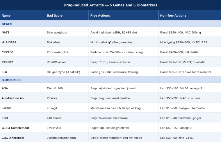
This article approaches that problem from two angles. The first focuses on six specific biomarkers you can measure through standard or specialty labs — markers that help identify whether a drug is driving your joint inflammation, how active that inflammation is, and whether things are genuinely improving. The second examines five genes that shape individual susceptibility to drug-induced arthritis, explaining what each variant does, how to test for it, and what to do when the result is unfavorable. Both paths point toward better decisions — and in this condition, better decisions almost always produce better outcomes.
Summary
This article examines drug-induced arthritis through two evidence-based lenses: the biomarkers your blood can reveal right now, and the genetic variants that may have made you more vulnerable from the start.
- Six biomarkers — ANA, anti-histone antibodies, hsCRP, ESR, complement C3/C4, and CBC with differential — each explained with testing costs, what the result means, and specific action plans with and without supplements. - Five genes — including the NAT2 slow-acetylator variant directly implicated in drug-induced lupus — with real-world protocols depending on your genetic result. - A summary of Ben Lynch's Dirty Genes, one of the most practically useful frameworks for understanding how gene variants interact with drug metabolism and joint inflammation — including the concept of genetic stacking and why epigenetic environment often matters more than the variant itself. - Five complementary approaches with meaningful human clinical evidence for inflammatory joint conditions, including mindfulness-based stress reduction, tai chi, massage therapy, photobiomodulation, and Sarah Ballantyne's Autoimmune Protocol.
Six Biomarkers to Track for Drug-Induced Arthritis
No single blood test confirms drug-induced arthritis on its own. What these six biomarkers do together is build a picture — of immune activation, ongoing inflammation, and whether a drug is the most plausible driver. Tracking them across time, before and after changing a medication, often tells a clearer story than any single result in isolation.
1. Antinuclear Antibodies (ANA)
Why it matters: ANA is the gateway test for drug-induced lupus, the most well-characterized form of drug-induced arthritis. Antinuclear antibodies are immune proteins that mistakenly target the body's own cell nuclei. In drug-induced lupus, ANA is positive in over 90% of confirmed cases. By itself, ANA positivity is not diagnostic — it also appears in healthy adults, particularly women over 50 — but a new positive result coinciding with starting a high-risk drug, especially at a significant titer, is a clinically meaningful signal that should not be dismissed.
How to measure it
Standard blood draw, analyzed by indirect immunofluorescence (IIF) on Hep-2 cells, which is the most sensitive method. Cost typically ranges from $30 to $100 through standard labs. Results appear as a titer (1:80, 1:160, 1:320 or higher) combined with a pattern. In drug-induced lupus, the homogeneous pattern is most common, and titers at or above 1:160 carry clinical weight. Test in the morning after fasting for consistency.
If ANA is elevated: the plan without supplements
The first step is identifying the suspected culprit drug and having a direct conversation with your prescribing physician about whether it can be safely stopped or swapped. Drugs most commonly associated with ANA elevation include hydralazine, procainamide, isoniazid, minocycline, quinidine, and checkpoint inhibitors used in cancer therapy. After stopping the drug, ANA often decreases over weeks to months, though it may not fully normalize. Maintain a structured symptom journal tracking joint stiffness, fatigue, and any rash alongside your ANA retest schedule — typically every 2–3 months after drug cessation.
If ANA is elevated: the plan with supplements
After drug discontinuation, supporting immune regulation is the next layer. Vitamin D3 targeting serum levels of 50–70 ng/mL is well-supported for modulating the B-cell and T-cell activity that drives ANA production. Dose: 2,000–5,000 IU daily with vitamin K2 (100–200 mcg) and a fat-containing meal for absorption. Omega-3 fatty acids (EPA + DHA combined, 2–4g daily from high-quality fish oil) reduce B-cell overactivation relevant to autoantibody production. Cycling: no strict cycling is needed for either; however, D3 levels should be checked via serum 25-OH-D every 6 months to avoid accumulation. Side effects: high-dose D3 without K2 carries modest cardiovascular calcification risk with prolonged use; fish oil at higher doses has a mild anticoagulant effect.
2. Anti-Histone Antibodies
Why it matters: Among all markers for drug-induced lupus, anti-histone antibodies are the most diagnostically specific. Histones are proteins that package DNA in the cell nucleus. Antibodies against them appear in 75–95% of drug-induced lupus cases while appearing in only 20–30% of idiopathic (spontaneous) lupus cases. This asymmetry makes them uniquely valuable for differential diagnosis. A strongly positive anti-histone result in someone who recently started hydralazine, procainamide, isoniazid, or minocycline is among the highest-confidence indicators that the drug is responsible — not an underlying autoimmune condition that requires indefinite immunosuppression.
How to measure it
Ordered either as a standalone test or as part of an extended autoantibody panel. ELISA-based assay. Cost: $50 to $200 depending on whether it is bundled in a lupus panel. Ask for it by name; it is not always included automatically. Results are typically reported as positive or negative with a quantitative index.
If anti-histone antibodies are positive: the plan without supplements
A positive result paired with joint symptoms warrants prompt drug discontinuation review — this is not a wait-and-see marker. Work with your rheumatologist to confirm the clinical picture and identify alternatives to the offending medication. Prognosis after stopping the drug is generally good: joint symptoms typically resolve within weeks to a few months, and anti-histone antibodies decline over 6–12 months in most cases, though they can persist longer without causing active symptoms. Document the timeline carefully — this record is valuable if you ever encounter a prescriber who considers the same drug class in the future.
If anti-histone antibodies are positive: the plan with supplements
NAC (N-acetylcysteine) at 600–1,200 mg/day restores glutathione levels and supports hepatic detoxification of reactive drug metabolites, which is particularly relevant for NAT2 slow acetylators (covered in the genetics section below). Curcumin with piperine — 500–1,000 mg curcumin combined with 5–10 mg piperine, twice daily with food — has been studied for reducing immune complex-driven inflammation. Cycling for NAC: use for 8 weeks, pause for 2 weeks, to avoid theoretical downregulation of endogenous glutathione production during prolonged continuous use. Side effects: NAC can cause nausea at higher doses, especially on an empty stomach; start at 300 mg and titrate up. Curcumin inhibits CYP1A2 and may slow the metabolism of certain co-administered drugs.
3. High-Sensitivity C-Reactive Protein (hsCRP)
Why it matters: CRP is the body's real-time inflammation signal. In drug-induced arthritis, an elevated hsCRP confirms that joint symptoms reflect genuine systemic inflammation rather than mechanical or structural issues. It also serves as a responsive monitoring tool — when a drug is stopped and hsCRP falls toward baseline, that trajectory provides some of the most practical evidence that the drug was responsible. Optimal level: below 1 mg/L. Levels of 1–3 mg/L indicate low-grade inflammation; above 3 mg/L signals active inflammatory disease; above 10 mg/L suggests significant ongoing pathology. Peter Attia has consistently emphasized hsCRP as one of the most actionable and underutilized markers in routine medicine — it is affordable, widely available, and responds measurably to intervention.
How to measure it
Standard blood draw; request the high-sensitivity (hs) version specifically — regular CRP is not sensitive enough to detect the low-grade chronic inflammation that matters here. Cost: $15 to $50. Test fasting, in the morning, and ideally not within two weeks of any illness or vaccination that would independently elevate CRP.
If hsCRP is elevated: the plan without supplements
Beyond drug cessation, foundational anti-inflammatory lifestyle changes drive meaningful CRP reductions: a Mediterranean-pattern diet eliminating refined seed oils and excess sugar, 7–9 hours of consistent sleep (poor sleep is a reliable, independent CRP elevator through IL-6 induction), and daily low-intensity movement — 30 minutes of walking has demonstrated CRP-lowering effects within 4–6 weeks in multiple trials. Stress reduction matters biochemically: chronic psychological stress activates the HPA axis and sustains elevated CRP independently of the drug reaction itself.
If hsCRP is elevated: the plan with supplements
Omega-3 fatty acids (EPA + DHA at 2–4g/day) are the best-supported supplement for hsCRP reduction, with consistent effect sizes across multiple RCTs. Magnesium glycinate or malate at 300–400 mg at night addresses the magnesium deficiency present in over 50% of Western adults, which is independently associated with elevated CRP. Berberine at 500 mg twice daily with meals has shown CRP-lowering effects in multiple trials, particularly in metabolic contexts. Cycling for berberine: 8 weeks on, 4 weeks off — extended continuous use may alter gut microbiome composition. Side effects: berberine can cause GI discomfort and interacts with metformin and some anticoagulants; omega-3 at higher doses has a modest antiplatelet effect.
4. Erythrocyte Sedimentation Rate (ESR)
Why it matters: Where hsCRP captures rapid, acute changes in inflammation, ESR reflects slower-moving, more chronic systemic inflammatory burden. In drug-induced arthritis, a significantly elevated ESR — above 40–50 mm/hr — points toward active systemic involvement and helps flag cases where multiple joints or organs are affected. ESR is less specific than CRP but has the advantage of being part of virtually every routine panel globally, making it easy to track longitudinally. Reference targets: men under 50 should ideally be below 15 mm/hr; women below 20 mm/hr; values above 40 mm/hr in either sex at any age warrant investigation.
One critical caveat worth knowing: ESR can remain elevated for several months after stopping the causative drug, even when the patient is clinically improving. This is a known phenomenon and should not be interpreted as ongoing active disease when CRP is normalizing and symptoms are resolving.
How to measure it
Standard blood draw. Cost: $15 to $40. Often included in routine inflammatory panels without needing to be requested separately.
If ESR is elevated: the plan without supplements
Persistently elevated ESR three or more months after drug cessation warrants rheumatology review to rule out an underlying autoimmune condition that the drug may have unmasked rather than caused from scratch. Gentle, consistent movement is important — immobility and deconditioning worsen ESR through changes in plasma viscosity and inflammatory cytokine accumulation. Structured breathing exercises (diaphragmatic breathing, 5 minutes 3x/day) reduce autonomic nervous system activation, which independently lowers ESR in several small human studies.
If ESR is elevated: the plan with supplements
Boswellia serrata extract (400 mg standardized to 65% boswellic acids, 2–3 times daily) has demonstrated reductions in both ESR and joint inflammatory markers in RCTs for inflammatory arthritis. Vitamin D3 optimization as described above. Ginger extract (standardized extract at 500 mg twice daily with food) showed statistically significant ESR reductions in a rheumatoid arthritis RCT and has plausible mechanisms relevant to immune-complex-driven inflammation. Cycling for boswellia: can be used continuously for 12-week intervals with a 2-week pause; generally well-tolerated. Side effects: ginger at sustained high doses may modestly increase bleeding time; boswellia may cause mild GI symptoms initially.
5. Complement C3 and C4
Why it matters: The complement system is a cascade of proteins that support immune defense and help clear immune complexes from the blood. In conditions where immune complexes form — as in lupus — these proteins are consumed, causing blood levels to fall. Here is the clinically essential distinction: in drug-induced lupus, complement levels are typically normal, because the drug-triggered immune reaction follows a different mechanistic route than idiopathic SLE, where complement consumption is a hallmark finding. This makes C3 and C4 a powerful differential diagnosis tool. If your complement is low alongside positive ANA and anti-histone antibodies, the picture may be more consistent with true underlying SLE that was activated or unmasked by the drug — not a self-limited drug reaction.
How to measure it
Blood draw, reported as complement C3 (normal range approximately 90–180 mg/dL) and C4 (normal range approximately 16–47 mg/dL). Cost: $50 to $150 for the C3/C4 panel. Often ordered alongside ANA as part of an autoimmune workup.
If complement is low: the plan without supplements
Low complement alongside drug-induced arthritis symptoms requires urgent rheumatology evaluation. This shifts the clinical question from "will this resolve when the drug is stopped?" to "has a primary autoimmune disease been present subclinically?" A full autoimmune workup including anti-dsDNA, anti-Smith antibodies, antiphospholipid antibodies, and urinalysis with microscopy is appropriate. The management pathway changes significantly depending on what that workup reveals — do not attempt to self-manage low complement with supplements alone.
If complement is low: supportive measures while working with a specialist
Omega-3 fatty acids (EPA + DHA at 2–4g/day) have direct complement-modulating and anti-inflammatory properties relevant to immune complex handling. Avoid supplements that potently stimulate immune activity — high-dose echinacea, astragalus, and similar immune stimulants can worsen immune complex deposition when complement is already impaired. Hydroxychloroquine (prescription) remains the standard of care for drug-induced lupus that does not fully resolve with drug cessation and for mild underlying SLE — its anti-inflammatory and complement-protective properties are well established in rheumatology practice.
6. Complete Blood Count (CBC) with Differential
Why it matters: Drug-induced arthritis, particularly when it overlaps with drug-induced lupus, rarely affects only the joints. The CBC reveals whether the drug reaction has extended to the blood cells. Lymphopenia (lymphocyte count below 1,000/μL) is one of the most consistent hematological findings in drug-induced lupus. Mild normocytic anemia reflecting chronic inflammation is also common. Thrombocytopenia (platelets below 100,000/μL) is less frequent but signals more severe systemic involvement requiring closer medical oversight. Tracking these values before, during, and after a medication change gives a broader picture of the immune system's response than biomarkers of inflammation alone.
How to measure it
Included in virtually every basic blood panel. Cost: $20 to $60 standalone; often already ordered in routine care. No special preparation needed beyond standard fasting protocols.
If CBC shows lymphopenia or anemia: the plan without supplements
Document the CBC trend in direct relation to the medication timeline. Lymphopenia that emerged after starting the drug and resolves after stopping it is clinically meaningful evidence supporting drug causation. Focus on sleep quality and stress reduction — both independently affect lymphocyte counts. Avoid additional immunosuppressants unless clearly indicated and medically supervised; they can worsen existing lymphopenia.
If CBC shows lymphopenia or anemia: the plan with supplements
Zinc picolinate at 15–30 mg/day supports lymphocyte maturation and function. Vitamin D3 at therapeutic serum levels (50–70 ng/mL) has a direct and well-documented role in lymphocyte health and production. For anemia associated with chronic inflammation, iron supplementation is generally not the right first step and can worsen oxidative stress at inflamed joints; instead, prioritize iron-rich whole foods (grass-fed meat, liver) with vitamin C to enhance absorption. Check serum B12 and folate if the MCV is elevated — drug interactions and dietary displacement can cause megaloblastic patterns that mimic inflammatory anemia. Side effects for zinc: sustained high-dose use depletes copper; balance with 1–2 mg of copper or a full-spectrum trace mineral supplement when supplementing zinc beyond 4–6 weeks.
Moving from what your blood can reveal right now to what your DNA may have influenced long before you ever took the drug, the following five genes explain why individual vulnerability to drug-induced arthritis varies so dramatically from person to person.
Five Genes That Shape Vulnerability to Drug-Induced Arthritis
Genetics does not make drug-induced arthritis inevitable, but it sets the risk landscape in ways that are both measurable and, to a meaningful degree, addressable. Understanding your gene variants allows you to approach drug selection, monitoring, and nutritional support with considerably more precision.
1. NAT2 — The Drug Metabolism Gate
What it does: NAT2 encodes the enzyme N-acetyltransferase 2, which detoxifies a specific class of drugs through acetylation — attaching an acetyl group to the drug molecule and marking it for safe elimination. When NAT2 functions efficiently (rapid acetylators), drugs like hydralazine, procainamide, isoniazid, and dapsone are processed quickly and cleared. When NAT2 is slow, these drugs and their reactive breakdown products accumulate in tissues, triggering immune reactions that can manifest as joint inflammation, skin rash, and systemic symptoms characteristic of lupus.
This is one of the most clinically validated pharmacogenomic relationships in medicine. NAT2 shows striking population-level variation: approximately 50–60% of Europeans are slow acetylators, compared to roughly 10–15% of East Asians. Key slow-acetylator variants include NAT2*5 (rs1801280), NAT2*6 (rs1799930), and NAT2*7 (rs1799931). Having two slow-acetylator alleles — a homozygous slow phenotype — carries the highest risk for drug-induced reactions.
How to test
Pharmacogenomic panels from services like Invitae, GeneDx, or hospital-based clinical pharmacogenomics programs. Cost: $150–$400 for a comprehensive panel. Consumer genetics platforms (23andMe, AncestryDNA) report some NAT2 variants but do not always cover the full clinical range — cross-check results with a pharmacist or clinical geneticist for a complete phenotype assignment.
If NAT2 is slow: the plan without supplements
The most impactful intervention is drug selection. If you are a slow acetylator and a prescriber is considering hydralazine (for blood pressure), isoniazid (for tuberculosis), or procainamide (for arrhythmia), discuss alternatives. For hypertension: ACE inhibitors, ARBs, calcium channel blockers, and most beta-blockers do not rely on NAT2 metabolism. For TB: rifampicin-based regimens are processed differently and carry lower risk in slow acetylators. If a NAT2-dependent drug cannot be avoided, dose reduction combined with regular ANA and hsCRP monitoring every 3 months is essential — catching immune activation early changes the outcome significantly.
If NAT2 is slow: the plan with supplements
Riboflavin (vitamin B2) at 25–50 mg/day supports the FAD-dependent aspects of acetyl-CoA metabolism that feed into the acetylation pathway. NAC at 600–1,200 mg/day supports glutathione synthesis, which helps neutralize the reactive drug metabolites that accumulate preferentially in slow acetylators. Pantothenic acid (B5) at 500 mg/day supports acetyl-CoA production upstream. Cycling: NAC should be cycled (8 weeks on, 2 weeks off) for long-term use; B vitamins at these doses can be taken continuously. Side effects: NAC causes nausea in some people at doses above 600 mg on an empty stomach; start low and titrate. B5 is exceptionally well-tolerated.
2. HLA-DRB1 — The Immune Recognition Filter
What it does: HLA-DRB1 encodes a human leukocyte antigen protein involved in presenting peptide fragments to the immune system's T-cells. Certain alleles create a biological environment in which drug-modified peptides are more readily recognized as threats, triggering auto-reactive immune cascades. Specific alleles linked to drug-induced lupus include HLA-DRB1*04:01 (associated with procainamide-induced reactions) and HLA-DRB1*03:01 (appearing more frequently in hydralazine-induced cases). HLA-DRB1 is also directly relevant to checkpoint inhibitor-induced inflammatory arthritis, where specific alleles predict both susceptibility and severity of immune-related adverse events during immunotherapy for cancer.
How to test
HLA typing through specialized immunology or transplant medicine laboratories, or as part of pharmacogenomic panels at academic medical centers. Cost: $150–$500 depending on resolution and number of loci tested.
If HLA-DRB1 carries a risk allele: the plan without supplements
Knowing your HLA profile before starting high-risk medications allows for a genuinely informed drug selection conversation. For those already on such drugs, a proactive ANA and anti-histone monitoring protocol — every 3–6 months — is prudent rather than waiting for symptoms to develop. For patients receiving checkpoint inhibitors in oncology settings, early HLA typing helps the treating team calibrate when to use corticosteroids versus when to hold immunotherapy, as checkpoint inhibitor-induced arthritis with certain HLA backgrounds can be more severe and persistent.
If HLA-DRB1 carries a risk allele: the plan with supplements
Supporting immune tolerance mechanisms: vitamin D3 targeting 60–70 ng/mL serum levels directly expands regulatory T-cells (Tregs) that dampen auto-reactive immune responses driven by risk HLA alleles. Omega-3 EPA specifically (not just a combined EPA/DHA supplement — seek one with EPA predominance) has been shown in human studies to shift the T-cell inflammatory profile relevant to HLA-associated reactivity. Palmitoylethanolamide (PEA) at 600 mg twice daily is an emerging option for modulating mast cell and immune activation in autoimmune joint conditions with a favorable safety profile. Side effects: PEA is generally well-tolerated; no significant drug interactions identified to date in the clinical literature.
3. CYP2D6 — The Drug Concentration Variable
What it does: CYP2D6 encodes one of the liver's most consequential drug-metabolizing enzymes, responsible for processing approximately 25% of all prescribed medications. With over 100 known variants, CYP2D6 creates four clinically meaningful phenotypes: poor metabolizers (two non-functional alleles), intermediate metabolizers, normal metabolizers, and ultra-rapid metabolizers. Poor metabolizers accumulate certain drugs to unusually high plasma concentrations — concentrations that can trigger immunotoxicity even at doses considered standard for the general population.
In the context of drug-induced arthritis, relevant CYP2D6 substrates with arthritic or immune-reactive potential include certain antiarrhythmics, some antipsychotics, several antidepressants, and codeine. If you are a CYP2D6 poor metabolizer, a standard prescription may behave pharmacologically like a significant overdose in your system.
How to test
Included in most commercial pharmacogenomic panels. Cost: $200–$500 as part of a comprehensive pharmacogenomics report. Some hospital systems now offer this testing before initiating complex drug regimens, and some insurance plans cover it for oncology patients.
If CYP2D6 shows poor metabolizer status: the plan without supplements
Drug dose adjustment is the primary intervention: poor metabolizers typically require 25–50% lower doses of CYP2D6-processed drugs to achieve the same plasma concentrations as normal metabolizers. Carry a printed pharmacogenomics summary to all medical appointments — several clinical decision support tools exist specifically for prescribers, including the FDA Table of Pharmacogenomic Biomarkers and CPIC (Clinical Pharmacogenomics Implementation Consortium) guidelines, both freely accessible online.
If CYP2D6 shows poor metabolizer status: the plan with supplements
Milk thistle (silymarin) at 140–420 mg standardized extract daily supports overall hepatic detoxification capacity. Note that silymarin can itself modestly inhibit some CYP enzymes; use it only when not taking narrow-therapeutic-index drugs simultaneously and confirm with your pharmacist. Cruciferous vegetables — or supplemental indole-3-carbinol (I3C) at 200–400 mg/day — upregulate complementary CYP detoxification pathways. Side effects: I3C can cause GI discomfort at higher doses and may interact with estrogen-metabolizing medications; milk thistle is generally well-tolerated but should be used thoughtfully alongside complex drug regimens.
4. PTPN22 — The Autoimmune Threshold Shifter
What it does: PTPN22 encodes a phosphatase enzyme that acts as a regulatory brake on T-cell and B-cell activation. The R620W variant (rs2476601) results in a protein that is overly effective at suppressing normal T-cell receptor signaling — which paradoxically causes a failure to eliminate auto-reactive T-cells during immune development. This variant is associated with significantly elevated risk for rheumatoid arthritis, lupus, type 1 diabetes, and other autoimmune conditions in multiple genome-wide association studies.
In the drug-induced arthritis context, PTPN22 R620W likely lowers the threshold at which a drug can tip the immune system into a self-reactive state. Someone carrying this variant may develop arthritis at a lower drug dose, more quickly, or with effects that persist much longer after the drug is stopped compared to someone without it.
How to test
23andMe and AncestryDNA both report the rs2476601 variant. Clinical genetics labs and comprehensive autoimmune risk panels also include it. Cost: $99–$300 consumer testing; included in clinical autoimmune risk panels.
If PTPN22 R620W is present: the plan without supplements
The primary goal with this variant is keeping the immune system's background activation as low as possible before and during any medication that carries arthritis risk. This means prioritizing consistent, high-quality sleep (7.5–9 hours; poor sleep directly elevates T-cell auto-reactivity), maintaining a healthy body weight (adipose tissue is an inflammatory cytokine source that compounds genetic autoimmune risk), and regular moderate aerobic exercise (one of the most consistent modulators of regulatory T-cell function in human studies). Avoid prolonged antibiotic courses where alternatives exist — microbiome disruption amplifies autoimmune risk in genetically susceptible individuals.
If PTPN22 R620W is present: the plan with supplements
Vitamin D3 at high-normal levels (60–70 ng/mL serum) is the highest-priority supplement here — vitamin D directly promotes the Treg expansion that counterbalances the PTPN22-driven auto-reactive pathway. Probiotics containing Lactobacillus rhamnosus GG and Bifidobacterium longum support gut epithelial integrity and reduce the systemic immune activation that interacts with genetic susceptibility (4–12 weeks of continuous use, evaluating response). Quercetin at 500 mg/day inhibits the NF-κB pathway that PTPN22 dysregulation tends to leave overactive. Side effects: quercetin may modestly inhibit thyroid peroxidase at doses above 1,000 mg/day; stay at 500 mg for long-term use. Probiotics are generally safe; introduce them gradually if GI-sensitive.
5. IL-6 Pathway Variants — The Inflammatory Amplifier
What it does: IL-6 (interleukin-6) is one of the central cytokines driving synovial joint inflammation. It stimulates the acute-phase response, fuels pain and stiffness in joint tissues, and drives the chronicity that makes inflammatory arthritis so disabling. The IL-6 promoter variant -174G>C (rs1800795) affects how much IL-6 is produced in response to immune stimuli. The GG genotype produces more IL-6 under stress and infection; the CC genotype produces less. A second variant in the IL-6 receptor gene, Asp358Ala (rs2228145), affects how efficiently cells respond to IL-6 signaling.
In drug-induced arthritis, a high-IL-6 genetic background means that when a drug triggers immune activation, the joint inflammatory response is amplified. This is why two patients on the same drug can have dramatically different clinical severity — one with mild joint aching and another with severely painful, swollen joints requiring urgent intervention.
How to test
Consumer SNP testing (23andMe reports rs1800795) or clinical inflammation genetics panels. Cost: $99–$300 consumer; higher for clinical panels.
If IL-6 variant suggests elevated inflammatory response: the plan without supplements
Lifestyle factors with direct, measurable effects on IL-6 production include: time-restricted eating (12–16 hour overnight fasting window) which reduces adipose-derived IL-6 production, adequate sleep (IL-6 surges measurably during sleep deprivation), progressive resistance training (trained skeletal muscle produces less IL-6 at rest than sedentary muscle — the muscle-as-anti-inflammatory-organ concept is well-supported), and brief cold water exposure (cold showers or brief cold immersion) which reduces peripheral IL-6 signaling in several small human studies.
If IL-6 variant suggests elevated inflammatory response: the plan with supplements
Boswellia serrata (400 mg standardized to 65% boswellic acids, 2–3 times daily) has specific 5-LOX inhibiting activity that complements direct IL-6 pathway modulation. Trans-resveratrol at 250–500 mg/day inhibits NF-κB and has shown reductions in serum IL-6 in multiple human trials with reasonable tolerability. Magnesium glycinate or malate (300–400 mg at night) reduces IL-6 in magnesium-deficient states, which is extremely common in the general population. Cycling for resveratrol: 8–12 week intervals with a 2-week pause are recommended; resveratrol can modestly inhibit CYP3A4 and may slow metabolism of some co-administered medications. Side effects: resveratrol at doses above 1,000 mg/day may have mild estrogen-mimicking activity; stay at 500 mg for long-term maintenance.
Now, sitting behind all these individual gene variants is a deeper and more practically important question: can you actually change how these genes behave in practice? The research says yes — and the best lay-accessible exploration of that question is a book that challenges what most clinicians tell their patients about genetic risk.
What Dirty Genes Reveals About Drug Reactions and Joint Inflammation
Dirty Genes by Dr. Ben Lynch (2018) is built on a premise that is straightforward but consistently underrepresented in clinical practice: genes are not destiny. They function more like dimmer switches than binary on/off switches, and the setting of those switches is largely determined by nutrition, environmental inputs, toxin load, and chronic stress. Lynch calls genes that are performing suboptimally "dirty" — whether from inherited variants or from environmental suppression — and argues that both types are meaningfully addressable.
For drug-induced arthritis specifically, Dirty Genes is unusually relevant because it directly engages the detoxification, methylation, and immune regulation genes most implicated in drug reactions.
1. NAT2 Is One of Lynch's Primary Clinical Targets
Lynch dedicates substantial attention to the NAT2 slow-acetylator phenotype, explaining not just the genetic mechanism but the nutritional framework for supporting it. His core position: slow acetylators do not need to avoid all NAT2-dependent drugs indefinitely — they need to support their acetylation capacity so the pathway functions more effectively. This involves specific B vitamins (riboflavin, pantothenic acid), reduction of inputs that further burden the pathway (alcohol, processed foods, certain pesticide exposures), and proactive monitoring when NAT2-dependent drugs cannot be avoided.
2. MTHFR and Methylation Indirectly Affect Drug Toxicity
MTHFR is Lynch's most thoroughly discussed gene. Its connection to drug-induced arthritis is indirect but real: the methylation cycle produces S-adenosylmethionine (SAMe), which feeds into multiple detoxification and acetylation reactions. MTHFR variants — particularly C677T (rs1801133) — reduce this production. Lynch recommends methylfolate (not folic acid, which requires MTHFR to convert) and methylcobalamin (B12 as methylcobalamin or hydroxocobalamin, not cyanocobalamin) to support this pathway. His critical caution: start at very low doses, as fast-ramping methyl donors can cause anxiety, irritability, and overstimulation in some genotypes — particularly those with slow COMT.
3. NOS3 and Joint Circulation
NOS3 encodes endothelial nitric oxide synthase, governing blood vessel dilation and local tissue perfusion. Lynch connects NOS3 dysfunction to poor joint microcirculation and reduced clearance of inflammatory debris from joint spaces. Supporting NOS3 with dietary nitrates (beetroot, leafy greens), riboflavin, and consistent aerobic exercise improves joint perfusion — directly relevant for joints already inflamed from drug exposure that need efficient waste clearance to recover.
4. MAOA Variants and Inflammatory Pain Amplification
MAOA governs how quickly monoamine neurotransmitters are broken down. Lynch explains that certain MAOA variants extend the duration of inflammatory pain signaling at the neurological level — not because MAOA causes arthritis, but because it shapes how intensely the central nervous system processes incoming pain from inflamed joints. This explains why some patients with drug-induced arthritis describe pain intensity that seems disproportionate to imaging findings, and why stress management and sleep directly affect perceived pain severity in these individuals.
5. The Concept of Genetic Stacking
One of Lynch's most clinically important ideas is genetic stacking: possessing multiple variants that each modestly impair a shared pathway creates a cumulative deficit far greater than any single variant alone. Someone with both NAT2 slow-acetylator status, MTHFR C677T, and NOS3 dysfunction has stacked vulnerability to drug-induced arthritis — and requires a layered solution addressing all three, not one supplement targeting a single gene.
6. Epigenetics Routinely Overrides Genetic Variants in Practice
Lynch's most consistently challenging-to-conventional-medicine argument: epigenetic changes driven by diet, stress, sleep, and toxin exposure often have greater real-world clinical impact than the genetic variants themselves. Two people with the same NAT2 slow-acetylator genotype can have dramatically different actual acetylation capacity, depending on how well they have managed their nutritional and environmental inputs. This is the mechanism behind why the same genetic variant produces widely different clinical outcomes across individuals.
7. The Four-Week Environmental Clean-Up Before Targeting Specific Genes
Lynch recommends a foundational four-week protocol before pursuing gene-specific supplementation: eliminate alcohol and highly processed foods, optimize sleep, address common nutrient depletions (D3, B12, magnesium, zinc), and reduce chemical exposures. His reasoning: attempting to correct a specific gene variant when the entire cellular environment is under systemic stress produces unreliable and sometimes counterproductive results. Clean the environment first, then target specific variants.
8. Why Consumer SNP Tests Require Functional Confirmation
Lynch recommends against over-interpreting consumer SNP results in isolation. His preferred approach combines variant data with functional markers — organic acids panel, plasma amino acids, serum nutrient levels — that show whether a gene is actually underperforming in real time, rather than assuming that a variant always means clinically significant dysfunction. The same variant in two individuals may have completely different functional expression.
9. The Danger of Aggressive Over-Supplementation
A consistently cited caution in Dirty Genes: pushing a gene variant too hard in the corrective direction can create as many problems as the original variant. Aggressive methyl-donor supplementation when COMT is slow, for example, can cause methyl-trapping, anxiety, and excess excitatory neurotransmitter activity. Lynch's principle applies universally here — start low, build slowly, monitor symptoms, and treat the response rather than the theory.
10. Gut Health as Epigenetic Medicine
Lynch closes his framework with the gut microbiome as an epigenetic modifier. Short-chain fatty acids produced by microbial fermentation of dietary fiber act as histone deacetylase inhibitors — essentially modulating gene expression in ways that reduce inflammatory pathway activation. Prebiotic fiber diversity — consuming 30 or more distinct plant foods per week — is one of his lowest-cost, highest-impact recommendations for improving gene expression at the cellular level, including in the immune and detoxification pathways most relevant to drug-induced arthritis.
For those managing both the practical pain of drug-induced arthritis and the longer-term goal of reducing inflammatory vulnerability, several complementary modalities have accumulated meaningful human clinical evidence that is worth knowing.
Complementary Approaches with Clinical Evidence
Mindfulness-Based Stress Reduction (MBSR)
MBSR is an 8-week structured program developed by Jon Kabat-Zinn combining meditation, body scan, and gentle movement. Its relevance for drug-induced arthritis operates on two levels: chronic psychological stress directly elevates IL-6 and CRP, accelerating the same inflammatory processes that drive joint damage; and the subjective pain experience in inflammatory arthritis is substantially shaped by central sensitization, which mindfulness practice has measurable effects on. For people navigating uncertain diagnoses, medication changes, and ongoing joint pain simultaneously, the stress burden is a clinical variable, not just background noise.
A randomized controlled trial in patients with inflammatory arthritis conditions found that MBSR produced significant reductions in pain catastrophizing, perceived pain intensity, and inflammatory markers compared to usual care. Effect sizes were modest but consistent and clinically meaningful — particularly for the psychological amplification of pain that often accompanies drug-induced arthritis while the underlying reaction is still active.
How to apply it: The standard 8-week MBSR program is available through hospital wellness centers, online through Palouse Mindfulness (free), or via apps including Waking Up and Insight Timer. A daily practice of 20 minutes, 5 days per week, produces the most consistent outcomes. During acute flares, shorter body-scan practices (10 minutes) targeting joint awareness without judgment are practical and accessible. MBSR does not treat the drug reaction directly but reduces its neurological and physiological toll meaningfully.
Tai Chi
Tai chi is a slow-movement practice that has been extensively studied in Western clinical settings for joint health and inflammatory conditions. For drug-induced arthritis, its primary value is in maintaining joint range of motion, periarticular muscle strength, and proprioception — all of which deteriorate during extended periods of joint inflammation and reduced activity. Unlike more intense exercise forms, tai chi can typically be practiced during subacute flares without worsening inflammation, making it suitable for the active phase of recovery.
A meta-analysis of RCTs examining tai chi across inflammatory arthritis conditions found statistically significant improvements in pain scores, physical function, and quality of life compared to usual care controls, with no reported adverse events across all included trials. The Arthritis Foundation specifically endorses tai chi as a first-line complementary modality for inflammatory joint conditions.
How to apply it: Begin with a modified beginner's program — Yang style, 24-form is widely accessible. Dr. Paul Lam's Tai Chi for Arthritis program offers a clinically adapted version specifically designed for people with joint limitations and is available through YouTube and his website. Target frequency: 3–4 sessions per week, 20–30 minutes each. Do not push through sharp joint pain; drug-induced arthritis flares can cause joint instability in severe cases, and tai chi should feel like gentle, controlled flow rather than effort.
Massage Therapy
Therapeutic massage is relevant for drug-induced arthritis through multiple mechanisms: improving lymphatic drainage (supporting clearance of immune complexes from joint spaces), reducing secondary muscle guarding around inflamed joints (which compounds mechanical pain independent of the immune reaction), and activating parasympathetic nervous system tone (reducing cortisol and downstream cytokine production). For people transitioning off arthritis-inducing medications, regular massage during the recovery period supports both the physical and neurological components of recovery.
A randomized trial examining moderate-pressure massage in patients with inflammatory joint conditions found significant reductions in pain intensity and improvements in grip strength compared to light-touch control conditions. Swedish massage and lymphatic drainage massage have the most relevant evidence base; deep tissue massage should be used with caution over acutely inflamed joints.
How to apply it: Frequency: once or twice weekly during active flares, transitioning to weekly maintenance as inflammation subsides. Always communicate current flare status to the therapist and avoid deep pressure directly over acutely warm or swollen joints. Lymphatic drainage massage, performed by a certified manual lymphatic drainage therapist, is particularly appropriate in cases where immune complex deposition is suspected. Cost: $60 to $150 per session; some insurance plans cover therapeutic massage with a rheumatologist's referral.
Low-Level Laser Therapy (Photobiomodulation)
Low-level laser therapy (LLLT), also known as photobiomodulation, uses specific wavelengths of red and near-infrared light (typically 630–850 nm) to stimulate mitochondrial function, reduce oxidative stress in joint tissues, and downregulate NF-κB-driven inflammation at the cellular level. For drug-induced arthritis, its appeal is that it targets joint tissue inflammation directly — without adding pharmacological load at a time when reducing medication burden is often a clinical goal.
A systematic review and meta-analysis of LLLT in rheumatoid arthritis (8 randomized controlled trials) found statistically significant short-term reductions in pain and morning stiffness compared to sham laser, with effects maintained at follow-up periods. Evidence for drug-induced arthritis specifically is extrapolated from this broader inflammatory arthritis literature — a reasonable extrapolation given the shared synovial inflammation mechanisms.
How to apply it: Clinical LLLT is administered by physiotherapists, sports medicine physicians, or rehabilitation specialists with appropriate equipment. Sessions typically last 5–20 minutes over affected joints, three times per week for 4–6 weeks initially, then as needed. Home devices (red light panels or handheld devices at 630–850 nm) are available from $50 to $500 and can supplement clinical sessions for ongoing maintenance. Key parameters to confirm: power density of 5–50 mW/cm² and energy dose of 1–4 J/cm² per joint area. Begin treatment in the subacute phase rather than over acutely hot, swollen joints.
The Autoimmune Protocol (AIP)
The Autoimmune Protocol, developed by Dr. Sarah Ballantyne in The Paleo Approach (2014) and refined through subsequent clinical research, is a dietary and lifestyle framework specifically designed for autoimmune conditions. Its relevance for drug-induced arthritis is direct: drug-induced lupus and drug-induced inflammatory arthritis share core mechanistic features with idiopathic autoimmune diseases — immune dysregulation, intestinal permeability, systemic inflammation, and a microbiome that fails to maintain adequate tolerance signaling — even when the triggering agent is exogenous.
A pilot RCT examining the AIP in an autoimmune inflammatory condition found significant reductions in inflammatory markers and clinical disease scores at 6 weeks, with the dietary intervention being safe and well-tolerated. The AIP involves an elimination phase removing foods associated with immune activation and intestinal permeability (grains, legumes, dairy, eggs, nightshades, nuts, seeds, refined sugars, seed oils, alcohol), followed by systematic reintroduction to identify personal triggers.
How to apply it: The elimination phase is demanding and works best with preparation. Ballantyne's website (ThePaleoMom.com) provides structured meal plans and reintroduction protocols. For drug-induced arthritis specifically, the most impactful initial elements are eliminating refined seed oils, gluten-containing grains, and highly processed foods — these three carry the strongest evidence for driving intestinal permeability and systemic immune activation. A modified approach focused on anti-inflammatory whole foods (organ meats, bone broth, fermented vegetables, fatty fish, diverse plants) captures the majority of benefit with considerably less restriction than the full elimination phase. Measure hsCRP and ESR at baseline and again at 6 weeks to objectively evaluate your individual response.
Conclusion
Drug-induced arthritis is one of those conditions where the standard clinical response — stop the drug and monitor — is necessary but leaves too many questions unanswered. It does not explain why you reacted when others on the same medication did not. It does not tell you what your future drug choices should look like. And it does not give you much to work with while the immune reaction is still active and the inflammation is still making daily life harder than it needs to be.
The six biomarkers covered here — ANA, anti-histone antibodies, hsCRP, ESR, complement C3/C4, and CBC with differential — give you a measurable, actionable foundation. They can confirm the drug connection, track recovery, and flag complications early. The five genes covered — NAT2, HLA-DRB1, CYP2D6, PTPN22, and IL-6 variants — provide the deeper biological context that explains your individual risk and informs smarter decisions going forward.
The most useful next step depends on where you are right now. If you are in an active flare or recent drug reaction, start with the biomarkers: they are affordable, widely available, and clinically actionable within days. If you are recovering and want to understand your long-term vulnerability, explore pharmacogenomic testing starting with NAT2. If you are managing a chronic situation and looking for additional leverage, the complementary approaches and dietary strategies described here offer layers of support that are unlikely to cause harm and reasonably likely to help.
Bring this information to a rheumatologist or functional medicine physician who can contextualize it against your specific drug history, symptom pattern, and overall health picture. Better information generates better questions — and better questions, in this condition, reliably lead to better outcomes.
Musculoskeletal: Joint Conditions
Autoimmune: Inflammatory Conditions Connective Tissue Conditions