This article contains AI generated content.
· UpdatedDuchenne Muscular Dystrophy Genes And Biomarkers: 6 Genes And 5 Biomarkers To Track
If you're reading this because a son, a nephew, a student, or a patient has just been diagnosed with Duchenne muscular dystrophy, you've probably already noticed a gap. On one side there's the clinical explanation — "a mutation in the dystrophin gene" — delivered in a ten-minute appointment. On the other side is everything you actually want to know: which parts of this are fixed, which parts vary from boy to boy, what the numbers on the lab report mean over time, and what, realistically, can be influenced.
Generic advice doesn't close that gap. "Eat well and stay active" is true and almost useless on its own, because it doesn't tell you why one boy with a dystrophin deletion loses the ability to walk at age 9 and another, with a seemingly similar mutation, is still walking at 13. That difference is not random. It's written, in part, into a small set of modifier genes that interact with the primary mutation — and it's tracked, in part, through a handful of blood tests and imaging markers that most families never have fully explained to them.
This article goes deeper into both halves of that picture. It starts with the genetics — the dystrophin gene itself, plus the modifier and compensatory genes that current research links to how the disease unfolds — and explains what, if anything, is currently actionable for each one, with honest labeling of where the evidence is strong and where it's still early. It then covers the biomarkers worth tracking over time, a set of research insights drawn from the last decade of gene-editing and gene-therapy work that has genuinely changed how clinicians think about this disease, and a short review of complementary approaches with real supporting evidence.
None of this replaces a neuromuscular specialist, and nothing here claims to reverse or cure the condition. What better information can do is help you ask sharper questions, understand what a genetic report or a lab result is actually telling you, and separate the therapies with real trial data from the ones that are still years away from proof.
Summary
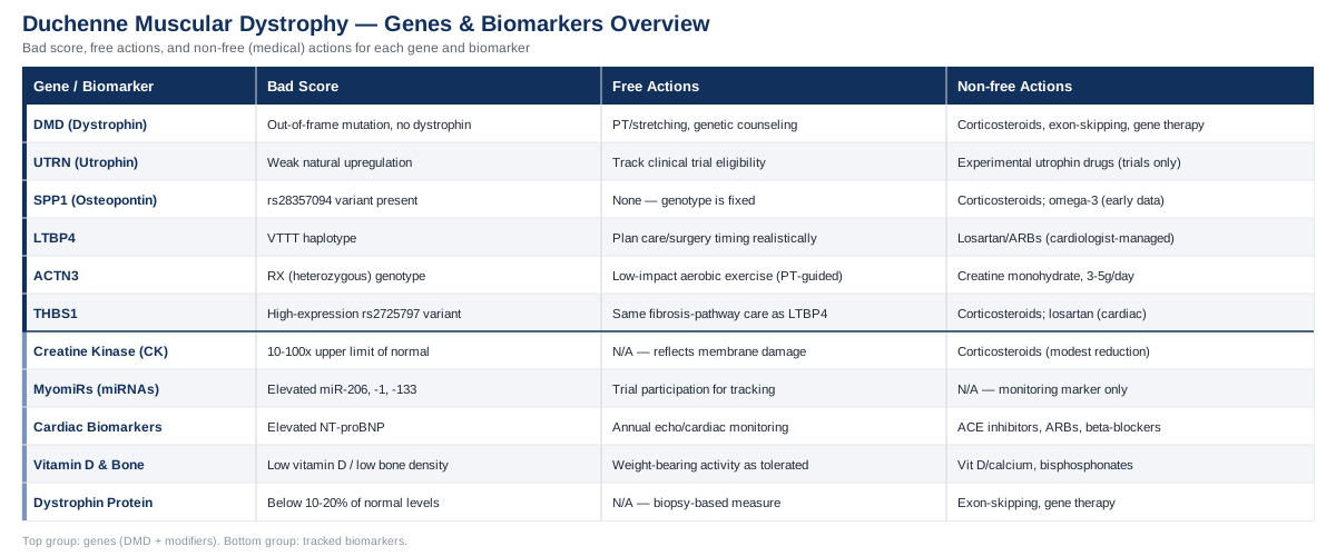
Duchenne muscular dystrophy is caused by mutations in a single gene, DMD, but the way the disease actually plays out in any given boy is shaped by more than that one gene. Below, you'll find a breakdown of the dystrophin gene itself plus five modifier and compensatory genes — UTRN, SPP1, LTBP4, ACTN3, and THBS1 — that help explain why disease course varies between boys with similar mutations, along with what current research does and does not support doing about each one. From there, the article moves to five biomarkers — creatine kinase, circulating myomiRs, cardiac markers, vitamin D and bone turnover markers, and dystrophin protein quantification — that give a running picture of muscle, heart, and bone status over time, each with realistic cost ranges and monitoring schedules. A dedicated section pulls out ten research developments, from the reading-frame rule to the 2025 gene-therapy safety signal, that have measurably shifted clinical thinking in the last several years. The article closes with a short, evidence-checked look at complementary approaches — inspiratory muscle training, manual stretching, music therapy, and photobiomodulation — that can support, but never substitute for, standard neuromuscular care.
The Genes Behind Duchenne — And How the Body Tries to Compensate
Duchenne muscular dystrophy is, at its core, a single-gene disease. But "single-gene" doesn't mean "single story." The primary mutation determines whether the disease happens at all; a small set of modifier genes helps determine how fast it progresses, how the muscle responds to treatment, and how much natural compensation the body can mount. Genome scientists like Ali Torkamani, who has spent years studying how common genetic variants modify disease risk and severity at scale, and clinicians like Gary Brecka, who popularized the idea that a genetic report is only useful once you know what to actually do with each variant, both make the same basic point: a gene result without an action plan is just trivia. The sections below try to avoid that trap for each of the six genes most relevant to Duchenne.
DMD (Dystrophin) — The Gene That Causes the Disease
The DMD gene sits on the X chromosome and is one of the largest genes in the human genome, with 79 exons coding for dystrophin, a protein that anchors the internal skeleton of a muscle fiber to its outer membrane. Without functional dystrophin, muscle membranes tear during normal contraction, triggering cycles of damage, inflammation, and incomplete repair that gradually replace muscle with fibrous and fatty tissue, as described in the GeneReviews clinical summary of dystrophinopathies.
About 65% of disease-causing mutations are large deletions, roughly 10% are duplications, and the remainder are smaller point mutations or splice-site changes. What matters clinically is not just the mutation's location but whether it keeps the genetic "reading frame" intact. Out-of-frame mutations usually abolish dystrophin production almost entirely, producing the more severe Duchenne phenotype; in-frame mutations often allow a shortened but partly functional protein, producing the milder Becker phenotype. This is why genetic testing — deletion/duplication analysis followed by sequencing if needed — is not optional detail but the single most important piece of information for planning care, confirming carrier status in mothers and sisters, and determining eligibility for exon-skipping or gene therapy.
Managing a Confirmed DMD Mutation: The Plan Without Supplements
There is no lifestyle or nutritional intervention that restores dystrophin production, and it's important to say that plainly rather than imply otherwise. The non-supplement plan is the medical and rehabilitative standard of care, and it's substantial:
- Corticosteroids (prednisone or deflazacort) remain the backbone of treatment, shown across decades of practice to preserve muscle strength and delay loss of ambulation, per the American Academy of Neurology practice guideline on corticosteroid treatment in Duchenne muscular dystrophy. Dosing and timing are individualized by a neuromuscular specialist and typically continue daily or on an alternating high-dose weekend schedule for years, with side effects (weight gain, growth suppression, behavioral changes, bone density loss, cataracts) monitored at every visit. - Exon-skipping antisense oligonucleotides — eteplirsen, golodirsen, viltolarsen, and casimersen — are FDA-approved for boys whose mutations are amenable to skipping exons 51, 53, 53, and 45 respectively, delivered as weekly infusions, as summarized in this review of exon-skipping therapies in neuromuscular disease. These only apply to specific mutation types and produce partial, not complete, dystrophin restoration. - Gene therapy (delandistrogene moxeparvovec, brand name Elevidys) delivers a shortened "micro-dystrophin" gene via a single infusion. It carries real safety considerations, including reported cases of acute liver injury, detailed in this NIH LiverTox monograph, and its availability has shifted as regulatory review has evolved — a reminder to confirm current status with a treating physician rather than relying on older information. - Physical therapy and stretching to prevent joint contractures, plus scheduled cardiac and respiratory monitoring, are non-negotiable parts of care regardless of which drug therapies apply. - Genetic counseling for mothers and sisters, since roughly two-thirds of cases are inherited from a carrier mother and the remainder arise from new mutations.
The Plan With Supplements or Equipment
None of the following changes the underlying genetics, and all should be discussed with the treating neuromuscular team before starting, especially because some interact with corticosteroids:
- Vitamin D and calcium are recommended for essentially all boys on chronic corticosteroids, given the added fracture risk. Typical vitamin D dosing is individualized to blood levels (often 600–2,000 IU daily), taken continuously rather than cycled, with periodic blood testing to avoid over-supplementation; excessive dosing can cause hypercalcemia. - Creatine monohydrate has the best supplement-level evidence in muscular dystrophy generally: a Cochrane systematic review found it modestly increases muscle strength and is well tolerated, described in this Cochrane review of creatine for muscle disorders. A common studied approach is 3–5 grams daily, taken continuously without cycling; mild gastrointestinal upset is the main side effect, and hydration should be maintained. - Assistive equipment — ankle-foot orthoses, night splints, standing frames, and later non-invasive ventilation and cough-assist devices — are equipment-based interventions with strong functional evidence for preserving mobility and respiratory function, prescribed and adjusted by a physiatrist or pulmonologist on a schedule tied to disease stage rather than a fixed calendar.
UTRN (Utrophin) — The Body's Natural Backup Gene
Utrophin is dystrophin's fetal-stage paralogue: structurally similar, binding many of the same membrane proteins, but normally switched off in mature muscle except at specialized junctions. In dystrophic muscle, utrophin is naturally upregulated 2–5 fold as part of the repair response, partially compensating for missing dystrophin, a mechanism reviewed in this analysis of utrophin modulator drugs for Duchenne and Becker muscular dystrophy. This natural compensation is the reason mouse models of dystrophin loss (mdx mice) have much milder disease than human patients — mice upregulate utrophin more efficiently than humans do.
If Natural Utrophin Compensation Is Insufficient: The Plan Without Supplements
There is currently no approved drug that reliably boosts utrophin in humans. The most advanced candidate, ezutromid, reached a Phase 2 trial (PhaseOut DMD) and showed increased utrophin expression and reduced muscle damage at 24 weeks, but failed to meet clinical efficacy endpoints at 48 weeks and was discontinued. Newer utrophin-modulating and gene-delivery approaches remain in earlier-stage research. The honest non-supplement plan here is simply staying current with clinical trial eligibility through a neuromuscular center, since this pathway is still experimental rather than actionable today.
The Plan With Supplements or Equipment
No supplement has been shown in controlled human trials to increase utrophin expression or muscle function through this pathway specifically. This is a case where the honest answer is "not yet" rather than offering an unproven regimen — a distinction worth taking seriously given how much online content promises otherwise.
SPP1 (Osteopontin) — A Fibrosis and Inflammation Modifier
SPP1 encodes osteopontin, a protein involved in inflammation and tissue remodeling. A common promoter variant (rs28357094) was shown, in a two-cohort study spanning Italian and international patients, to predict faster disease progression and reduced grip strength, and to modify how well patients responded to corticosteroids — detailed in the original Neurology study on SPP1 genotype and disease severity in Duchenne muscular dystrophy. This is one of the better-replicated modifier findings in the field, though a follow-up study found the related gene TGFBR2, not the SPP1 promoter variant itself, correlated with actual osteopontin protein expression in muscle tissue — a reminder that genotype-to-protein relationships in this area are still being worked out.
If the SPP1 Genotype Suggests Faster Progression: The Plan Without Supplements
There is no way to change this inherited variant, and no approved SPP1-targeted drug exists. What the genotype is currently useful for is clinical trial design and expectation-setting: researchers increasingly use SPP1 status as a covariate to reduce noise in trial results, and clinicians can use it to interpret an individual boy's trajectory in context rather than alarm. Standard-of-care anti-inflammatory treatment (corticosteroids, described above) remains the primary lever regardless of genotype.
The Plan With Supplements or Equipment
Because osteopontin sits in an inflammatory pathway, omega-3 fatty acids have been tested directly in DMD for their anti-inflammatory effect. A small, well-controlled pilot trial of a flavonoid-and-omega-3 combination in boys with muscular dystrophy showed preliminary safety and some biological signal, reported in this randomized double-blind placebo-controlled pilot trial. This is early-stage, small-sample evidence, not proof of functional benefit, and it should be framed that way. Typical studied dosing used continuous daily intake over several months rather than cycling, with no serious side effects reported beyond mild gastrointestinal symptoms in a minority of participants; fish-oil-based omega-3 can modestly affect bleeding risk, so it's worth mentioning before any planned surgery.
LTBP4 — The TGF-Beta Pathway Modifier
LTBP4 (latent TGF-beta binding protein 4) was first identified as a modifier in mouse models of muscular dystrophy and later confirmed in humans: a specific haplotype (IAAM) was associated with a roughly two-year later loss of ambulation compared with the alternative (VTTT) haplotype in corticosteroid-treated boys, per the Annals of Neurology study on LTBP4 genotype and age of ambulatory loss. LTBP4 works by regulating how much active TGF-beta is available to drive fibrosis — the replacement of muscle with scar-like tissue that limits function over time.
If the LTBP4 Genotype Is Associated With Faster Progression: The Plan Without Supplements
As with the other modifier genes, the variant itself can't be changed, but knowing it helps set realistic expectations for the pace of disease and can inform decisions about the timing of interventions like tendon-release surgery or transition to power mobility. Angiotensin receptor blockers such as losartan, which directly blunt TGF-beta signaling, have been tested in a randomized human trial for DMD-related cardiomyopathy and improved cardiac function, though without a measurable reduction in skeletal muscle fibrosis, according to this randomized trial of lisinopril and losartan in Duchenne cardiomyopathy. This is a prescription medication decision for a cardiologist, not a self-directed one.
The Plan With Supplements or Equipment
There is no supplement with confirmed human evidence of favorably shifting TGF-beta signaling in Duchenne muscle. Preclinical (animal) work on anti-fibrotic compounds is active, but it would be misleading to present any of it as a current, human-proven option.
ACTN3 — The "Speed Gene" With a Surprising Second Job
ACTN3 is best known in sports genetics as the "speed gene," where the R577X variant distinguishes power/sprint athletes (RR genotype) from those with reduced fast-twitch muscle protein (XX genotype). In Duchenne, the same variant turns out to matter for a different reason: the null (XX) genotype is linked to reduced fast-twitch muscle strength at baseline but appears to shift muscle metabolism toward a more oxidative, dystrophy-resistant profile, while heterozygous (RX) patients showed one-to-two years earlier loss of ambulation in the discovery cohort, per this Nature Communications study on ACTN3 as a genetic modifier of Duchenne muscular dystrophy. The relationship is genuinely more complex than "one version is simply better," and that nuance is worth keeping rather than flattening into a slogan.
If the ACTN3 Genotype Is Associated With a Less Favorable Course: The Plan Without Supplements
Exercise physiologists have explored whether the ACTN3-linked shift toward oxidative metabolism can be supported through carefully dosed, low-impact aerobic activity (stationary cycling, swimming) rather than eccentric, high-resistance exercise, which can worsen membrane damage in dystrophic muscle. This should be structured by a physical therapist familiar with neuromuscular disease, since the wrong type or intensity of exercise can accelerate damage rather than help.
The Plan With Supplements or Equipment
No supplement has been shown to alter ACTN3-related muscle metabolism in DMD specifically. Creatine monohydrate, discussed under the primary DMD gene section above, remains the best-evidenced general muscle-support supplement regardless of ACTN3 status, at the same 3–5 gram daily, non-cycled dosing.
THBS1 (Thrombospondin-1) — A Newer Fibrosis-Network Modifier
THBS1 encodes thrombospondin-1, which activates TGF-beta signaling by binding directly to LTBP4 in the extracellular matrix. A regulatory variant (rs2725797) linked to reduced THBS1 expression was found to be protective, associated with later loss of ambulation, in a genome-wide study described in this Annals of Neurology paper on long-range genomic regulators of THBS1 and LTBP4. This is a newer finding than the SPP1 or LTBP4 discoveries and, while it fits a coherent fibrosis-pathway story, it should still be considered earlier-stage evidence pending further replication.
If the THBS1 Genotype Suggests a Less Favorable Course: The Plan Without Supplements
Because THBS1 sits in the same TGF-beta/fibrosis network as LTBP4, the practical non-supplement levers are the same ones discussed above — corticosteroid therapy as first-line anti-fibrotic and anti-inflammatory treatment, with losartan considered by a cardiologist specifically for cardiac fibrosis rather than as a general anti-fibrotic strategy.
The Plan With Supplements or Equipment
As with LTBP4, there's no human trial testing a THBS1-targeted supplement in Duchenne muscular dystrophy. The general anti-inflammatory omega-3 evidence noted under SPP1 is the closest available human data touching this broader pathway, and it should be presented with the same caveats: small trial size, biomarker-level rather than functional outcomes, and no claim of altering the genetic modifier itself.
The overall picture across these six genes is consistent: one gene causes the disease, and a handful of others tilt its pace and severity through fibrosis, inflammation, and metabolic pathways. Genetic testing for the modifiers isn't yet part of routine care the way DMD mutation testing is, but it's increasingly used in clinical trial design, and that's likely to translate into more individualized treatment planning over the next several years. With the genetics mapped out, it's worth turning to the biomarkers that let a family and care team track how the disease — and its modifiers — are actually behaving over time.
Biomarkers Worth Tracking Alongside the Genetics
Genetics tells you the starting conditions; biomarkers tell you what's actually happening month to month. Cardiologists and lipidologists like Peter Attia, Thomas Dayspring, and Allan Sniderman have built entire careers on the idea that a single snapshot lab value means little without a trend line and a clear action threshold — the same logic applies directly to Duchenne muscular dystrophy monitoring, where several blood and imaging markers are already built into standard-of-care schedules.
Creatine Kinase (CK)
Why it matters: CK, and specifically the muscle-specific CK-MM isoform, leaks into the bloodstream when muscle membranes are damaged. In untreated Duchenne, CK is typically 10 to 100 times the upper limit of normal, and elevated CK-MM in a dried blood spot is now sensitive and specific enough to be used for newborn screening before symptoms appear, according to this study on CK-MM concentration in newborn dried blood spots.
How it's measured: A standard blood draw processed by any hospital lab, typically $15–$50 out of pocket if not covered by insurance, or a dried blood spot heel-stick for newborn screening panels where implemented.
What may improve it: CK levels naturally fall as muscle mass is progressively lost in later disease stages, which paradoxically means a falling CK in an older boy reflects less remaining muscle to damage, not improvement — a distinction worth understanding rather than misreading as good news. In earlier stages, corticosteroid therapy modestly reduces CK by limiting membrane damage.
Circulating myomiRs (miR-206, miR-1, miR-133)
Why it matters: Muscle-specific microRNAs, or myomiRs, are released from damaged and regenerating muscle fibers. miR-206 in particular shows close to 100% specificity for distinguishing DMD from healthy controls and sits at intermediate levels in the milder Becker form, tracked longitudinally in this longitudinal study of three microRNAs in Duchenne and Becker muscular dystrophy. These are increasingly used as trial-response markers because, unlike CK, they seem to correlate with ongoing regeneration activity rather than just cumulative damage.
How it's measured: Currently a research-lab test (quantitative PCR on blood serum or plasma), typically only available through academic neuromuscular centers or clinical trial participation rather than routine commercial labs; when available, costs are usually study-covered rather than out-of-pocket.
What may improve it: This remains a monitoring marker rather than a target to manipulate directly — its main value is showing whether a therapy (exon skipping, gene therapy, or an anti-inflammatory approach) is measurably changing muscle turnover.
Cardiac Biomarkers (NT-proBNP and Troponin)
Why it matters: Cardiomyopathy is a leading cause of mortality in Duchenne as respiratory and cardiac muscle become involved later in the disease course. Among standard cardiac biomarkers, NT-proBNP — unlike BNP or troponin alone — has been associated with mortality and correlates with left ventricular volume changes on cardiac MRI, per this review of imaging and serum biomarkers for cardiomyopathy in Duchenne muscular dystrophy. Troponin can be mildly and asymptomatically elevated in some boys, which complicates its use as a standalone warning sign.
How it's measured: A blood draw, typically $30–$100 depending on the specific panel and location, generally ordered alongside an annual or twice-yearly echocardiogram or cardiac MRI (echocardiograms run roughly $200–$1,000 before insurance; cardiac MRI is more, often $1,000–$3,000).
What may improve it: Guideline-directed heart failure therapy (ACE inhibitors, angiotensin receptor blockers, beta-blockers), started prophylactically in many centers before overt cardiomyopathy develops, is the primary evidence-based lever, managed by a cardiologist experienced in neuromuscular disease.
Vitamin D and Bone Turnover Markers
Why it matters: Boys with Duchenne face compounded bone fragility from reduced mechanical loading (less walking, less weight-bearing) plus the bone-thinning effects of long-term corticosteroid use, making low-trauma vertebral and long-bone fractures common. Screening guidance calls for routine vitamin D and bone density monitoring in essentially all patients on steroids, as summarized in this review of interventions to prevent corticosteroid-induced osteoporosis in Duchenne muscular dystrophy.
How it's measured: A 25-hydroxyvitamin D blood test (roughly $40–$100) plus periodic dual-energy X-ray absorptiometry (DXA) bone density scans (roughly $100–$300), typically annually once corticosteroids begin, along with spinal imaging to catch silent vertebral compression fractures.
What may improve it: Vitamin D and calcium supplementation, dosed to blood levels and taken continuously rather than in cycles, is first-line; bisphosphonate medications are added by an endocrinologist when bone density is already significantly reduced or a fracture has occurred, and are prescription-managed rather than self-directed.
Dystrophin Protein Quantification
Why it matters: This is the most direct biomarker of all — a muscle biopsy analyzed by Western blot or immunohistochemistry to measure how much dystrophin protein is actually present, used as the primary efficacy readout in exon-skipping and gene therapy trials, since restoring even 10–20% of normal dystrophin levels can meaningfully alter disease trajectory.
How it's measured: A muscle biopsy (typically performed under sedation in a clinical or research setting), which is invasive and generally reserved for diagnostic confirmation or clinical trial participation rather than routine monitoring; costs are highly variable and usually covered under trial protocols or, outside of trials, billed as a surgical procedure (often several hundred to a few thousand dollars depending on setting and anesthesia).
What may improve it: Currently, only exon-skipping therapies and gene therapy have direct trial evidence of increasing measurable dystrophin protein; no supplement or lifestyle intervention has been shown to do so.
Biomarkers and genetics tell complementary parts of the same story — one is the blueprint, the other is the read-out — and together they explain why the research landscape in Duchenne has moved so quickly over the past decade. That momentum is worth understanding in its own right, because it's reshaped what clinicians consider possible.
10 Research Insights Reshaping How Doctors Think About Duchenne
Duchenne muscular dystrophy has functioned as a proving ground for genetic medicine more broadly, a theme traced in detail in Siddhartha Mukherjee's The Song of the Cell, which follows the decades-long arc from identifying the dystrophin gene to attempting to correct it directly. The ten points below draw on that broader arc along with the specific trial and mechanistic evidence already cited above.
1. Duchenne Was the Proving Ground for the Entire Field of Gene Therapy
Because the disease is caused by a single, well-characterized gene affecting a large and accessible tissue (skeletal muscle), Duchenne became one of the first serious human testing grounds for viral gene delivery, well before gene therapy was viable for most other conditions.2. The Reading-Frame Rule Explains Why Some Mutations Are Milder Than Others
The single biggest predictor of severity isn't the size of the mutation but whether it preserves the gene's reading frame — in-frame deletions tend to produce the milder Becker phenotype, out-of-frame deletions the more severe Duchenne phenotype, as detailed in the GeneReviews entry on dystrophinopathies.3. Exon Skipping Deliberately Recreates the Milder Pattern
Exon-skipping drugs work by intentionally forcing the cell to skip an exon during RNA processing, converting an out-of-frame (Duchenne-type) mutation into an in-frame (Becker-type) one — turning a genetic rule discovered by observation into a designed treatment strategy, as reviewed in this clinical review of exon-skipping oligonucleotides.4. Micro-Dystrophin Is a Compromise, Not a Cure
Current gene therapy can't fit the full 2.2-million-base dystrophin gene into a viral vector, so it delivers a shortened "micro-dystrophin" construct retaining only key functional domains — a workable compromise, but explicitly not a full restoration of normal protein.5. Modifier Genes Explain the "Same Mutation, Different Outcome" Puzzle
SPP1, LTBP4, ACTN3, and THBS1 collectively demonstrate that clinical trajectory in Duchenne is not fully determined by the primary mutation, which is part of why clinical trials increasingly stratify or adjust for modifier genotype.6. Gene Editing Was Proven in Dogs Before It Reached Any Human
CRISPR-based correction restored dystrophin to as much as 92% of normal levels in cardiac muscle of a large-animal (canine) model, a critical proof-of-concept step documented in this Science paper on gene editing in a canine model of Duchenne muscular dystrophy, well before any human CRISPR trial for this condition began.7. The Body's Own Backup Gene Has Been Harder to Drug Than Expected
Utrophin upregulation looked like a clean therapeutic shortcut in mice, but the lead human drug candidate, ezutromid, failed to meet clinical endpoints despite biologically increasing utrophin — a reminder that a compelling mechanism doesn't guarantee a working drug.8. Newborn Screening Is Shifting Diagnosis From Age 5 to Day 5
CK-MM testing on the same dried blood spot already used for other newborn screening panels can flag Duchenne before symptoms appear, years earlier than the traditional path of a pediatrician noticing delayed walking, per this study on CK-MM newborn screening — a shift that matters because earlier diagnosis means earlier access to corticosteroids and trial eligibility.9. The Liver Has Become Part of the Gene-Therapy Safety Conversation
Reports of serious, and in rare cases fatal, acute liver injury following AAV-based gene therapy — serious enough to prompt a 2025 FDA clinical hold — have shifted the field's tone from pure enthusiasm to a more measured risk-benefit conversation, documented in the NIH LiverTox monograph on delandistrogene moxeparvovec.10. Corticosteroids Remain the Unglamorous Backbone of Treatment
Despite decades of headline-grabbing gene therapy and gene-editing research, the single intervention with the longest track record for preserving function in Duchenne is still a decades-old, inexpensive steroid, underscoring that the newest science and the most reliable science aren't always the same thing.These research threads are mostly playing out in specialist clinics and trial centers, which is exactly why it's worth knowing what's realistic to layer in at home — not as a replacement for that care, but as genuine support for it.
Complementary Approaches That Can Support — Not Replace — Medical Care
Breathing-Based Therapies (Inspiratory Muscle Training)
As dystrophin loss progressively weakens the diaphragm and intercostal muscles, respiratory decline becomes one of the most consequential aspects of Duchenne, which is why targeted breathing training has been studied directly in this population rather than borrowed from a general wellness context.A meta-analysis pooling multiple trials found that structured inspiratory muscle training was associated with measurable improvements in inspiratory pressure and helped maintain pulmonary function over time in boys with Duchenne, reported in this meta-analysis of inspiratory muscle training in Duchenne muscular dystrophy, though an earlier individual crossover trial found no clear benefit over a placebo device, so the evidence base, while generally positive, isn't uniform.
In practice, this looks like short daily sessions (often 10–20 minutes) using a handheld inspiratory muscle trainer set at a resistance calibrated by a respiratory therapist, reassessed periodically as lung function changes; it should be introduced under pulmonology guidance rather than as a stand-alone home program, particularly once ventilatory support becomes relevant.
Massage Therapy and Manual Stretching
Joint contractures — permanent tightening of muscles and tendons around the ankles, knees, and hips — are one of the most predictable complications of Duchenne, driven by muscle imbalance and reduced mobility, making manual therapies aimed at flexibility a natural and directly relevant complementary approach rather than a general relaxation technique.An international consensus conference on motor rehabilitation in muscular dystrophies formally recommends structured active and passive stretching, performed by therapists, caregivers, or self-managed by the patient after training, as a core part of contracture prevention, detailed in this consensus report on motor rehabilitation in muscular dystrophies and in this review on prevention and management of limb contractures in neuromuscular disease.
A realistic protocol involves daily passive stretching of the ankles, knees, hips, and wrists (often integrated into a bedtime routine), taught by a physical therapist and adjusted as range of motion changes; massage can be added for comfort and muscle tension but should avoid deep or aggressive techniques over very weak or fragile muscle tissue.
Music Therapy and Music-Based Interventions
Living with a progressive, life-limiting diagnosis carries a real psychological burden for both the child and the family, and music-based interventions have accumulated evidence for supporting mood, communication, and engagement during medical care in pediatric neurological populations, even though large Duchenne-specific trials are limited.A broad overview of music therapy in pediatric health care found consistent benefits for mood regulation, communication, and quality of life across chronic and neurological pediatric conditions, summarized in this overview of music therapy and music-based interventions in pediatric health care; evidence specific to Duchenne muscular dystrophy itself is still sparse, so this should be understood as a well-supported general pediatric approach rather than a disease-specific treatment.
Practically, this means involving a credentialed music therapist during physical therapy sessions, hospital stays, or procedures known to cause anxiety (infusions, imaging), rather than expecting music alone to influence disease course — its value here is genuinely about coping and engagement, not muscle biology.
Low-Level Laser Therapy / Photobiomodulation
Photobiomodulation uses specific wavelengths of red or near-infrared light aimed at reducing inflammation and supporting cellular repair, and it has drawn research interest in Duchenne because muscle inflammation and impaired regeneration are central to the disease process.The evidence here is still preclinical: superpulsed low-level laser therapy reduced muscle damage, inflammation, and fibrosis in the mdx mouse model when applied five times weekly for 14 weeks, and a related study found dose-dependent protective effects through modulation of dystrophin-associated pathways, summarized in this review explicitly titled "Photobiomodulation Therapy for Muscular Dystrophy: Time for a Trial?" — a title that itself signals human trials have not yet been established.
Given that the evidence base is currently animal-only, this is best understood as a promising research direction rather than a treatment to pursue today; anyone considering a laser device marketed for muscular dystrophy should ask specifically whether any human trial data exists, since as of now it does not.
Putting It All Together
The clearest takeaway from all of this is that Duchenne muscular dystrophy is genuinely a one-gene disease with a many-gene story. The dystrophin mutation determines the diagnosis; modifier genes like SPP1, LTBP4, ACTN3, and THBS1 help explain the variation in how it unfolds; and a defined set of biomarkers — CK, myomiRs, cardiac markers, bone health markers, and dystrophin quantification itself — turns that biology into something a family and care team can actually track over time. Nothing in this article changes the underlying genetics, and no supplement or complementary approach substitutes for corticosteroids, exon-skipping therapy, gene therapy where eligible, or structured cardiac, respiratory, and orthopedic monitoring.
What does change with better information is the quality of the conversation you can have with a neuromuscular specialist — which biomarkers to ask about at the next visit, which modifier-gene research is close to clinical relevance versus still preclinical, and which complementary approaches are worth layering in versus skipping entirely. If you haven't already, the next concrete step is a direct conversation with the treating neuromuscular team about current CK trends, the most recent cardiac and bone density results, and whether genetic testing has clarified exon-skipping or gene therapy eligibility — that conversation, more than any single fact in this article, is where real decisions get made.
Respiratory Endocrine & Metabolic
Musculoskeletal: Bone Conditions