This article contains AI generated content.
· UpdatedDysbaric Osteonecrosis: 7 Biomarkers and 5 Genes to Track
Introduction
If you dive professionally or have done so for years, you already know that decompression sickness is not the only risk lurking under pressure. Dysbaric osteonecrosis (DON) — a form of avascular necrosis triggered by repeated hyperbaric exposure — develops silently in the bones of the shoulder or hip, often without symptoms until significant structural damage has already occurred. Most divers who receive the diagnosis feel blindsided, partly because the condition is rarely discussed before it becomes a problem, and partly because standard pre-dive health evaluations were never designed to detect individual biological susceptibility.
The frustrating part is that not every diver develops it. Two divers with nearly identical exposure histories, dive tables, and habits can have radically different outcomes. That gap almost always comes down to individual biology — how efficiently your body manages gas bubbles, controls coagulation tendency, handles fat metabolism, and repairs bone microvasculature after each hyperbaric stress event. Generic advice to follow decompression protocols is correct but profoundly incomplete. It does nothing to explain why some people are more vulnerable, or what those individuals could actually do differently.
This article takes a more precise approach. Rather than repeating the standard prevention checklist, it explores the biological mechanisms that make certain individuals more susceptible — specifically through two complementary lenses: biomarkers you can measure with standard blood tests, and genetic variants you can identify through accessible testing. Together, these two frameworks give you something actionable: a signal to track, a mechanism to address, and concrete information to bring to a hyperbaric medicine specialist or physician.
Better information rarely leads to certainty, but it consistently leads to better decisions. Whether you are a professional diver assessing long-term risk, a clinician supporting patients post-diagnosis, or someone already managing DON who wants to understand the underlying biology, the frameworks below can meaningfully change how you approach this condition. The biomarker strategy comes first, because it offers the most measurable, actionable entry point. The genetic framework follows as a deeper layer of individual risk profiling.
Summary
Dysbaric osteonecrosis is not simply the result of bad luck or aggressive diving technique. For a meaningful subset of those affected, the root vulnerability lies in specific biomarkers and genetic variants that compromise vascular integrity, increase coagulation tendency, impair bone repair capacity, or dysregulate fat metabolism — all mechanisms directly implicated in how bone tissue dies under repeated pressure exposure.
This article covers 7 biomarkers worth tracking — including ApoB, Lp(a), homocysteine, and bone turnover markers — explaining what each reveals about your specific risk, how to measure it, what a concerning result looks like, and exactly what to do about it with and without supplementation. It also covers 5 genes with documented links to osteonecrosis susceptibility (MTHFR, NOS3, Factor V Leiden, APOE, and VDR), each with an actionable plan based on your result. Beyond that, you will find a synthesis of the most relevant insights from Peter Attia's Outlive framework applied to the DON context, and four complementary modalities — from photobiomodulation to microbiome-directed therapy — that have meaningful clinical evidence for bone repair and vascular support.
There are no cure claims here, and no shortcuts. What you will find is a more complete map of the terrain — one precise enough to act on.
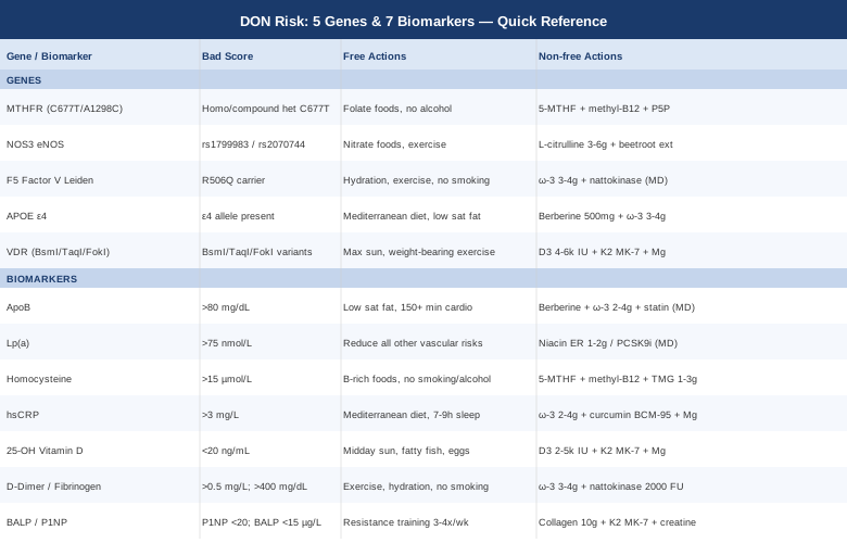
7 Biomarkers That Reveal Your True Risk for Dysbaric Osteonecrosis
The biology of dysbaric osteonecrosis converges on a few key pathways: lipid embolism, hypercoagulability, endothelial dysfunction, impaired nitric oxide signaling, and inadequate bone repair. These are not theoretical constructs — each is measurable through blood tests available in most medical laboratories. The seven biomarkers below are organized from the most vascular-centric to the most bone-specific, because in DON, vascular compromise almost always precedes and drives structural bone damage.
1. ApoB (Apolipoprotein B)
Why it matters for DON: ApoB is the protein carried by every atherogenic lipoprotein particle — LDL, VLDL, IDL, and Lp(a). Because each particle carries exactly one ApoB molecule, measuring ApoB gives the most accurate count of potentially harmful lipid particles circulating in your blood. In the context of DON, this matters because fat emboli are one of the leading hypotheses for how bone ischemia develops: lipid particles can aggregate under hyperbaric pressure, occlude the microvasculature of bone marrow, and trigger ischemic necrosis at the cellular level. Elevated ApoB also reflects a broader atherogenic state that weakens the small vessels feeding bone tissue over time.
Clinicians including Peter Attia and Thomas Dayspring have argued compellingly that ApoB outperforms LDL-C as a cardiovascular risk marker — and the same logic applies to microvascular bone risk. Standard lipid panels often miss the real picture because LDL-C can appear normal even when particle count and true vascular risk are both elevated.
How to measure it: ApoB is measured via a standard blood test, fasting or non-fasting, and is increasingly available on routine lipid panels, though it sometimes needs to be requested explicitly. Cost: $20–$60 depending on lab and country. Optimal range: below 80 mg/dL for higher-risk individuals; below 90 mg/dL for the general population. Some clinicians target below 60 mg/dL for those with established vascular disease.
If the score is bad, the plan without supplements: The most impactful dietary lever is reducing saturated fat — not dietary fat in general, but specifically saturated fats (red meat, dairy fat, palm oil, coconut oil) that raise LDL particle number. Replacing these with monounsaturated fats (olive oil, avocado) and omega-3-rich foods (fatty fish, walnuts) tends to reduce ApoB meaningfully within 6–12 weeks. Aerobic exercise at 150–200 minutes per week independently lowers ApoB. Eliminating or significantly reducing alcohol is also underrated as a lever — alcohol raises VLDL production and thus overall particle count. These combined changes can reduce ApoB by 10–20% in adherent individuals.
If the score is bad, the plan with supplements or medications: Berberine (500 mg twice daily with meals, cycled 8 weeks on / 2 weeks off) modestly reduces ApoB and LDL particle count via upregulation of LDL receptors. Omega-3 fatty acids at pharmacological doses (2–4 g EPA+DHA per day) primarily lower triglycerides and VLDL, offering an indirect ApoB benefit. For persistently elevated ApoB despite lifestyle change, statins remain the most evidence-supported pharmacological intervention and should be managed by a prescribing physician. Potential side effects of statins include myopathy and, rarely, liver enzyme elevation — baseline liver function monitoring is standard practice.
2. Lipoprotein(a) — Lp(a)
Why it matters for DON: Lp(a) is a lipoprotein particle that combines an LDL-like core with apolipoprotein(a), a protein structurally similar to plasminogen — the body's primary clot-dissolving enzyme. This structural similarity means Lp(a) can competitively inhibit fibrinolysis, resulting in clots that form more easily and dissolve more slowly. For DON, this is directly and mechanistically relevant: occlusion of bone microvasculature by small thrombi is one of the core pathways to bone ischemia, and elevated Lp(a) amplifies exactly this mechanism following any hyperbaric exposure event.
Lp(a) is up to 90% genetically determined and does not respond meaningfully to most lifestyle interventions. Knowing your level does not change the genetics, but it changes clinical decisions — particularly around diving frequency, how conservative your decompression profiles should be, and what other vascular risk factors need compensatory management.
How to measure it: A single Lp(a) blood test is sufficient — levels are genetically set and do not fluctuate substantially. Ideally measured in nmol/L rather than mg/dL, as the unit conversion is imprecise. Cost: $30–$80. High risk: above 75 nmol/L (some clinicians use 50 nmol/L as a lower threshold). This marker is often absent from standard panels — request it explicitly.
If the score is bad, the plan without supplements: Lifestyle interventions do not meaningfully lower Lp(a). The practical response is compensatory: reducing every other modifiable vascular risk factor aggressively, maintaining the lowest achievable ApoB, optimizing blood pressure, avoiding dehydration and prolonged immobility before and after dives, and never smoking. For divers, significantly elevated Lp(a) is a strong clinical argument for more conservative decompression profiles, longer surface intervals, and reduced frequency of deep exposures.
If the score is bad, the plan with supplements or medications: Niacin (extended-release form, 1–2 g/day under medical supervision) can reduce Lp(a) by 20–30%, though its cardiovascular outcomes benefit remains debated in clinical trials due to side effects including flushing and glucose dysregulation. Novel drugs including PCSK9 inhibitors and RNA-based therapies targeting apolipoprotein(a) are showing strong phase 2 and 3 results — this is a rapidly evolving area worth following with a cardiologist if your Lp(a) is significantly elevated.
3. Homocysteine
Why it matters for DON: Homocysteine is an amino acid produced during methionine metabolism. When methylation capacity is impaired — most commonly due to MTHFR gene variants, B vitamin insufficiency, or both — homocysteine accumulates in the blood and damages endothelial cells. That damage reduces nitric oxide availability, increases vascular stiffness, promotes platelet aggregation, and raises the overall thrombotic burden. For bone specifically, elevated homocysteine is independently associated with increased fracture risk, reduced bone density, and impaired bone matrix repair. In DON, the combination of endothelial fragility and diminished repair capacity is particularly consequential.
Multiple studies have explored the link between hyperhomocysteinemia and avascular necrosis, consistently pointing to endothelial dysfunction as the shared mechanistic thread.
How to measure it: Serum homocysteine, measured fasting. Cost: $25–$60. Optimal: below 10 µmol/L. Elevated: above 15 µmol/L. High risk: above 20 µmol/L. Some functional medicine practitioners target below 7 µmol/L for optimal vascular protection. Note that acute illness and dehydration transiently elevate homocysteine — test under normal conditions.
If the score is bad, the plan without supplements: Diet is the first intervention. Increasing B vitamin-rich foods (dark leafy greens for folate, eggs and meat for B12, poultry and legumes for B6) directly supports the methylation cycle. Reducing very high-protein diets without adequate B vitamin support lowers methionine load. Smoking cessation and alcohol reduction both meaningfully lower homocysteine — each is an independent driver of elevated levels.
If the score is bad, the plan with supplements: The targeted intervention is the methylation triad: methylfolate (5-MTHF, 400–1,000 mcg/day), methylcobalamin (B12, 500–1,000 mcg/day sublingual), and P5P (pyridoxal-5-phosphate, the active form of B6, 25–50 mg/day). Using the methyl forms specifically is important — standard folic acid may not be efficiently converted in individuals with MTHFR variants. Betaine (trimethylglycine, 1–3 g/day) provides an alternative methylation pathway and can reduce homocysteine independently of folate status. B6 toxicity is possible above 100 mg/day — stay below that threshold. Retest homocysteine every 3 months to confirm efficacy and calibrate dosing.
4. High-Sensitivity CRP (hsCRP)
Why it matters for DON: Chronic low-grade inflammation damages the endothelium, impairs bone repair signaling, and may worsen the microvascular compromise already present in DON. hsCRP is the most accessible and widely used marker of systemic inflammation in clinical practice. While non-specific by nature, persistently elevated hsCRP in the context of DON suggests that inflammatory activity is compounding the vascular and ischemic mechanisms driving disease progression or preventing recovery.
How to measure it: Standard blood test, ideally measured fasting and outside any acute illness (which transiently elevates CRP to levels that mask baseline). Cost: $15–$40. Target: below 1 mg/L (cardiovascular optimal). Moderate risk: 1–3 mg/L. High risk: above 3 mg/L. The "high-sensitivity" designation refers to the assay's ability to detect values below 1 mg/L — ensure that your lab is running hsCRP and not standard CRP.
If the score is bad, the plan without supplements: A Mediterranean diet pattern — olive oil as the primary fat, abundant vegetables, legumes, fatty fish, and limited refined carbohydrates — consistently reduces hsCRP in randomized trials. Eliminating ultra-processed foods and seed oils contributes meaningfully. High-quality sleep (7–9 hours nightly) and 150+ minutes of moderate aerobic exercise weekly both independently reduce systemic inflammation. Excess visceral adiposity is one of the strongest drivers of elevated hsCRP — even modest fat loss of 5–10% body weight has a disproportionate anti-inflammatory effect.
If the score is bad, the plan with supplements: High-dose omega-3 (2–4 g EPA+DHA/day) is the best-supported supplement for reducing hsCRP and has a strong safety profile at these doses. Curcumin in a bioavailable form — BCM-95 or phospholipid complex (500 mg twice daily) — has consistent anti-inflammatory evidence; cycle 8 weeks on, 4 weeks off to maintain efficacy. Vitamin D supplementation corrects deficiency-driven inflammation. Magnesium (glycinate or malate, 300–400 mg/day, ongoing) supports endothelial function and modestly reduces inflammatory markers. High-dose omega-3 can modestly raise LDL in a small subset — monitor ApoB if adding this intervention.
5. 25-OH Vitamin D
Why it matters for DON: Vitamin D is essential for calcium absorption, bone mineralization, and immune regulation within the bone marrow microenvironment. Beyond direct bone effects, vitamin D plays a significant role in endothelial function — deficiency is independently associated with increased cardiovascular risk and reduced vascular repair capacity. In DON, vitamin D status directly affects the bone's ability to respond to ischemic injury and mount an effective repair response. It is one of the most modifiable biomarkers on this list, and also one of the most commonly deficient — particularly in individuals at northern latitudes or those who spend substantial time underwater away from midday sun.
How to measure it: Serum 25-hydroxyvitamin D. Cost: $30–$70. Optimal: 40–60 ng/mL (100–150 nmol/L). Deficiency: below 20 ng/mL. Test in winter when baseline is lowest, and retest after 3 months of supplementation to confirm absorption and calibrate dose.
If the score is bad, the plan without supplements: 15–20 minutes of midday sun on large skin surface area (arms, legs, torso) generates approximately 1,000–2,000 IU/day in lighter-skinned individuals. Dietary sources — fatty fish, egg yolks, fortified foods — contribute modestly but are rarely sufficient to correct true deficiency alone.
If the score is bad, the plan with supplements: Vitamin D3 (cholecalciferol) at 2,000–5,000 IU/day is a well-tolerated starting dose; significantly deficient individuals may require 10,000 IU/day under medical supervision. Always co-administer vitamin K2 (MK-7 form, 100–200 mcg/day) to direct calcium into bone rather than arterial walls — this pairing is essential at higher D3 doses. Magnesium is a required cofactor for vitamin D conversion — if magnesium status is uncertain, supplement simultaneously. Retest at 3 months to calibrate. Toxicity is possible at sustained very high doses without monitoring — do not exceed 10,000 IU/day without 25-OH D oversight.
6. D-Dimer and Fibrinogen
Why it matters for DON: D-dimer is a fibrin degradation product that rises when the body is actively forming and breaking down clots. Fibrinogen is the structural precursor to fibrin and reflects coagulation tendency at baseline. Both are directly relevant to the thrombotic theory of DON: if microthrombi are occluding bone vasculature after decompression events, these markers reveal whether an individual is operating in a chronically hypercoagulable state. Persistently elevated D-dimer or fibrinogen suggests a pro-thrombotic environment that amplifies the vascular consequences of any bubble formation or fat embolism during hyperbaric exposure.
How to measure it: Both are available as standard blood tests. D-dimer is often ordered in acute settings but can serve as a baseline risk assessment. Fibrinogen appears on many expanded cardiac risk or coagulation panels. Cost: $20–$50 each. D-dimer optimal: below 0.5 mg/L FEU. Fibrinogen: 200–400 mg/dL; sustained levels above 400 mg/dL warrant clinical attention.
If the score is bad, the plan without supplements: Regular aerobic exercise significantly reduces fibrinogen over time through improved fibrinolytic activity. An omega-3-rich dietary pattern reduces platelet aggregation and modestly affects coagulation tendency. Hydration is underappreciated: dehydration concentrates coagulation factors and dramatically increases clotting risk — maintaining optimal hydration before, during, and after dives is clinically meaningful for anyone with elevated fibrinogen. Smoking cessation is among the most powerful single interventions for lowering fibrinogen — it is one of the strongest modifiable drivers of chronically elevated levels.
If the score is bad, the plan with supplements: High-dose EPA/DHA omega-3 (3–4 g/day) has clinically relevant antiplatelet and mild anticoagulant properties — disclose this to any prescribing physician, particularly before surgical procedures. Nattokinase (100–200 mg standardized, approximately 2,000 FU, twice daily on an empty stomach) is a fibrinolytic enzyme derived from fermented soy with growing evidence for reducing fibrinogen and clot burden; use with extreme caution in those already on anticoagulants. Cycle nattokinase 3 months on, 1 month off. Always disclose coagulation-active supplements to your hyperbaric medicine physician before diving — the interplay with decompression physiology requires clinical judgment.
7. Bone Alkaline Phosphatase (BALP) and P1NP
Why it matters for DON: These are the most clinically informative markers of active bone formation. Bone-specific alkaline phosphatase reflects osteoblast activity; P1NP (procollagen type 1 N-terminal propeptide) is considered the most sensitive and specific marker of new bone formation currently available, recommended by the International Osteoporosis Foundation as the preferred bone formation marker. In DON, tracking these markers reveals whether the affected bone is in active repair mode or operating in a state of suppressed remodeling. This is particularly useful during recovery from diagnosed DON or when evaluating whether an intervention is having a meaningful effect on bone biology. Pairing with CTX (C-terminal telopeptide, a resorption marker) completes the bone turnover picture.
How to measure it: BALP and P1NP are serum blood tests available at most reference laboratories, though they may need to be requested specifically. Cost: $40–$100. P1NP in active adults: optimal 20–60 µg/L. BALP: 15–40 µg/L. CTX (resorption): below 0.58 ng/mL in premenopausal women, below 0.70 ng/mL in men. Note that these markers have circadian variation — measure in the fasting morning state for reproducibility.
If the score is bad, the plan without supplements: Weight-bearing resistance exercise is the most powerful stimulus for osteoblast activity — 3–4 sessions per week of progressive resistance training has consistent evidence for raising P1NP and BALP over 12–24 weeks. Adequate dietary protein (1.2–1.6 g/kg body weight daily) is non-negotiable for osteoblast function. Eliminating proton pump inhibitors where not medically necessary (they impair calcium absorption), reducing excessive alcohol (direct osteoblast suppressor), and prioritizing sleep quality — bone formation peaks during slow-wave deep sleep stages — all meaningfully support bone turnover toward formation.
If the score is bad, the plan with supplements: Hydrolyzed collagen peptides (10 g/day from a type I/III collagen source, co-administered with vitamin C for synthesis support) have growing evidence for improving bone formation markers and bone matrix quality. Vitamin K2 as MK-7 (150–180 mcg/day) activates osteocalcin, a key bone matrix protein — this is the most bone-specific K2 application and is particularly relevant in DON. Creatine monohydrate (3–5 g/day, ongoing) supports muscle loading capacity and may independently increase bone density via enhanced mechanical loading. For significantly low P1NP suggesting severely blunted bone repair, a bone specialist consultation is warranted — pharmacological interventions exist but require careful evaluation of the specific DON presentation.
Together, these seven biomarkers provide a functional map of the vascular, inflammatory, coagulation, and bone-repair processes most relevant to DON. Tracking all of them at least annually — with more frequent monitoring for any values out of range — creates an early-warning and response system that no decompression table can replicate.
5 Genes That Shape Your Susceptibility to Dysbaric Osteonecrosis
Genetics does not determine destiny in DON — dive profile and decompression practice remain the dominant controllable risk factors. But for a meaningful subset of individuals, specific genetic variants in key pathways make the same hyperbaric exposure substantially more dangerous. Understanding these variants helps explain the seemingly random variability in who develops DON and who does not — and gives those individuals a more targeted biological strategy.
MTHFR (C677T and A1298C)
The variant: MTHFR encodes methylenetetrahydrofolate reductase, the enzyme that converts dietary folate into its active methylated form (5-methyltetrahydrofolate), which is then used to remethylate homocysteine into methionine. The C677T variant reduces enzyme activity by approximately 35% in heterozygotes and up to 70% in homozygotes. A1298C has a smaller individual effect but compounds C677T in compound heterozygotes.
How it connects to DON: Reduced MTHFR function means elevated homocysteine — directly connecting this gene to the third biomarker above. Elevated homocysteine damages endothelial cells, increases thrombotic tendency, and impairs the microvascular function of bone. MTHFR variants have been associated with avascular necrosis in multiple published studies, with endothelial damage and thrombophilia as the proposed shared mechanisms.
If the gene is bad, the plan without supplements: Prioritize folate-dense whole foods: dark leafy greens, legumes, and liver are among the richest sources. Minimize processed foods fortified with synthetic folic acid — the unconverted form may compete with methylfolate in MTHFR-impaired individuals. Avoid alcohol, which independently impairs folate absorption and methylation cycle function. Ensure protein intake is adequate (folate and B12 deficiency are more impactful in high-methionine, low-B-vitamin diets). Monitor homocysteine as the downstream readout of methylation capacity.
If the gene is bad, the plan with supplements: Methylfolate (5-MTHF, 400–800 mcg/day for heterozygotes; 800–1,600 mcg/day for homozygotes) is the targeted correction — bypassing the defective enzyme entirely. Methylcobalamin (B12, 500–1,000 mcg/day sublingual) works synergistically. P5P (active B6, 25–50 mg/day) supports the alternative transsulfuration pathway for homocysteine clearance. Start low with methylfolate — some individuals with MTHFR variants experience temporary overstimulation when beginning methyl donors. If this occurs, reduce dose and titrate up over 2–4 weeks. Ongoing supplementation is generally appropriate; retest homocysteine every 3–6 months to confirm efficacy.
NOS3 (eNOS) — rs1799983 and rs2070744
The variant: NOS3 encodes endothelial nitric oxide synthase (eNOS), the enzyme responsible for producing nitric oxide (NO) in blood vessel walls. NO is essential for vasodilation, inhibiting platelet aggregation, and maintaining endothelial integrity. Two common variants — the Glu298Asp substitution (rs1799983) and the T-786C promoter variant (rs2070744) — have been associated with reduced eNOS expression and lower baseline NO production.
How it connects to DON: Reduced NO production impairs vasodilation in the microcirculation of bone, narrowing the safety margin when gas bubbles or fat emboli transiently compromise bone perfusion. This is a structural vascular vulnerability that does not change with dive technique alone. NOS3 variants have been studied in the context of avascular necrosis and ischemic bone conditions, and the biological plausibility is strong: when the vasodilatory response is blunted, even a modest occlusive event can produce ischemia that a healthier endothelium would have survived.
If the gene is bad, the plan without supplements: Dietary nitrates provide a powerful salvage pathway that produces NO through a mechanism that bypasses eNOS entirely. Eating one to two servings of nitrate-rich vegetables daily — beetroot, arugula, spinach, celery, radishes — can raise NO levels meaningfully even in NOS3 variant carriers. Aerobic exercise upregulates eNOS expression over time even in the presence of these variants. Crucially, avoid antibacterial mouthwash, which destroys the oral bacteria responsible for converting dietary nitrates to nitrites — this preserves the dietary nitrate pathway. Cold exposure transiently stimulates NO production through alternative mechanisms.
If the gene is bad, the plan with supplements: L-citrulline (3–6 g/day) raises plasma arginine — the substrate for eNOS — more effectively than direct arginine supplementation because citrulline bypasses first-pass hepatic clearance. Beetroot extract (500 mg concentrated extract daily, equivalent to approximately 300 mg dietary nitrate) provides the direct NO pathway bypass. Pycnogenol (French maritime pine bark extract, 150–200 mg/day) has evidence for upregulating eNOS expression; cycle 3 months on, 1 month off. Note that NO-boosting interventions can lower blood pressure — monitor if already on antihypertensives and disclose to your prescribing physician.
F5 (Factor V Leiden) — rs6025
The variant: The Factor V Leiden mutation (R506Q) creates a form of Factor V resistant to inactivation by activated protein C (APC), the body's natural anticoagulant brake. This results in prolonged Factor Va activity and a hypercoagulable state. Heterozygotes have a 4–8-fold increased risk of venous thrombosis; homozygotes have an approximately 80-fold increase. It is the most common inherited thrombophilia in people of European ancestry, present in 3–8% of the population.
How it connects to DON: The thrombotic pathway to DON — microthrombi occluding bone vasculature following decompression — is directly amplified by Factor V Leiden. Carriers have an impaired ability to brake clot formation, making the vascular consequences of bubble formation or fat emboli more severe and more sustained. Among the genetic variants on this list, Factor V Leiden has the most direct mechanistic connection to the thrombotic theory of DON and arguably the strongest argument for changing clinical management of affected divers.
If the gene is bad, the plan without supplements: Vigorous hydration before and after dives reduces blood viscosity — particularly important for F5 Leiden carriers. Avoiding dehydration is not optional for this population. Consistent aerobic exercise, healthy body weight, smoking abstinence, and avoiding prolonged pre-dive immobility all matter more for Leiden carriers than for the general diving population. Women carrying F5 Leiden should avoid estrogen-containing oral contraceptives, which interact dramatically with this variant to multiply thrombotic risk.
If the gene is bad, the plan with supplements: Antiplatelet and fibrinolytic support with medical oversight: omega-3 (3–4 g EPA+DHA/day), nattokinase (as described in the D-dimer section, with the same contraindication caveats regarding anticoagulants), and possibly low-dose aspirin (81 mg/day) with physician agreement. None of these correct the underlying F5 Leiden defect — they partially offset it. F5 Leiden homozygotes considering continued professional diving should obtain a formal consultation with a hematologist and a hyperbaric medicine specialist. This is one genetic result that should directly and substantially modify the clinical risk conversation.
APOE (ε2, ε3, ε4 Alleles)
The variant: APOE encodes apolipoprotein E, central to lipid transport and clearance. The three major alleles (ε2, ε3, ε4) differ substantially in their metabolic effects. APOE ε4 carriers tend to have higher LDL-C, higher ApoB, higher Lp(a), and more inflammatory responses to dietary fat compared to ε3 carriers. They also show impaired lipid clearance following high-fat meals.
How it connects to DON: Fat embolism is a leading mechanistic hypothesis for the bone ischemia in DON. APOE ε4 carriers present a lipid metabolism profile that could increase the size and frequency of fat emboli following hyperbaric exposure, while also raising baseline vascular and microvascular risk. Compounding this, APOE ε4 is associated with increased neuroinflammation and microvascular dysfunction — downstream pathways that likely worsen outcomes across ischemic conditions including bone.
If the gene is bad, the plan without supplements: APOE ε4 carriers are disproportionately sensitive to dietary saturated fat — even a modest increase in saturated fat intake raises ApoB more in ε4 carriers than in ε3 carriers. A Mediterranean dietary pattern is particularly important for this genotype: high in monounsaturated fats and omega-3s, low in saturated fat. Monitoring ApoB quarterly rather than annually is warranted. Regular aerobic exercise is a stronger cardiovascular risk-reduction tool for ε4 carriers than for most other genotypes.
If the gene is bad, the plan with supplements: The ApoB-lowering supplementation strategy (berberine, high-dose omega-3, possible pharmacological intervention) applies with greater urgency in ε4 carriers. Research suggests ε4 carriers may require higher omega-3 doses to achieve the same anti-inflammatory effect as ε3 carriers — targeting 3–4 g EPA+DHA/day rather than 2 g. Phosphatidylcholine (from soy or sunflower lecithin, 1,200–2,400 mg/day) supports lipid emulsification and hepatic clearance — the evidence base is limited in the DON context specifically, but the biological plausibility for fat embolism risk reduction is reasonable.
VDR (Vitamin D Receptor) — BsmI, TaqI, FokI
The variant: VDR encodes the nuclear receptor for vitamin D. Common SNPs including BsmI (rs1544410), TaqI (rs731236), and FokI (rs2228570) influence receptor sensitivity and downstream gene expression. The key practical implication: two individuals with identical serum 25-OH D levels can have substantially different biological vitamin D activity depending on their VDR genotype.
How it connects to DON: VDR variants influence bone mineral density, bone repair rate, immune regulation in bone marrow, and osteoblast responsiveness to vitamin D signaling. In DON, where the bone must mount a repair response to ischemic damage, reduced VDR sensitivity can delay healing and reduce the apparent effectiveness of vitamin D supplementation unless serum levels are pushed higher. Studies examining VDR polymorphisms in avascular necrosis populations have found associations with disease presentation and bone density outcomes in multiple ethnic cohorts.
If the gene is bad, the plan without supplements: The compensatory strategy for impaired VDR sensitivity is to push circulating 25-OH D levels to the upper end of the optimal range — typically 50–70 ng/mL — so that even a less-responsive receptor achieves adequate vitamin D gene activation. This requires maximizing sun exposure and tracking serum levels closely. Weight-bearing exercise and adequate dietary calcium provide the bone-mechanical stimulus that functions in parallel with VDR signaling pathways.
If the gene is bad, the plan with supplements: Target 50–70 ng/mL serum 25-OH D rather than the lower boundary of "normal" — this typically requires 4,000–6,000 IU/day D3, with K2 (MK-7, 150–200 mcg/day) essential at these doses to prevent arterial calcium deposition. Magnesium (300–400 mg/day glycinate or malate) is a required cofactor for VDR function and vitamin D conversion — deficiency dramatically limits vitamin D effectiveness regardless of supplementation dose. Retest serum levels at 3 months, then every 6 months once stable. Do not exceed 10,000 IU/day without serum 25-OH D monitoring.
Understanding the genetic picture alongside the biomarker data gives a layered view of risk that neither approach alone can provide. A person with elevated Lp(a), Factor V Leiden, and an NOS3 variant is facing a very different risk profile than someone with elevated hsCRP alone — and their management strategy should reflect that.
What Peter Attia's "Outlive" Can Teach Divers About Vascular and Bone Longevity
Peter Attia's Outlive: The Science and Art of Longevity (2023) is not a diving medicine textbook, but its central framework — identifying the upstream drivers of disease long before clinical manifestation — maps directly onto the challenge of DON prevention and recovery. The book synthesizes decades of research on cardiovascular risk, metabolic health, and bone longevity, with an emphasis on biomarker-driven, individualized prevention rather than population-level guidelines. The following ten insights from Attia's framework are the most directly applicable to the DON context.
1. ApoB Is the Number That Matters Most for Vascular Risk
Attia argues that ApoB should replace LDL-C as the primary lipid management target in clinical practice. Every unit reduction in ApoB represents a proportional reduction in the number of particles capable of depositing in or occluding vessels — including the small bone microvasculature central to DON. He recommends lifelong tracking starting in the early 30s for all adults.2. Lp(a) Is a Silent, Underdiagnosed, High-Stakes Risk Factor
Attia advocates for universal Lp(a) testing — once in a lifetime is enough to know your genetic set-point. High Lp(a) changes everything downstream: it means other modifiable vascular risk factors must be managed even more aggressively to compensate for what cannot be changed.3. Zone 2 Cardio Is the Most Powerful Metabolic Intervention Available
Attia emphasizes 3–4 hours per week of Zone 2 (low-intensity, fully conversational aerobic exercise) as the single highest-yield intervention for mitochondrial health, insulin sensitivity, and endothelial function. For DON prevention, this translates to sustained microvascular protection across all tissues — including the bone vascular supply that matters most.4. VO2 Max Predicts Longevity More Than Almost Any Other Measurable Metric
Higher VO2 max means better oxygen delivery to all tissues, including ischemic or marginally perfused bone. Attia targets the top 25th percentile for age and sex as a minimum meaningful goal. Training to improve VO2 max also upregulates eNOS expression over time — directly relevant for NOS3 variant carriers.5. Bone Mineral Density Must Be Tracked Early — Not After the Fracture
Attia recommends baseline DEXA scans in the early 40s, or earlier for high-risk individuals, treating bone as a metabolic organ that requires active management rather than passive clinical attention. In DON, structural bone damage occurs at sites that may not appear on standard DEXA until advanced — but tracking density trajectory and bone turnover markers from a healthy baseline creates a meaningful reference point.6. Sleep Is the Non-Negotiable Foundation of Tissue Repair
Attia gives significant weight to sleep architecture — particularly deep slow-wave sleep — for growth hormone release, tissue repair, and bone remodeling. For DON patients, this means sleep optimization is not a lifestyle suggestion but a genuine physiological repair mechanism. Chronic sleep deprivation suppresses osteoblast activity and elevates cortisol — both directly worsen bone health.7. Protein Intake Is Chronically Underestimated in Active Adults
Attia advocates for 1.6–2.2 g of protein per kg of body weight daily for active individuals, distributed across meals. Adequate protein is required for collagen synthesis, osteoblast function, and the muscle mass that mechanically drives bone remodeling. Most people with or at risk for DON are eating substantially less than this.8. Insulin Resistance Quietly Damages the Vasculature Years Before Diagnosis
Elevated fasting insulin and impaired glucose regulation damage endothelial cells through advanced glycation end-products (AGEs) and oxidative stress — pathways that worsen bone microvascular function and repair capacity. Attia recommends periods of continuous glucose monitoring (CGM) to identify insulin resistance patterns that are entirely invisible to fasting glucose tests alone.9. Time-Restricted Eating May Improve Metabolic Flexibility Without Caloric Restriction
Attia discusses the potential of aligning eating windows with circadian biology to improve lipid profiles, reduce systemic inflammation, and enhance autophagy — cellular maintenance processes relevant to vascular wall integrity. For divers, metabolic optimization in the offseason represents a meaningful window of preventive intervention.10. Psychological Stress Is a Direct Physiological Risk Variable
Attia frames chronic psychological stress as a driver of cortisol elevation that suppresses bone formation, raises fibrinogen, promotes endothelial dysfunction, and worsens insulin sensitivity. Chronic occupational stress in professional divers is a genuine physiological risk modifier that deserves the same systematic clinical attention as ApoB or homocysteine.Complementary Approaches With Clinical Relevance
The modalities below do not replace medical management of dysbaric osteonecrosis. For those looking to support bone repair, manage chronic pain, or reduce systemic risk factors alongside their clinical treatment, several have meaningful human evidence worth knowing.
Low-Level Laser Therapy (Photobiomodulation)
Photobiomodulation (PBM) uses red and near-infrared light (typically 630–1,070 nm) to stimulate cellular energy production through mitochondrial cytochrome c oxidase. In musculoskeletal conditions, this translates to reduced local inflammation, accelerated tissue repair, and improved microcirculation — mechanisms directly relevant to bone ischemia recovery in DON. The most compelling application is supporting osteoblast activity and angiogenesis at affected bone sites, potentially accelerating the limited repair response that damaged bone can mount.
A systematic review published in Photobiomodulation, Photomedicine, and Laser Surgery evaluated PBM for bone healing and found consistent evidence of enhanced osteoblast activity, improved vascularization in treated areas, and accelerated bone repair in human and high-quality animal studies. Several PubMed-indexed reviews document the cellular mechanisms. DON-specific human RCTs are not yet available, but the bone repair mechanism is directly applicable.
Practical application: clinical-grade devices delivering 5–10 J/cm² at 810–850 nm applied over the affected joint (most commonly hip or shoulder in DON), 3–5 sessions per week for 8–12 weeks, administered by a trained physiotherapist or rehabilitation specialist. Home devices vary widely in therapeutic power — clinical units deliver substantially higher irradiance. Contraindicated over active cancer sites; otherwise the safety profile is excellent.
Tai Chi
Tai chi is a low-impact movement practice integrating slow flowing postures, balance training, and breath regulation. For DON patients, its relevance lies in two areas: maintaining proprioception and joint-loading capacity around compromised joints without high-impact stress, and its documented systemic effects on bone density and inflammation through sustained weight-bearing movement. Joint integrity and balance are frequently compromised in hip or shoulder DON — tai chi addresses both without the load of conventional resistance training during recovery phases.
Meta-analyses of randomized controlled trials examining tai chi and bone mineral density show consistent beneficial effects at the lumbar spine and femoral neck, with the mechanism combining mechanical loading with cortisol reduction. Multiple rheumatology guidelines recognize tai chi as beneficial for managing pain and function in hip and shoulder joint conditions, and it compares favorably to other conservative interventions in head-to-head studies.
For DON patients: a beginner program of 20–30 minutes, five times per week, with a certified instructor for the first three months to ensure correct joint mechanics. Particularly relevant for hip DON, where gait patterns and balance are affected. Not appropriate during acute inflammatory episodes — begin after stabilization and in coordination with the treating orthopedic or hyperbaric medicine specialist.
Mindfulness-Based Stress Reduction (MBSR)
MBSR is an 8-week structured program — combining mindfulness meditation, body scan practices, and gentle movement — originally developed by Jon Kabat-Zinn at the University of Massachusetts Medical School. Its relevance to DON extends beyond pain management: consistent MBSR practice reduces cortisol, and chronically elevated cortisol suppresses osteoblasts, raises fibrinogen, and promotes endothelial dysfunction — all of which are active pathological mechanisms in DON. Addressing the stress-physiology layer is not a soft complement to biomarker management; it is a direct biological intervention.
The evidence for MBSR in chronic musculoskeletal pain is among the most robust in the mind-body literature. A landmark randomized trial published in JAMA Internal Medicine by Cherkin and colleagues found MBSR significantly more effective than usual care for chronic pain, with benefits sustained at 52 weeks — establishing a meaningful evidence baseline for its application in conditions like DON where chronic pain and psychological burden intersect. Relevant studies are indexed on PubMed.
For practical use: the 8-week MBSR course is available through certified instructors in person and online. After the structured program, 20–30 minutes of daily practice (body scan, breath awareness, or gentle movement) maintains the physiological benefits. Cortisol-reducing effects become consistent after 6–8 weeks of regular practice — this is a commitment, not a quick intervention, but the downstream physiological effects are clinically meaningful for the DON population.
Breathing-Based Therapies
Breathing practices are directly relevant to the DON population for a mechanistic reason that often goes unrecognized: diaphragmatic breathing and controlled exhale-focused techniques modulate intrathoracic pressure variation, which drives venous return from the extremities and affects blood viscosity and coagulation activation — pathways directly implicated in thrombotic bone ischemia. Beyond this, slow breathing at 4–6 cycles per minute activates the parasympathetic nervous system, reduces cortisol, lowers fibrinogen, and improves heart rate variability — a validated surrogate for cardiovascular and vascular resilience.
Coherence breathing (approximately 5–6 full breath cycles per minute, with equal inhale and exhale) has been studied in cardiovascular and inflammatory contexts and shown to improve heart rate variability and reduce inflammatory markers in multiple controlled studies. The research is not DON-specific, but the mechanistic pathway through vascular function and coagulation regulation is evidence-grounded. Relevant indexed studies demonstrate the autonomic and vascular effects.
Practical application: 10–15 minutes of slow diaphragmatic breathing (targeting 5–6 full cycles per minute) daily, ideally in the morning and before diving activity. Box breathing (4-second inhale, 4-second hold, 4-second exhale, 4-second hold) is accessible for beginners. For divers specifically, conscious breath practice may also calibrate ventilation patterns that influence gas exchange during ascent — a secondary but not trivial benefit for those managing DON risk.
Conclusion
Dysbaric osteonecrosis sits at the intersection of environmental exposure and individual biology. Decompression protocols matter enormously — but they do not explain why susceptibility varies so widely among divers with comparable exposure histories, and they provide no biological roadmap for those already managing the condition. The biomarkers and genetic variants covered in this article offer a more complete picture: one that can guide more targeted risk management, earlier detection of biological vulnerabilities, and more informed conversations with clinicians who specialize in hyperbaric medicine.
The practical first step is measurement. Get ApoB, Lp(a), homocysteine, hsCRP, 25-OH vitamin D, a coagulation panel including D-dimer and fibrinogen, and P1NP. If genetic testing is accessible, MTHFR, NOS3, Factor V Leiden, APOE, and VDR represent the highest-yield panel for this specific condition. No single abnormal result defines your outcome, but each one points toward a correctable mechanism — and each correctable mechanism is one fewer unmanaged vulnerability going into hyperbaric exposure.
Bring these results to a physician with genuine expertise in hyperbaric medicine and vascular health — someone who can integrate the data into your actual diving risk profile rather than evaluating each value in isolation. This article gives you the questions and the biological framework to work with. A qualified specialist helps you turn that framework into decisions that matter.
Cardiovascular Endocrine & Metabolic
Musculoskeletal: Bone Conditions Joint Conditions
Cardiovascular: Vascular Conditions