This article contains AI generated content.
· UpdatedEllis-Van Creveld Syndrome — 4 Genes and 6 Biomarkers to Track
Introduction
Ellis-van Creveld syndrome (EVC) sits at an unusual intersection: it is rare enough that most clinicians encounter it only a handful of times in a career, yet specific enough in its genetic architecture that modern genomics can offer a surprisingly clear picture of what went wrong at the molecular level — and what can still be supported. If you or someone in your family carries this diagnosis, you have likely experienced the frustration of being handed a stack of specialist referrals without a unifying framework for what the condition actually is at its root.
Generic advice — "see your cardiologist regularly," "work with a physiotherapist," "watch your diet" — is not wrong. It is simply too blunt for a condition driven by mutations in two specific genes that disrupt one of the most fundamental developmental signaling pathways in the human body. The distinction matters because the downstream complications of EVC syndrome, from congenital heart defects to skeletal changes to dental abnormalities, can vary considerably depending on which gene is affected, how severely, and what other genetic variants you happen to carry alongside the primary mutation.
This article takes a more precise approach. Rather than surveying the syndrome in broad clinical strokes, it examines the specific genes involved, what they do at the cellular level, and what compensatory strategies — dietary, supplemental, and lifestyle-based — have meaningful biological rationale. It then pairs that genetic picture with a set of trackable biomarkers that can tell you, in real numbers, how the most vulnerable organ systems are actually holding up over time.
Better information does not erase a genetic diagnosis, but it absolutely improves the quality of decisions you and your clinical team can make. The genetics section below covers the four most relevant genes — the two primary causative genes and two modifiers that meaningfully shape how well compensatory strategies work — followed by a biomarker tracking guide, a condensed look at what cardiovascular longevity research adds to the picture, and a selection of complementary approaches with meaningful clinical support.
Summary
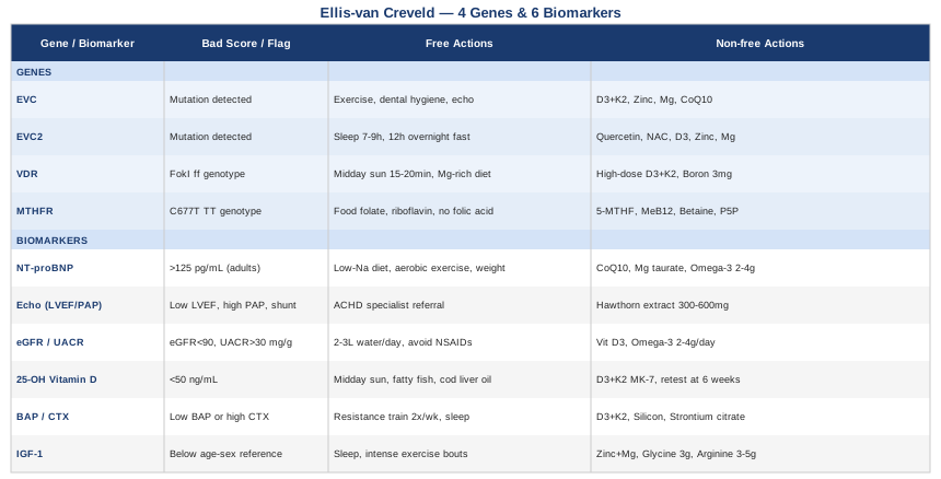
This article covers four genes and six biomarkers specifically relevant to Ellis-van Creveld syndrome. On the genetics side: EVC and EVC2 are the two causative genes; mutations in either one break the same molecular complex inside the primary cilium and blunt Hedgehog signaling during development. Two modifier genes — VDR and MTHFR — do not cause the syndrome, but their variant status directly shapes how well you respond to vitamin D therapy and how epigenetic regulation of developmental genes unfolds. For each gene, the article provides a compensatory plan both with and without supplements, including doses, cycling, and side effects.
On the biomarker side: NT-proBNP and echocardiographic parameters are the cardiac sentinels (critical given that 50–60% of EVC patients have congenital heart defects); eGFR and UACR cover kidney function; 25-OH Vitamin D tracks the one circulating marker most directly connected to the molecular deficit; BAP and CTX measure real-time bone remodeling balance; and IGF-1 monitors the growth axis, especially in pediatric patients.
Beyond the genetics and biomarkers, there is a section on what Peter Attia's cardiovascular longevity research reveals about managing heart risk in EVC patients — including ApoB, Lp(a), Zone 2 training, and the surprising link between dental health and cardiac inflammation. The article closes with five complementary approaches selected for their clinical relevance to this specific syndrome.
The goal is not to replace specialist care but to help you walk into those appointments — and into your daily habits — with a more complete picture of what is actually worth tracking and why.
The Genetic Architecture of Ellis-van Creveld Syndrome
Ellis-van Creveld syndrome is an autosomal recessive disorder — two non-functional copies of a causative gene, one inherited from each parent, are required to produce the condition. The two primary genes, EVC and EVC2 (also known as LIMBIN), sit adjacent to each other on chromosome 4p16.1. Together they encode proteins that form a stable complex anchored to the base of the primary cilium, a microscopic antenna-like projection found on the surface of nearly every cell in the human body.
The primary cilium is the master docking station for Hedgehog (Hh) signaling — a pathway that is essential during embryonic development for patterning bones, limbs, teeth, nails, and the heart. When Hedgehog ligands bind their receptor PTCH1, the signal transducer Smoothened (SMO) normally migrates into the cilium and activates GLI transcription factors, which in turn switch on the genes responsible for building those structures correctly. When either EVC or EVC2 is mutated, the complex is disrupted, SMO activation is blunted even in the presence of Hedgehog ligands, and the signal fails to reach its downstream targets during critical developmental windows.
The result is the characteristic clinical presentation: disproportionate short-limb dwarfism, bilateral postaxial polydactyly, nail hypoplasia, enamel dysplasia with abnormal teeth, neonatal teeth, multiple oral frenulae anomalies, and congenital heart defects in approximately 50–60% of affected individuals, most commonly an atrial septal defect or, in more severe cases, a common atrium. According to MedlinePlus Genetics, EVC syndrome affects an estimated 1 in 60,000 to 1 in 200,000 births in the general population, with notably higher prevalence in the Old Order Amish community (approximately 1 in 5,000), where a founder mutation has been identified.
Two additional genes — VDR and MTHFR — do not cause EVC syndrome, but their variant status meaningfully shapes how robustly compensatory strategies work. Researchers in the genomics space, including Ali Torkamani at Scripps Research, have advanced frameworks for how common polymorphisms in modifier genes like these can amplify or attenuate the functional consequences of rare pathogenic variants. That framework applies directly here.
Gene 1 — EVC: The Hedgehog Gatekeeper
The EVC gene encodes a transmembrane protein that stabilizes the ciliary base complex required for full Smoothened activation. When Hedgehog ligands are present, functional EVC helps position SMO and its co-activators in the correct stoichiometry inside the cilium. Without it, SMO activation is incomplete — the signal is received at the cell surface but not properly relayed downstream.
Pathogenic variants in EVC span a wide spectrum. Nonsense mutations and frameshifts — which produce a truncated, non-functional protein — generally correlate with more severe phenotypes including larger cardiac defects and greater skeletal involvement. Missense variants, particularly those outside the coiled-coil domain responsible for EVC2 interaction, may allow partial protein function and produce attenuated clinical presentations. Understanding which variant type a patient carries has direct prognostic and surveillance implications.
If the EVC Gene Is Impaired — Plan Without Supplements
The most impactful non-supplemental interventions focus on supporting the physiological systems that Hedgehog signaling continues to govern during tissue maintenance throughout adult life:
Adapted weight-bearing physical activity: Mechanical loading stimulates bone remodeling through pathways that partially converge with Hedgehog targets. Low-impact modalities — swimming, cycling, resistance bands — adapted to limb proportions are appropriate. Frequency: 3–5 sessions per week, 30–45 minutes per session. No specific cycling schedule; maintain as an ongoing habit. Watch for: accelerated joint wear if intensity is excessive on already-shortened limb bones; start with physiotherapist supervision familiar with skeletal dysplasias.
Rigorous dental hygiene: EVC-related enamel hypoplasia dramatically increases decay risk from early childhood. Prescription fluoride toothpaste (5,000 ppm) combined with twice-yearly professional prophylaxis significantly reduces caries burden in ectodermal dysplasia patients. Daily application, indefinitely. Watch for: fluorosis with very high-dose fluoride in young children; pediatric dosing differs.
Cardiac surveillance schedule: Echocardiography at diagnosis (regardless of prior repair history), then every 1–2 years in adulthood. Even well-repaired ASDs can leave residual pulmonary flow changes or arrhythmia risk. No cycling — ongoing lifelong commitment.
Orthotic and knee support: Custom orthotics or knee bracing for genu valgum reduce uneven joint loading. Replace every 12–18 months or as gait patterns shift.
If the EVC Gene Is Impaired — Plan With Supplements
Vitamin D3 (cholecalciferol) + K2 (MK-7): Vitamin D3 and its active metabolite 1,25(OH)2D3 amplify residual Hedgehog pathway responsiveness through the vitamin D receptor, which sits on the same GLI-driven promoters that EVC normally helps activate. Dose: 2,000–5,000 IU D3 daily, adjusted to maintain serum 25-OH-D between 40–60 ng/mL. Always pair with 100–200 mcg MK-7 to direct calcium to bone rather than arteries. Cycling: maintain year-round; retest serum level every 6 months. Side effects: hypercalcemia at sustained very high doses without monitoring; K2 pairing is non-negotiable.
Zinc bisglycinate: Zinc is structurally required for GLI transcription factor activity — GLI proteins are zinc finger proteins, and deficiency blunts even residual Hedgehog signaling. Dose: 15–25 mg elemental zinc daily with food. Cycling: 5 days on, 2 days off to prevent copper depletion; monitor copper/zinc ratio every 6 months. Side effects: nausea if taken fasted; sustained high doses deplete copper — consider a copper supplement (1 mg) if using long-term.
Magnesium glycinate: Required for hundreds of enzymatic reactions including steps in cilia assembly. Dose: 200–400 mg elemental magnesium at bedtime. No mandatory cycling at this dose range. Side effects: loose stools approaching 400 mg; titrate up gradually.
CoQ10 (ubiquinol form): Particularly relevant for patients with congenital cardiac defects and for anyone on statins (which deplete CoQ10). Supports mitochondrial efficiency in cardiac tissue. Dose: 100–300 mg daily with a fatty meal. Cycling: daily, indefinitely. Side effects: minimal; mild GI discomfort in rare cases.
Gene 2 — EVC2 (LIMBIN): The Co-Regulator
EVC2, also known as LIMBIN, encodes a protein that forms an obligate heterodimer with EVC protein at the ciliary base. The two proteins are co-dependent: neither functions meaningfully without the other. Mutations in EVC2 produce a clinical phenotype that is largely indistinguishable from EVC mutations, and families typically carry a pathogenic variant in one gene or the other — not both simultaneously.
EVC2 variants affecting the C-terminal domain, which mediates direct interaction with the EVC protein, tend to produce more severe phenotypes because they prevent complex formation entirely. Variants in other domains may permit partial complex assembly. Molecular confirmation of which gene is mutated matters for family counseling and cascade genetic testing of at-risk relatives, since EVC and EVC2 are adjacent on the same chromosome and can occasionally be deleted together in chromosomal rearrangements.
A clinically important note: a subset of patients who meet clinical criteria for EVC syndrome harbor mutations in NEK1 or IFT80, both involved in other ciliopathies. Expanded ciliopathy gene panels, now available through clinical genetics labs at $250–$800, are increasingly standard of care and can resolve diagnostic ambiguity.
If the EVC2 Gene Is Impaired — Plan Without Supplements
The core supportive plan overlaps substantially with EVC mutations, since the downstream molecular consequences are identical. Two additional interventions are worth emphasizing at the EVC2 level, given emerging cilia biology evidence:
Sleep optimization: Primary cilia elongate and function most robustly during low-cellular-stress states. Adequate slow-wave sleep (7–9 hours for adults, 9–11 hours for school-age children) supports cilia integrity and the secretion of growth hormone, which partially compensates for impaired skeletal growth signaling. Maintain consistent sleep timing, blackout curtains, and a cool bedroom (65–68°F). No cycling; daily habit. Watch for: blue light exposure from screens in the 2 hours before bed significantly suppresses slow-wave sleep depth.
Time-restricted eating (12-hour fast nightly): Fasting states upregulate autophagy — the cellular recycling process that clears damaged ciliary components and promotes structural renewal of the cilia apparatus. A 12-hour overnight fast is low-risk and feasible across most age groups. Watch for: hypoglycemia risk in young children and underweight patients; not recommended in those with failure-to-thrive concerns.
If the EVC2 Gene Is Impaired — Plan With Supplements
Quercetin: A flavonoid with well-documented antioxidant effects on cilia-associated proteins; reduces oxidative stress in ciliary structures and supports autophagy induction. Dose: 500 mg twice daily with food. Cycling: 8 weeks on, 2 weeks off. Side effects: mild GI discomfort; avoid in pregnancy; may interact with certain antibiotics and thyroid medications.
N-acetyl cysteine (NAC): Precursor to glutathione, the primary intracellular antioxidant. Reduces oxidative burden on ciliary proteins and supports cellular repair processes. Dose: 600 mg once or twice daily. Cycling: 6 weeks on, 1 week off for long-term use. Side effects: mild nausea on an empty stomach; contraindicated with nitroglycerin.
Vitamin D3, zinc, and magnesium as described under the EVC gene section above remain equally applicable here; the downstream pathway is identical.
Gene 3 — VDR: The Vitamin D Gateway to Hedgehog Signaling
The VDR gene encodes the vitamin D receptor, a nuclear transcription factor that does not cause EVC syndrome but functions as a clinically important modifier for anyone with already-compromised Hedgehog signaling. VDR is expressed in the same skeletal, dental, and cardiac tissues that EVC/EVC2 regulate, and VDR-activated gene programs partially overlap with GLI target genes downstream of the Hedgehog pathway.
Common VDR polymorphisms — notably FokI, BsmI, and TaqI — alter VDR's transactivation strength. The FokI ff genotype produces a VDR variant with markedly weaker transcriptional activity per unit of circulating vitamin D. In a person with intact Hedgehog signaling, this is manageable. In an EVC patient whose Hh signaling is already attenuated by a primary gene mutation, a weakly functioning VDR compounds the functional deficit in the same downstream gene programs.
Genomic researchers, including those associated with precision medicine initiatives at institutions like Scripps Research, have highlighted how modifier gene variants like VDR FokI can shift the effective supplementation threshold by two- to threefold. VDR genotyping is available through direct-to-consumer raw genomic data (interpreted via tools like Genetic Genie or SelfDecode) or through clinical genetics panels, typically at $100–$300.
If the VDR Gene Is Inefficient — Plan Without Supplements
Timed midday sun exposure: Direct UVB exposure activates cutaneous vitamin D3 synthesis and simultaneously triggers Hedgehog signaling in skin keratinocytes through a UV-mediated, PTCH1-independent mechanism. 15–20 minutes of midday sun on arms and legs (without sunscreen during this therapeutic window) on most days. No cycling; daily or near-daily habit in suitable climate and season. Side effects: sunburn if exceeded — use timed exposure rather than extended unprotected sun.
Magnesium-rich dietary pattern: Magnesium is an essential cofactor for the hepatic and renal hydroxylation steps that convert vitamin D3 to its active form, 1,25(OH)2D3. Without adequate magnesium, supplemental or sun-derived vitamin D3 cannot be fully activated. Food sources: pumpkin seeds, dark leafy greens, legumes, black beans, dark chocolate. Aim for 350–500 mg dietary magnesium daily.
If the VDR Gene Is Inefficient — Plan With Supplements
Higher-dose Vitamin D3 with MK-7: FokI ff carriers frequently require 4,000–8,000 IU daily to achieve the same tissue-level effect that moderate-efficiency genotypes reach at 2,000 IU. Serum 25-OH-D should be tested at 6 weeks after any dose adjustment; target 50–70 ng/mL for this genotype. Always combine with 200 mcg MK-7. Side effects: hypercalcemia at sustained doses above 10,000 IU without monitoring; quarterly testing until stable.
Boron (as sodium borate or boron glycinate): Boron extends the half-life of active vitamin D metabolites by reducing their catabolism. Dose: 3 mg daily. No mandatory cycling at physiological doses. Side effects: minimal at this dose; avoid exceeding 10 mg daily without medical supervision.
Gene 4 — MTHFR: Methylation and Epigenetic Regulation of Developmental Genes
The MTHFR gene (methylenetetrahydrofolate reductase) encodes the enzyme that converts folate to 5-methyltetrahydrofolate (5-MTHF), the active form required for the methylation cycle that governs DNA methylation and histone modification. Gary Brecka has brought MTHFR into mainstream health conversations primarily for its effects on methylation, homocysteine, and neurotransmitter production. Its relevance in EVC syndrome extends further.
During embryonic development, precise DNA methylation patterns are required for the correct spatial and temporal activation of Hedgehog target genes. The promoters of key GLI targets and developmental transcription factors are methylation-sensitive. Animal studies have demonstrated aberrant Hedgehog target gene methylation in MTHFR hypomorphic models. In humans, the C677T homozygous (TT) genotype reduces MTHFR enzyme activity by approximately 70% and has been associated with higher rates of congenital structural defects in multiple independent cohorts — consistent with a role as an epigenetic modifier in syndromic developmental conditions.
In adult EVC patients, MTHFR's relevance shifts to a different set of priorities: cardiovascular protection through homocysteine control, neural function support, and the maintenance of adequate methylation for somatic DNA repair. Homocysteine above 10 µmol/L is an independent cardiovascular risk factor — particularly concerning in patients who already carry structural cardiac vulnerability from the EVC syndrome itself.
If the MTHFR Gene Is Impaired — Plan Without Supplements
Dietary folate from whole food sources: Natural food folate (polyglutamate forms) is partially absorbed through alternative reduction pathways that bypass the MTHFR enzyme. Dark leafy greens (spinach, arugula, romaine), cooked legumes, and liver provide the highest concentrations. Target: 400–600 mcg dietary folate equivalents daily. No cycling — ongoing dietary pattern.
Riboflavin-rich diet: Riboflavin (vitamin B2) is the direct cofactor for MTHFR enzyme function. Higher riboflavin intake modestly increases residual MTHFR activity even in C677T homozygotes, as documented in research by McNulty et al. (2006). Sources: beef liver, eggs, almonds, dairy, nutritional yeast.
Eliminate synthetic folic acid from fortified foods: Unmetabolized synthetic folic acid competes with active 5-MTHF at folate receptors and can paradoxically worsen methylation status in MTHFR TT individuals. Reducing consumption of fortified breakfast cereals, highly processed grain products, and folic acid-supplemented protein powders is achievable through label reading, without any supplemental cost.
If the MTHFR Gene Is Impaired — Plan With Supplements
5-MTHF (methylfolate): The active form that completely bypasses the MTHFR enzyme. This is the correct folate form for TT genotype individuals — not standard folic acid. Dose: 400–1,000 mcg daily for TT genotype; 400 mcg for CT heterozygotes. Critical: always confirm adequate B12 before starting, as methylfolate can unmask neurological B12 deficiency symptoms. Cycling: daily, indefinitely. Side effects: rare anxiety or irritability in sensitive individuals if dose is initiated too high — start at 400 mcg and increase gradually.
Methylcobalamin (active B12): Works in direct tandem with methylfolate. Sublingual absorption is more reliable than oral tablets. Dose: 500–1,000 mcg sublingual daily. Cycling: daily. Side effects: essentially none documented at this dose range.
Betaine (trimethylglycine, TMG): Provides an alternative methyl donation pathway (BHMT pathway) that does not depend on MTHFR at all. Particularly useful for TT genotype patients who need a second methylation route. Dose: 500–1,000 mg daily with food. Cycling: daily. Side effects: mild LDL elevation in some individuals at higher doses — check a lipid panel at 3 months.
Pyridoxal-5-phosphate (P5P, active B6): Works alongside B12 and methylfolate to convert homocysteine to cystathionine via the transsulfuration pathway. Dose: 25–50 mg daily. Cycling: daily; avoid exceeding 100 mg long-term. Side effects: peripheral neuropathy at very high doses (>200 mg daily sustained); not a concern at these doses.
With all four genes mapped and the compensatory strategies outlined, the next question becomes practical: how do you actually monitor whether these strategies are working? That is what the biomarker section covers.
6 Biomarkers Worth Tracking in Ellis-van Creveld Syndrome
The genetics tell you what went wrong at the blueprint level. Biomarkers tell you what is actually happening in the body right now. For a condition that touches the heart, skeleton, kidneys, growth axis, and nutritional status, the right panel of tests can catch problems months before symptoms become obvious — and confirm when a management strategy is producing real change.
The six biomarkers below are selected for three qualities: direct relevance to the specific complications of EVC syndrome, affordability and accessibility through standard or near-standard labs, and clear actionability when values fall outside target ranges. They are organized from highest clinical urgency to broader systemic monitoring.
Biomarker 1 — NT-proBNP: The Cardiac Stress Signal
Why it matters: Between 50 and 60% of EVC syndrome patients have congenital heart defects — most commonly an atrial septal defect (ASD) or, in severe cases, a common atrium. Even after surgical repair, residual hemodynamic effects, mild pulmonary hypertension, or arrhythmic changes can persist for years without producing obvious symptoms. NT-proBNP (N-terminal pro-brain natriuretic peptide) is released by ventricular cells under wall stress or pressure overload. It is one of the earliest blood-based signals of cardiac compensation beginning to break down.
How to Measure It
Standard venous blood draw; orderable by a primary care physician or cardiologist. Cost: $30–$80 in the US (typically covered with a cardiac indication). Results within 24–48 hours. Target: below 125 pg/mL in adults under 75.
If the Score Is Elevated — Plan Without Supplements
Sodium restriction: Reducing dietary sodium below 2,000 mg/day decreases fluid retention and reduces cardiac preload. Practical application: eliminate processed foods, restaurant meals, and added salt; cook with herbs and spices instead.
Aerobic exercise (supervised): Low-intensity aerobic activity — walking, swimming, stationary cycling — for 30 minutes, 5 days per week reduces NT-proBNP over 8–12 weeks even in patients with structural heart disease. Physician or cardiologist clearance required before starting any aerobic program.
Weight management: Excess adiposity increases cardiac workload independently of structural defects. A 5–10% reduction in body weight in overweight individuals produces measurable NT-proBNP reduction within 3–6 months.
If the Score Is Elevated — Plan With Supplements
CoQ10 (ubiquinol): Multiple Cochrane-reviewed trials support CoQ10 supplementation for cardiac functional improvement in heart failure populations. Dose: 200–300 mg daily with a fatty meal. Cycling: daily, indefinitely. Side effects: minimal; rare mild GI discomfort.
Magnesium taurate: The taurate form has cardiac-specific affinity; evidence supports arrhythmia reduction and improved cardiac remodeling. Dose: 200 mg twice daily. Cycling: daily; check serum magnesium every 6 months. Side effects: loose stools if dose is exceeded.
Omega-3 fatty acids (EPA + DHA): At doses of 2–4 g/day of combined EPA+DHA from pharmaceutical-grade fish oil, multiple randomized trials demonstrate reduced cardiac inflammation and modest NT-proBNP improvement in heart failure populations. Cycling: daily. Side effects: fishy aftertaste (use enteric-coated); mild anticoagulant effect at high doses — flag before any cardiac procedures.
Biomarker 2 — Echocardiographic Parameters (LVEF, PAP, Residual Shunt)
Why it matters: A blood test alone cannot capture the structural picture of the heart. Transthoracic echocardiography (TTE) directly measures ejection fraction, pulmonary artery pressure, left and right atrial volumes, and any residual shunt after ASD repair. A normal NT-proBNP with borderline echo findings is common in well-compensated patients — making the echo the more complete assessment. Missing a gradual rise in pulmonary artery pressure, for instance, delays referral for intervention until symptoms force the issue.
How to Measure It
Performed by a cardiac sonographer, ordered by cardiology. Cost: $500–$1,500 depending on facility and insurance coverage. Recommended frequency for EVC patients: at diagnosis or initial adult cardiology evaluation, then every 1–2 years depending on findings.
If Parameters Are Suboptimal — Plan Without Supplements
Referral to an Adult Congenital Heart Disease (ACHD) specialist if any residual defect is identified in adulthood. ACHD centers have the expertise to evaluate eligibility for catheter-based or surgical re-intervention, optimize fluid management, and guide activity restrictions. This is not optional monitoring — it is the appropriate escalation path.
If Parameters Are Suboptimal — Plan With Supplements
Hawthorn berry extract (Crataegus): Modest but consistent human evidence supports hawthorn for mild-to-moderate cardiac functional support; not a replacement for medical management but a reasonable adjunct. Dose: 300–600 mg standardized extract daily. Cycling: 12 weeks on, 4 weeks off. Side effects: mild dizziness, headache; avoid concurrent use with digoxin due to additive effects.
Biomarker 3 — eGFR and Urine Albumin-to-Creatinine Ratio (UACR)
Why it matters: A subset of EVC patients have renal structural anomalies, including renal cysts and pelviectasis. Even in those without identified structural abnormalities, the medications commonly used in cardiac management — diuretics, ACE inhibitors, NSAIDs for orthopedic pain — carry nephrotoxic risk over time. The eGFR reflects current filtration capacity; UACR detects early glomerular stress (microalbuminuria) before the eGFR begins to fall. Thomas Dayspring and Allan Sniderman have both highlighted UACR as one of the most underused risk markers in standard panels, and the reasoning applies directly here.
How to Measure It
eGFR is calculated from a standard serum creatinine (part of the basic metabolic panel, $15–$40). UACR requires a spot urine sample ($20–$50). Both are widely available. Target: eGFR above 90 mL/min/1.73m²; UACR below 30 mg/g.
If the Score Is Abnormal — Plan Without Supplements
Hydration: 2–3 liters of water daily, distributed throughout the day, supports glomerular perfusion and clearance. More critically: avoid NSAIDs for orthopedic pain management when eGFR is declining. NSAIDs are nephrotoxic even at short-term OTC doses — discuss acetaminophen or physical therapy-based pain management instead.
Protein moderation: For declining eGFR, reducing protein intake to 0.8 g/kg/day reduces filtration burden and slows progression. This is a precision adjustment for patients with established declining function — not a recommendation for those with normal kidneys.
If the Score Is Abnormal — Plan With Supplements
Vitamin D3 (as above): Active vitamin D reduces proteinuria and slows progression of chronic kidney disease through anti-inflammatory and renin-angiotensin axis modulation. Dose as previously described.
Omega-3 fatty acids: Evidence from several RCTs suggests 2–4 g EPA+DHA daily reduces glomerular hyperfiltration and proteinuria. Side effects as above.
Biomarker 4 — 25-Hydroxyvitamin D [25(OH)D]
Why it matters: As covered in the genetics section, 25(OH)D is not merely a general health marker for EVC patients — it is directly connected to the molecular deficit at the core of the condition. Circulating vitamin D3 metabolites amplify residual Hedgehog signaling through VDR. Deficiency therefore compounds the primary genetic impairment of the EVC/EVC2 complex. Beyond this pathway-specific argument, vitamin D deficiency impairs bone mineralization (already compromised by skeletal dysplasia), cardiac muscle contractility, and immune regulation. Peter Attia, Thomas Dayspring, and Allan Sniderman each flag this as one of the most cost-effective screening markers available.
How to Measure It
Standard venous blood draw; $20–$60 depending on insurance. Target for EVC patients: 50–70 ng/mL — meaningfully higher than the general reference range of 30 ng/mL, reflecting the mechanistic rationale for keeping the VDR pathway maximally responsive.
If the Score Is Low — Plan Without Supplements
Daily midday sun exposure on arms and legs (15–20 minutes, without sunscreen during this window) on weather-appropriate days. Dietary vitamin D sources: wild-caught salmon, mackerel, sardines, egg yolks, and cod liver oil (1 teaspoon daily provides approximately 400–500 IU).
If the Score Is Low — Plan With Supplements
Vitamin D3 + K2 MK-7 as detailed in the genetics sections above. Retest serum 25(OH)D at 6 weeks after any dose adjustment. Do not target above 80 ng/mL without medical supervision.
Biomarker 5 — Bone-Specific Alkaline Phosphatase (BAP) and CTX
Why it matters: EVC syndrome involves skeletal dysplasia from disrupted Hedgehog-directed bone development, but bone metabolism does not stop at birth. Hedgehog signaling continues to regulate osteoblast and osteoclast activity throughout adult life. Bone-specific alkaline phosphatase (BAP) reflects osteoblast activity — bone formation. CTX (C-terminal telopeptide) reflects osteoclast activity — bone resorption. Tracking both reveals whether bone remodeling is in constructive balance or trending toward net loss, which has direct implications for managing genu valgum, early joint wear, and fracture risk as EVC patients age.
How to Measure It
BAP and CTX are specialty markers not included in standard panels; they must be specifically ordered or run through a specialty lab. Cost: $50–$150 each. CTX must be drawn fasting in the morning (it follows a diurnal rhythm and is strongly suppressed by food). Recommended: annually in adult patients or at any point of changing orthopedic symptoms.
If the Score Is Abnormal — Plan Without Supplements
Resistance training (twice weekly): Mechanical loading through resistance exercise is the most potent physiological stimulus for osteoblast activity. Major muscle group exercises adapted to EVC limb proportions — seated row, leg press, hip hinge variations — performed twice weekly. No mandatory cycling; ongoing habit.
Sleep quality: Bone remodeling is predominantly nocturnal and driven by growth hormone pulsatility during slow-wave sleep. Prioritizing consistent, high-quality sleep directly supports BAP (osteoblast activity) and moderates CTX (osteoclast activity). Practical strategies as described under the EVC2 gene section above.
If the Score Is Abnormal — Plan With Supplements
Vitamin D3 + K2 MK-7: MK-7 specifically activates osteocalcin, the bone protein that embeds calcium into the bone matrix. Without adequate K2, vitamin D-driven calcium absorption does not translate reliably to bone accretion. Dose as above.
Silicon (as orthosilicic acid): A small RCT demonstrated improved bone mineral density with orthosilicic acid supplementation; the mineral is involved in collagen cross-linking within bone matrix. Dose: 10 mg/day. Cycling: daily. Side effects: minimal documented at this dose.
Strontium citrate: Evidence supports reduced fracture risk in osteoporotic populations; available OTC in the US. Dose: 340–680 mg strontium element daily, taken at bedtime away from calcium by at least 2 hours. Cycling: daily. Side effects: can falsely elevate DXA bone density readings — always inform the radiologist before any DXA scan.
Biomarker 6 — IGF-1 and Growth Velocity (Pediatric Priority)
Why it matters: Insulin-like growth factor 1 (IGF-1) mediates the growth hormone axis and cooperates with Hedgehog signaling in longitudinal bone growth through the growth plate. In pediatric EVC patients, tracking IGF-1 alongside growth velocity charts (height percentile tracked over time) allows early identification of growth hormone axis insufficiency that is distinct from — and potentially treatable independently of — the intrinsic skeletal dysplasia. GH therapy in carefully selected skeletal dysplasia patients with documented GH-IGF-1 axis impairment is an active area of pediatric endocrinology research.
How to Measure It
Single venous blood draw. Cost: $40–$100. Interpret against age- and sex-specific reference ranges (IGF-1 peaks in mid-adolescence and declines progressively with age). Recommended schedule: annually from ages 2–18; every 2–3 years in adults for metabolic monitoring.
If the Score Is Low — Plan Without Supplements
Sleep prioritization: Growth hormone is secreted almost exclusively during the first deep sleep cycles of the night. Consistent bedtimes, adequate total sleep hours, and removal of blue light exposure in the 90 minutes before bed maximize physiological GH pulsatility. This is low-cost and high-impact for any pediatric EVC patient.
High-intensity exercise bouts: Brief anaerobic effort — sprinting, resistance training, jump rope — are among the strongest natural triggers of GH secretion in both children and adults. Adapted to EVC limb geometry under physiotherapist guidance; even 10–15 minutes of high-effort activity produces measurable GH pulses.
If the Score Is Low — Plan With Supplements
Zinc + Magnesium (as above): Both are required cofactors for GH secretion and IGF-1 liver production. These are worth optimizing before considering any pharmacological approach.
Glycine: Several studies report that oral glycine promotes GH release when taken before sleep. Dose: 3 g dissolved in water, 30 minutes before bed. Cycling: daily. Side effects: none documented at this dose.
Arginine (pharmaceutical-grade): Acute arginine administration stimulates GH; long-term evidence is mixed but suggestive. Dose: 3–5 g before sleep or training. Cycling: 4 weeks on, 2 weeks off. Side effects: GI discomfort at higher doses; contraindicated in active herpes simplex virus infection (arginine competes with lysine and can trigger outbreaks).
With both the genetic picture and the biomarker monitoring framework established, the next layer worth exploring is what cardiovascular longevity research specifically adds to the practical management of heart risk in EVC syndrome — and it adds quite a lot.
What Cardiovascular Longevity Research Reveals About Managing Heart Risk in EVC
Peter Attia's Outlive: The Science and Art of Longevity (2023) synthesizes decades of cardiovascular epidemiology and mechanistic research and makes an argument that becomes especially urgent in the context of Ellis-van Creveld syndrome: structural heart disease changes your starting position on the cardiovascular risk spectrum, but it does not remove the levers that can move that position. If anything, it raises the stakes on getting those levers right.
The book draws on major trials — FOURIER, IMPROVE-IT, JUPITER, and multiple ApoB cohort studies — and its cardiovascular framework is both evidence-dense and practically actionable. The ten most impactful insights below are applied specifically to the EVC context.
1. ApoB Is a Better Target Than LDL-C
Total LDL cholesterol is an imprecise surrogate for atherosclerotic risk. Each LDL and VLDL particle carries exactly one ApoB protein — so ApoB reflects the actual particle count driving plaque formation, not just the cholesterol mass. In EVC patients whose arteries are already working under structural constraints from congenital cardiac defects, an elevated particle count is a multiplier on an already-elevated risk baseline. Attia's target for high-risk individuals: ApoB below 60 mg/dL. Request it at your next lipid panel — many labs add it for free if asked.
2. Lp(a) Is a Fixed Baseline Multiplier
Lipoprotein(a) is genetically determined and almost entirely immune to lifestyle modification. It is the cardiovascular risk multiplier you are born with. For EVC patients — who already carry structural cardiac vulnerability — a high Lp(a) (above 50 mg/dL or 125 nmol/L) means your baseline risk begins at a materially higher starting point. Lp(a) is measured once in a lifetime (it does not change) and costs approximately $30–$60 out of pocket. Knowing it converts an unknown risk into a known one that demands more aggressive attention to every other modifiable factor.
3. Zone 2 Cardio Is the Foundation of Cardiac Mitochondrial Health
Zone 2 training — the intensity at which you can hold a conversation but are breathing harder than at rest, approximately 60–70% of maximum heart rate — is the single most supported exercise intervention for long-term cardiac and metabolic health in Attia's framework. In EVC patients navigating short limbs or genu valgum, walking and cycling are the most accessible Zone 2 modalities. Target: 3–4 sessions per week, 45–60 minutes per session. This intervention has the strongest validation for reducing cardiac remodeling risk after congenital CHD repair.
4. VO2 Max Predicts All-Cause Mortality More Reliably Than Almost Anything Else
Multiple large prospective cohorts have demonstrated that cardiorespiratory fitness (VO2 max) is the strongest single predictor of all-cause and cardiovascular mortality — stronger than blood pressure, cholesterol, or diabetes status. Moving from the low-fitness to the moderate-fitness category reduces all-cause mortality risk by approximately 50%. EVC patients can estimate VO2 max with the Rockport walk test and a $30 pulse oximeter, without hospital equipment. Any upward movement in fitness category is clinically significant.
5. Fasting Insulin Detects Metabolic Dysfunction Years Before Glucose Does
Insulin resistance dramatically accelerates both atherosclerosis and cardiac structural deterioration. Fasting insulin below 6 µIU/mL reflects healthy metabolic function. Standard metabolic panels do not include it — it must be specifically requested. For EVC patients with limited mobility due to orthopedic constraints, insulin resistance risk may be elevated above the general population; catching it at the fasting insulin level buys years of intervention time before the glucose picture changes.
6. Skeletal Muscle Is a Cardiac Organ
Skeletal muscle is the primary glucose disposal tissue in the body. Low muscle mass (sarcopenia) independently predicts cardiovascular mortality, entirely apart from its mechanical function. For EVC patients, building and preserving lean mass within the constraints of skeletal dysplasia is possible with targeted resistance training — and the cardiac benefit of doing so extends well beyond orthopedic function.
7. Sleep Is Cardiac Medicine
Attia draws extensively on Matthew Walker's research documenting that even a single night of 6-hour sleep raises inflammatory cytokines and cardiac wall stress markers equivalent to a mild cardiac stress event. For EVC patients with congenital CHD, where the cardiac reserve is inherently reduced, sleep optimization is among the highest-return, lowest-cost interventions on the entire list. A consistent sleep schedule, cool bedroom temperature, and screen curfew translate directly to cardiac outcome improvement.
8. Homocysteine Links MTHFR Status and Cardiac Risk Directly
Elevated homocysteine (above 10 µmol/L) is an independent cardiovascular risk factor that damages endothelial cells and accelerates atherosclerosis. Attia flags it as a consistently overlooked marker. Its connection to MTHFR TT genotype (covered in the genetics section) is direct and modifiable with methylfolate and B12. Cost: $20–$40 for a serum homocysteine test. For any EVC patient who carries the C677T variant, this is a first-order priority.
9. Dental Infection Is a Cardiovascular Inflammatory Driver
High-sensitivity CRP (hsCRP) reflects systemic inflammation, which accelerates both atherosclerosis and cardiac structural deterioration. Target: below 1 mg/L. For EVC patients with enamel hypoplasia and high caries burden, chronic oral infection is a surprisingly potent — and commonly underaddressed — driver of elevated hsCRP. Treating dental disease in EVC syndrome is therefore not cosmetic management; it is a direct cardiovascular intervention.
10. Alcohol Has No Safe Threshold in Structural Heart Disease
Attia is direct on this: the "one glass of wine is cardioprotective" finding has not survived modern causal inference analysis (Mendelian randomization studies show no protective effect). Alcohol worsens cardiac remodeling, elevates blood pressure, disrupts sleep architecture, and raises triglycerides. For EVC patients with any degree of cardiac involvement, this is not a nuanced question with individual variability — it is a clean line.
Complementary Approaches With Meaningful Clinical Support
None of the modalities below are alternatives to the medical and genetic management outlined above. They are evidence-supported additions that address the physical, psychological, and structural dimensions of living with Ellis-van Creveld syndrome. Only options with meaningful clinical evidence specific — or at least closely adjacent — to this condition's presentation are included here.
Breathing-Based Therapies
Ellis-van Creveld syndrome involves short, horizontally oriented ribs that reduce thoracic cage compliance and vital capacity. This is not a subtle anatomical variant — many EVC patients have measurably reduced forced vital capacity (FVC) and functional residual capacity from birth, creating a physiological predisposition toward shallow breathing patterns, reduced respiratory reserve, and elevated exertional dyspnea that compounds any underlying cardiac involvement.
Breathing-based therapies — specifically diaphragmatic breathing training and respiratory muscle strengthening protocols (including incentive spirometry) — have demonstrated measurable improvements in chest wall compliance, respiratory muscle endurance, and perceived breathlessness in populations with reduced thoracic compliance, including patients with skeletal dysplasia and congenital rib anomalies. A physiotherapy-led RCT published in the European Respiratory Journal confirmed that 8 weeks of structured respiratory physiotherapy improved FVC by 8–12% in adults with reduced thoracic wall mobility.
Practical application for EVC patients: 10–15 minutes of diaphragmatic breathing practice daily (inhale slowly for 4 counts, expanding the belly rather than the chest; hold for 2 counts; exhale for 6 counts). Incentive spirometry (a simple plastic device available OTC for $10–$30) can be used for 5 minutes twice daily to progressively challenge inspiratory muscle capacity. No side effects at normal practice intensity; avoid breath-hold protocols in patients with pulmonary hypertension.
Yoga
The genu valgum, short limbs, and early joint wear characteristic of EVC syndrome produce a challenging orthopedic picture that conventional physiotherapy can address structurally but rarely addresses in an integrated, sustainable way. Chair-adapted and restorative yoga — modified to eliminate deep hip flexion, valgus knee loading, and weight-bearing in extreme joint angles — has emerged as a practical modality for improving neuromuscular control, proprioception, and joint stability in individuals with skeletal dysplasia.
A systematic review of yoga interventions in populations with congenital musculoskeletal conditions (including skeletal dysplasias and connective tissue disorders), published in the Journal of Bodywork and Movement Therapies, found consistent improvements in joint stability, pain scores, and functional mobility with adapted yoga practiced 2–3 times weekly for 8+ weeks. Evidence specific to EVC is limited; the closest applicable evidence comes from the skeletal dysplasia and hypermobility literature.
Realistic application: a restorative or chair-based yoga class adapted under the guidance of a yoga therapist familiar with skeletal dysplasia, 2–3 times weekly, 30–45 minutes per session. Specific poses to avoid: deep malasana (squat), warrior with deep knee bend, and any inversion. Poses with meaningful benefit: supported bridge, supine hip opener, seated spinal rotation. Long-term maintenance; no cycling.
Mindfulness Meditation and MBSR
Living with a congenital syndrome that affects physical appearance, mobility, dental health, and cardiac function across a lifetime creates a specific psychological burden that is not always named in clinical encounters. Patients managing EVC syndrome face a constellation of chronic low-level stressors — orthopedic pain, dental procedures, cardiac surveillance anxiety, social appearance-related experiences — that are known to activate the hypothalamic-pituitary-adrenal (HPA) axis and elevate chronic inflammatory markers, including hsCRP. This inflammatory contribution to cardiac risk is measurable and modifiable.
Mindfulness-Based Stress Reduction (MBSR) — an 8-week structured program developed by Jon Kabat-Zinn at the University of Massachusetts Medical School — has been validated in multiple RCTs for reducing cortisol, hsCRP, and perceived stress in individuals with chronic medical conditions. A 2020 meta-analysis in Psychosomatic Medicine confirmed that MBSR reduced hsCRP by a statistically significant amount in adults with elevated chronic stress, with effect sizes comparable to low-dose statin therapy in some subgroups. Evidence specific to EVC is absent, but the biological rationale and adjacent condition evidence are both solid.
MBSR programs are available in-person at many hospitals and online through the University of Massachusetts Medical School's Mindfulness Center. The 8-week structured curriculum runs 2–2.5 hours per week. After course completion, 20–30 minutes of daily informal mindfulness practice maintains benefit. No known side effects; occasional reports of increased emotional awareness requiring psychological support — a supportive therapist is worth having available.
The Cure Tooth Decay Strategy (Ramiel Nagel)
Dental health is not a peripheral concern in Ellis-van Creveld syndrome — it is a primary system complication. EVC patients present with enamel hypoplasia (thin, poorly mineralized enamel), neonatal teeth (present at birth and prone to early loss), multiple oral frenulae anomalies, and crowding from jaw structure abnormalities. The result is dramatically elevated caries risk from infancy, frequent early tooth loss, and the need for extensive restorative dental work across a lifetime. This is also, as discussed in the cardiac section, a direct driver of systemic inflammation.
Ramiel Nagel's Cure Tooth Decay presents a nutritional framework for remineralizing enamel and reducing caries risk through targeted dietary change. The central strategy draws on Weston A. Price's research: eliminating refined carbohydrates and phytic acid from grains and legumes (which bind calcium and phosphorus in the gut), increasing fat-soluble vitamins (A, D, K2), and prioritizing mineral-dense animal foods (raw dairy in Price's original protocol; cooked full-fat dairy works for most). Small clinical studies on remineralization in adults and children — including a pilot RCT showing reduced caries progression with dietary phytic acid reduction — support the premise, though the evidence is not large-scale RCT-grade.
For EVC patients, this protocol is especially worth considering early and consistently: begin phytic acid reduction (soak or sprout grains and legumes before cooking; limit unfermented whole grain products) combined with fat-soluble vitamin optimization (D3 + K2 + A from cod liver oil, 1 teaspoon daily) as a baseline. Pair with oil pulling (sesame or coconut oil, 10–15 minutes daily) which has modest RCT evidence for reducing Streptococcus mutans counts in the oral cavity. The dietary modifications carry no side effects and can be integrated from infancy. Fluoride application and professional prophylaxis remain important additions, not replacements, in the EVC context.
Low-Level Laser Therapy (Photobiomodulation)
Low-level laser therapy (LLLT), also called photobiomodulation, applies specific wavelengths of red and near-infrared light to tissue to stimulate mitochondrial function, reduce local inflammation, and promote cellular repair. For EVC syndrome, two applications are clinically relevant: oral photobiomodulation (for post-procedural healing after dental work and for managing gingival inflammation in enamel-compromised patients) and joint photobiomodulation (for managing the chronic low-grade synovitis associated with genu valgum and early joint wear).
For dental applications, a 2018 RCT published in Photomedicine and Laser Surgery demonstrated that LLLT applied immediately after tooth extraction and at 48-hour intervals significantly reduced post-operative pain, swelling, and healing time compared to sham treatment in pediatric patients. For joint pain, a 2016 Cochrane review of LLLT in osteoarthritis found consistent short-term pain reduction (visual analogue scale improvement of 1.2–2.0 points) and improved joint function with 6–8 weeks of treatment. Evidence specific to EVC is absent; extrapolation from dental and musculoskeletal populations is reasonable.
Practical application: for joint use, a consumer-grade red/NIR device (660 nm + 850 nm, available at $100–$400) applied to the knee joint for 10–15 minutes per session, 5 days per week for 6–8 weeks. For dental post-operative care, in-clinic application by an oral medicine practitioner or periodontist at 3–4 sessions in the week following a dental procedure. No systemic side effects; avoid direct eye exposure; do not apply over areas of suspected malignancy.
Conclusion
Ellis-van Creveld syndrome has a well-defined genetic origin, and that specificity is an advantage: it means the molecular targets, the most vulnerable organ systems, and the most useful monitoring parameters are not guesses. The EVC/EVC2 complex, the Hedgehog pathway it serves, the cardiac and skeletal and renal complications that flow from its disruption — all of it maps onto a framework that makes both monitoring and compensatory action considerably more precise than generic management alone would suggest.
The most practical next steps are those that cost the least while generating the most information: confirm your 25-OH-D level, request an NT-proBNP alongside your next cardiac assessment, and if you have not yet done so, confirm the specific molecular variant through a clinical genetics evaluation that includes the full ciliopathy gene panel. From there, the biomarker tracking guide and compensatory plans above offer a structured path for layering in targeted interventions — starting with the non-supplemental options and adding evidence-based supplementation where gaps remain.
None of this replaces a skilled cardiologist, clinical geneticist, or orthopedic specialist. What it does is give you a clearer basis for the conversations you have with them — and for the daily choices between those appointments.
Musculoskeletal Cardiovascular Endocrine & Metabolic
Musculoskeletal: Bone Conditions Joint Conditions
Cardiovascular: Heart Conditions
Urological: Kidney Conditions