This article contains AI generated content.
· UpdatedEmery-Dreifuss Muscular Dystrophy Genes Biomarkers — 6 Genes and 7 Biomarkers to Track
Introduction
Living with Emery-Dreifuss Muscular Dystrophy, or caring for someone who does, puts you in a particular kind of uncertainty. The early contractures in the ankles, elbows, and neck, the slowly progressing humeroperoneal weakness, and then — often without much warning — the cardiac complications that carry the highest risk to life. EDMD does not follow the same trajectory as other muscular dystrophies, and its severity varies enormously depending on which gene is involved. If you have found that most available information either oversimplifies the condition or becomes too technical to apply, that frustration is legitimate.
What makes EDMD especially difficult to navigate is that the dimension most people focus on — the muscle symptoms — is rarely the most dangerous part. The cardiac complications, including arrhythmias, conduction defects, and dilated cardiomyopathy, can appear before muscle weakness is severe and can progress silently until they cause a life-threatening event. Generic muscular dystrophy advice rarely addresses this cardiac dimension with the specificity it deserves. And knowing you have EDMD is rarely enough: knowing which specific gene variant is involved changes the risk profile significantly and should drive monitoring intensity and clinical decision-making.
This article takes a more targeted approach. Rather than restating what EDMD is, it focuses on two practical layers: the biomarkers you should be tracking and what to do when results come back off, and the specific genetic variants associated with EDMD and what each implies about clinical strategy. Both layers are actionable, both are grounded in current evidence, and both can meaningfully improve the conversations you have with your care team.
Grounded hope in a rare disease context means this: the tools to monitor EDMD have improved substantially, the understanding of which genetic mutations carry the highest cardiac risk is now solid enough to be clinically useful, and there are evidence-backed strategies — from targeted supplementation to specific monitoring protocols — that can make a real difference in outcomes. This article walks through seven biomarkers that matter most for tracking this condition, six key genes and what each implies, a distillation of Peter Attia's cardiac monitoring framework applied to EDMD, and a set of complementary approaches with actual clinical evidence behind them.
Summary
This article covers the seven most important biomarkers to track in Emery-Dreifuss Muscular Dystrophy — including several cardiac markers that are routinely missed until something goes wrong — alongside the six main genes that drive the condition, each carrying a different risk profile and requiring a different monitoring intensity. For each biomarker and each gene, you will find specific action plans: what to do first without supplements, and what to add with supplements or equipment, including dosing, cycling, and side effect notes. Beyond the clinical tracking layer, ten insights from Peter Attia's Outlive explain what proactive cardiac surveillance looks like at its best — applied directly to the EDMD context. The article closes with three complementary approaches — yoga for contracture management, breathing therapy for autonomic support, and MBSR for quality of life — each with at least some grounding in clinical evidence. If you are looking for a map that actually reflects how EDMD behaves, not how muscular dystrophy in general behaves, this is designed to give you that.
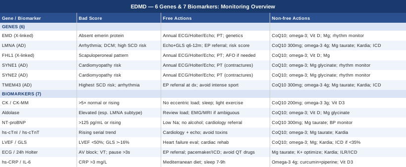
7 Biomarkers to Track in Emery-Dreifuss Muscular Dystrophy
Most EDMD patients are monitored primarily for muscle progression. What is consistently underemphasized — and what this section addresses directly — is that cardiac monitoring is not secondary care in EDMD. It is primary. The leading cause of death in EDMD is not muscle weakness but cardiac arrhythmia or heart failure, and the good news is that several reliable biomarkers can detect problems early enough to act on. The seven markers below span both muscle and cardiac domains, organized from the most accessible to the more specialized, with practical plans for each.
1. Creatine Kinase (CK / CK-MM)
Creatine kinase is the standard marker of skeletal muscle membrane damage, and in EDMD it tells a story that differs from most other muscular dystrophies. CK levels in EDMD are typically only mildly to moderately elevated — usually 2 to 10 times the upper limit of normal — which contrasts sharply with Duchenne MD, where CK can reach 50 to 100 times normal. This modest elevation reflects the fact that EDMD disrupts nuclear architecture and mechanosignaling rather than causing raw membrane breakdown. Tracking CK trends over months and years matters more than any single reading; a sudden upward spike warrants investigation, and a gradually rising trend across a year signals accelerated muscle stress that can be addressed.
How to Measure It
Standard blood test, ordered by a GP or neurologist. Request total CK with CK-MM fractionation for the most muscle-specific result. Cost: $20 to $50, routinely covered by insurance with a neuromuscular indication. Recommended frequency: every 6 to 12 months as part of routine neuromuscular monitoring.
If the Score Is Bad — The Plan Without Supplements
If CK is trending upward or consistently above five times normal, eliminate activities involving heavy eccentric muscle loading: downhill running, slow-negative phases in resistance training, and sudden high-force activities. Prioritize sleep — the majority of muscle repair signaling occurs during slow-wave sleep, and chronic poor sleep independently and measurably elevates CK. Add regular low-intensity movement such as walking or gentle cycling at a conversational pace to maintain circulation without imposing additional membrane stress. Avoid chronic NSAID use, which impairs prostaglandin-mediated muscle repair pathways.
If the Score Is Bad — The Plan with Supplements or Equipment
Coenzyme Q10 at 200 to 300 mg daily — taken with a fat-containing meal for absorption, divided across two doses — supports mitochondrial energy production in muscle cells and is particularly relevant in myopathies where cellular energy metabolism is under stress. Omega-3 fatty acids at 3 to 4 g per day of combined EPA and DHA reduce membrane-level inflammation and slow damage accumulation. Vitamin D3, if serum 25-OH-D is below 40 ng/mL, should be supplemented at 2,000 to 5,000 IU daily alongside Vitamin K2 at 100 to 200 mcg to support calcium regulation in muscle tissue. Side effects: CoQ10 is well-tolerated; occasional digestive discomfort at higher doses. Omega-3 at these doses may mildly reduce platelet aggregation — relevant if surgery is planned. Cycling: omega-3 and Vitamin D3 are used continuously. CoQ10 can be cycled 8 weeks on / 2 weeks off if cost is a constraint, though continuous use is preferred for active muscle disease.
2. Aldolase
Aldolase is a glycolytic enzyme found in high concentrations in muscle tissue. It can be elevated in EDMD even when CK is only borderline abnormal, making it a useful complement — particularly in LMNA-associated EDMD where the pattern of muscle involvement differs from EMD-EDMD. Elevated aldolase alongside only mildly elevated CK can help distinguish EDMD subtypes and gauge the degree of active inflammatory activity in affected muscle. Because it reflects a slightly different aspect of muscle metabolism than CK, having both values gives your neurologist a more complete and actionable picture of what is happening at the tissue level.
How to Measure It
Blood test, often included in comprehensive muscle enzyme panels but not in standard metabolic panels — request it specifically. Cost: $30 to $60. Frequency: annually or whenever CK results are ambiguous or inconsistent with reported symptoms.
If the Score Is Bad — The Plan Without Supplements
Persistently elevated aldolase warrants a review of physical activity load and a discussion with your neurologist about whether updated electromyography or muscle MRI should be performed to assess active inflammation or progression. The anti-inflammatory lifestyle foundation applies here as well: consistent sleep, reduced ultra-processed food intake, and manageable daily stress levels are the first interventions to optimize before adding anything else.
If the Score Is Bad — The Plan with Supplements or Equipment
The same core stack as for elevated CK: omega-3 at 3 to 4 g/day EPA+DHA, CoQ10 at 200 to 300 mg/day, Vitamin D3 to sufficiency. Add magnesium glycinate at 300 to 400 mg before bed — magnesium is required for glycolytic enzyme activity, and deficiency is common in people with chronic neuromuscular disease. An RBC magnesium test (not serum magnesium, which is insensitive to tissue deficiency) can confirm whether repletion is needed. Side effect: magnesium glycinate at these doses is well-tolerated; the glycinate form avoids the laxative effect of magnesium oxide or magnesium citrate at high doses.
3. NT-proBNP / BNP (Brain Natriuretic Peptide)
This is arguably the most critical blood biomarker for EDMD patients, and consistently the most undertested in routine neuromuscular care. NT-proBNP rises when the heart ventricles are under mechanical strain — a direct signal of early heart failure or impaired cardiac function, often detectable months or years before clinical symptoms appear. In EDMD, dilated cardiomyopathy can develop gradually and silently, and NT-proBNP provides an early warning that clinical examination and ECG alone cannot. Peter Attia, in Outlive and throughout his podcast, consistently identifies NT-proBNP as one of the most underused biomarkers in preventive cardiology. For EDMD patients, tracking this proactively is not optional — it belongs in the core monitoring panel from the time of diagnosis.
How to Measure It
Standard blood test. Cost: $50 to $150, usually covered with a cardiac monitoring indication. Age-adjusted normal: below 125 pg/mL for patients under 75. Values between 125 and 300 pg/mL indicate borderline strain and warrant follow-up. Above 300 pg/mL signals significant ventricular overload requiring prompt cardiology evaluation. Frequency: every 6 to 12 months in all confirmed EDMD; every 3 to 6 months if LMNA mutation is confirmed or previous values were borderline.
If the Score Is Bad — The Plan Without Supplements
An elevated NT-proBNP in EDMD should trigger a cardiology referral and an updated echocardiogram. On the lifestyle side: reduce dietary sodium to below 2,000 mg/day (processed and restaurant foods are the primary source), eliminate alcohol entirely — which is directly cardiotoxic and worsens arrhythmia burden — and limit fluid intake if your cardiologist advises. Avoid strenuous unmonitored exercise until cardiac function is formally reassessed. If sleep apnea is suspected, request a sleep study — nocturnal hypoxemia is a significant and underappreciated driver of elevated BNP in at-risk patients.
If the Score Is Bad — The Plan with Supplements or Equipment
Coenzyme Q10 at 300 mg/day has the strongest evidence in this domain: the Q-SYMBIO trial, a randomized controlled study in heart failure patients, demonstrated significant reductions in major cardiac events with CoQ10 300 mg/day versus placebo. Magnesium taurate at 300 to 400 mg/day supports cardiac conduction stability and has been associated with reduced arrhythmia burden in observational data. Omega-3 at 3 to 4 g/day EPA+DHA reduces triglycerides and cardiac inflammatory load. Equipment: a home blood pressure monitor used daily, a pulse oximeter for overnight spot checks, and a wearable rhythm monitor with irregular beat alerts (Apple Watch, Garmin with ECG features) provides continuous data between clinical visits. When NT-proBNP is significantly elevated, medications — ACE inhibitors, beta-blockers, SGLT2 inhibitors — are the primary treatment; the above are adjuncts and do not replace them.
4. High-Sensitivity Cardiac Troponin (hs-cTnI or hs-cTnT)
High-sensitivity cardiac troponin detects subclinical cardiomyocyte injury at concentrations far below what standard troponin assays capture. In EDMD — particularly in LMNA-associated disease — chronic low-level troponin elevation can precede overt cardiomyopathy by years. Thomas Dayspring, one of the leading voices in preventive cardiology biomarker interpretation, has emphasized hs-cTnI as a marker that deserves serial tracking in any patient with structural or genetic cardiac risk. A single normal result is reassuring; a rising trend across three to four consecutive measurements is a warning signal even when absolute values remain within the laboratory normal range.
How to Measure It
Blood test; requires a laboratory that runs high-sensitivity assays — specify hs-cTnI or hs-cTnT explicitly when ordering, as standard troponin lacks the necessary sensitivity. Cost: $30 to $100 depending on the laboratory. Frequency: annually at minimum; every 6 months if baseline is elevated or shows an upward trend over consecutive measurements.
If the Score Is Bad — The Plan Without Supplements
Elevated hs-cTnI warrants cardiology referral and an updated echocardiogram. Avoid cardiotoxic exposures: alcohol, recreational stimulants, high-dose NSAIDs, and strenuous unmonitored exercise. Treating acute febrile illness aggressively is relevant — fevers drive troponin spikes in tissue that is already under metabolic stress. Monitor for symptoms of pericarditis or myocarditis (chest pain, shortness of breath following viral illness) and report them to your physician promptly.
If the Score Is Bad — The Plan with Supplements or Equipment
Core supplement stack: omega-3 at 3 to 4 g/day, CoQ10 at 300 mg/day, magnesium taurate at 300 mg/day. Consider adding L-carnitine at 2 g/day — evidence in ischemic cardiac conditions supports carnitine's role in supporting mitochondrial energy transfer in cardiac tissue, which is relevant when cardiomyocytes are under metabolic stress. Side effect: fishy odor at high doses, manageable by switching to acetyl-L-carnitine form or dividing the dose across the day. Cycling: continuous use is appropriate in cardiac monitoring contexts. Equipment: a personal ECG device such as the Kardia Mobile 6L (~$150 device plus subscription) enables weekly home rhythm checks and produces physician-readable ECG strips between clinical Holter monitors.
5. Left Ventricular Ejection Fraction (LVEF) and Global Longitudinal Strain (GLS)
These echocardiographic parameters function as the most important quantitative cardiac measures in EDMD and should be tracked serially with the same discipline as any blood biomarker. LVEF — the percentage of blood ejected from the left ventricle per beat — is the standard pump function measure, with values below 50% indicating systolic dysfunction. But LVEF has a critical limitation: it detects dysfunction late. Global Longitudinal Strain (GLS) measures myocardial deformation during each heartbeat, detecting subtle dysfunction while LVEF is still in the normal range. Normal GLS is more negative than −20% (for example, −22%). A GLS of −17% or −18% with preserved LVEF is an early warning that LVEF-only monitoring would miss entirely. In EDMD, catching this early window is the difference between preventive treatment and emergency management.
How to Measure It
Transthoracic echocardiogram with strain imaging capability, performed by a cardiologist or cardiac sonographer experienced in GLS. Not all echocardiography laboratories report GLS routinely — request it explicitly when scheduling. Cost: $500 to $1,500 depending on institution and insurance. Frequency: every 1 to 2 years in all confirmed EDMD; every 6 to 12 months in LMNA-EDMD, TMEM43-EDMD, or when values are already trending abnormal.
If the Score Is Bad — The Plan Without Supplements
LVEF below 40% or GLS worse than −16% in EDMD should prompt a formal heart failure evaluation. Medication discussion — ACE inhibitors or ARBs, beta-blockers, and SGLT2 inhibitors — is appropriate and evidence-supported. Cardiac rehabilitation (supervised low-to-moderate intensity aerobic exercise) has demonstrated improvements in cardiac function even in established cardiomyopathy and is safe under medical supervision. Unmonitored high-intensity exercise should stop. For LMNA-EDMD with declining LVEF and coexisting arrhythmia risk factors, ICD evaluation should enter the conversation proactively.
If the Score Is Bad — The Plan with Supplements or Equipment
CoQ10 at 300 mg/day, omega-3 at 3 to 4 g/day, magnesium taurate at 300 to 400 mg/day. Equipment in this range: daily home BP monitoring, a wearable rhythm monitor, and weekly home ECG with a Kardia Mobile device. If LVEF is below 35% and LMNA mutation is confirmed, current cardiomyopathy guidelines often support ICD implantation given the sudden arrhythmic death risk — this is a specialist decision that should be made proactively, not only after a dangerous event.
6. ECG and 24-Hour Holter Monitoring (PR Interval, QRS Duration, Arrhythmia Burden)
Among all monitoring tools for EDMD, cardiac rhythm monitoring is arguably the most critical for preventing sudden death. EDMD — particularly LMNA-EDMD — carries a significantly elevated risk of sudden cardiac death from complete heart block, sustained ventricular tachycardia, or ventricular fibrillation, and these events frequently occur in patients whose skeletal muscle symptoms are still mild. The PR interval (normal less than 200 ms) and QRS duration (normal less than 120 ms) on a standard 12-lead ECG are the first detectable signs of conduction system disease. A 24-hour Holter monitor captures what a resting ECG misses: brief runs of atrial fibrillation, non-sustained ventricular tachycardia, sinus node dysfunction, and the precise degree of AV block across a full day and night.
How to Measure It
Standard 12-lead ECG: $50 to $200. 24-hour Holter monitor: $200 to $500. Both should be requested at every cardiology visit in EDMD. Frequency: ECG and Holter at minimum annually; every 6 months if any conduction abnormality is already present. For high-risk patients with LMNA or TMEM43 mutations, prior non-sustained VT, or abnormal Holter findings, extended Holter (7 to 14 days) or an implantable loop recorder (ILR) provides substantially greater sensitivity for paroxysmal arrhythmias.
If the Score Is Bad — The Plan Without Supplements
Any evidence of second- or third-degree AV block, frequent non-sustained VT runs, or significant pauses greater than 3 seconds should trigger urgent electrophysiology referral. Pacemaker or ICD implantation has been life-saving in EDMD — this decision should not be deferred. Strictly avoid QT-prolonging medications, a broad list that includes certain antihistamines, macrolide antibiotics, azole antifungals, and some antidepressants — cross-check every new prescription against a validated QT database. Maintain normal electrolytes: hypokalemia and hypomagnesemia are independent arrhythmia triggers that are straightforward to screen and correct.
If the Score Is Bad — The Plan with Supplements or Equipment
Magnesium taurate at 300 to 400 mg/day supports cardiac electrical stability and has been associated with reduced arrhythmia burden in observational data. Potassium optimization — maintaining serum potassium between 4.0 and 5.0 mEq/L through avocados, leafy greens, legumes, and supplementation if needed — is equally important for rhythm stability. Equipment: the Kardia Mobile 6L personal ECG device allows daily rhythm capture at home, with a physician-readable single-lead ECG strip that can be transmitted to your electrophysiology clinic. Wearables with irregular rhythm detection (Apple Watch, Garmin with ECG) add continuous passive monitoring between deliberate recordings. When the clinical risk threshold is crossed, an ILR or ICD replaces the monitoring question with a treatment question — a shift that should be managed in close partnership with an experienced electrophysiologist.
7. High-Sensitivity C-Reactive Protein (hs-CRP) and Interleukin-6 (IL-6)
EDMD is increasingly understood as not only a structural problem but a signaling problem. Lamin A/C dysfunction — particularly in LMNA-EDMD — activates inflammatory cascades through the NF-κB pathway, producing systemic low-grade inflammation that accelerates both muscle and cardiac decline. Research on LMNA-related laminopathies supports this inflammatory activation as a contributor to disease progression. hs-CRP (optimal below 1.0 mg/L, acceptable below 3.0 mg/L) and IL-6 quantify this inflammatory burden and can be tracked serially to gauge whether lifestyle and nutritional interventions are producing a measurable biological effect.
How to Measure It
hs-CRP: standard blood test, $20 to $50, widely available. IL-6: less commonly ordered, $50 to $150, may need to be specifically requested. Frequency: every 6 to 12 months, consistently measured during stable periods — not during active infection, injury, or within 48 hours of intense physical activity, all of which transiently spike both markers.
If the Score Is Bad — The Plan Without Supplements
Elevated hs-CRP warrants a dietary audit. Adopt a Mediterranean-style anti-inflammatory eating pattern centered on extra virgin olive oil, fatty fish at least three times per week, abundant non-starchy vegetables, minimal ultra-processed food, and low refined carbohydrate intake. Prioritize consistent sleep at 7 to 9 hours — poor sleep is one of the most reliable and modifiable drivers of sustained CRP elevation. Reduce sedentary time through light daily movement (10 to 15 minutes of walking after meals is effective and accessible). Address chronic psychological stress not as a lifestyle nicety but as a direct IL-6 driver: sustained cortisol elevation measurably raises interleukin-6 output.
If the Score Is Bad — The Plan with Supplements or Equipment
Omega-3 fatty acids at 4 g per day EPA+DHA is the best-supported anti-inflammatory supplement available, with multiple randomized controlled trials confirming its hs-CRP-lowering effect across cardiovascular risk populations. Curcumin with piperine at 500 to 1,000 mg curcumin and 5 to 10 mg piperine daily inhibits NF-κB-mediated inflammation — directly relevant to the mechanism driving inflammatory signaling in LMNA-EDMD. Side effect: curcumin at these doses can mildly prolong bleeding time; discuss with your cardiologist if anticoagulants are prescribed. Cycling: both omega-3 and curcumin can be used continuously at these doses. Vitamin D3 at 2,000 to 4,000 IU daily (when serum 25-OH-D is below 40 ng/mL) independently reduces IL-6, a consistent finding across clinical trials in deficient populations.
Tracking these seven biomarkers gives you the clearest possible picture of where things stand right now. The genetics section below adds the layer that explains why — and what the specific mutation implies about long-term risk and the intensity of monitoring your situation requires.
The 6 Genes Driving Emery-Dreifuss Muscular Dystrophy
Understanding which gene is mutated in EDMD is not an academic exercise. It determines your cardiac risk profile, the likely pace and pattern of progression, and how aggressively monitoring and intervention should be pursued. The six genes below account for the vast majority of confirmed EDMD cases. Researchers like Ali Torkamani at Scripps Research, who focuses on translating genomic data into actionable clinical insights for rare disease, consistently reinforce the principle that the specific variant matters as much as the diagnosis itself. Knowing your gene is the starting point; knowing your specific mutation type within that gene is the next level of precision.
EMD — The Emerin Gene (X-Linked EDMD1)
The EMD gene on chromosome Xq28 encodes emerin, an inner nuclear membrane protein that anchors the nuclear lamina to the cytoskeleton and plays a key role in gene expression regulation and mechanical stress response. Most pathogenic EMD mutations result in complete absence of emerin protein, which can be confirmed by immunostaining of a muscle biopsy sample. The condition follows X-linked recessive inheritance: males are primarily affected, and female carriers can have clinically significant cardiac involvement that should not be dismissed as benign.
The muscle phenotype in EMD-EDMD typically includes early-onset contractures in the Achilles tendons, elbows, and posterior cervical muscles, followed by slowly progressive humeroperoneal weakness. CK is usually mildly elevated. Cardiac involvement — typically atrial arrhythmias and conduction defects — is common and can become severe over time, though typically later and somewhat less aggressively than in LMNA-EDMD.
If the Gene Is Bad — The Plan Without Supplements
Annual cardiac monitoring including ECG, 24-hour Holter, and echocardiogram is appropriate from the time of confirmed diagnosis, regardless of symptom severity. Physical therapy targeting contracture management and functional muscle preservation should begin early — before contractures become fixed. Genetic counseling for all family members is essential; female carriers need their own cardiac surveillance program. Avoid high-impact contact sports and activities involving repeated acute muscle stress. Discuss with your cardiologist whether GLS imaging should be included from the outset rather than waiting for LVEF to change.
If the Gene Is Bad — The Plan with Supplements or Equipment
No supplement reverses an EMD null mutation at the genetic level, but reducing downstream metabolic stress on nuclear membrane-compromised muscle and cardiac tissue is feasible. CoQ10 at 200 to 300 mg/day, omega-3 at 3 to 4 g/day, Vitamin D3 to sufficiency, and magnesium glycinate at 300 to 400 mg/day form the relevant support foundation. For female carriers with confirmed cardiac involvement, a wearable rhythm monitor adds a continuous surveillance layer between clinical Holter appointments. Frequency and cycling as described in the biomarker section above.
LMNA — The Lamin A/C Gene (Autosomal Dominant EDMD2)
LMNA on chromosome 1q22 encodes lamins A and C — the primary structural proteins of the inner nuclear lamina. LMNA-EDMD is the most common and best-characterized form of the disease. More than 450 pathogenic variants have been identified, and the specific mutation type — missense versus truncating versus splice-site — meaningfully affects cardiac prognosis. The cardiac prognosis in LMNA-EDMD is substantially worse than in EMD-EDMD: arrhythmias and dilated cardiomyopathy appear earlier, progress faster, and carry a significantly higher risk of sudden cardiac death. A validated LMNA sudden death risk score — incorporating variables including non-sustained ventricular tachycardia, LVEF below 45%, male sex, truncating or splice-site mutation, and first documented non-sinus rhythm — has been developed to guide ICD implantation decisions. Research on this validated clinical risk tool is available on PubMed and should be reviewed with your electrophysiologist.
If the Gene Is Bad — The Plan Without Supplements
Aggressive cardiac surveillance from diagnosis: ECG and 24-hour Holter every 6 to 12 months, echocardiogram with GLS annually. If conduction abnormalities develop or LVEF declines below 45%, electrophysiology referral should not be delayed. Formal calculation of the LMNA sudden death risk score is appropriate — it is a validated decision-support tool, not a rough estimate, and it directly informs the ICD conversation. Physical activity planning should involve cardiology: zone 2 aerobic training at 60 to 70% of maximum heart rate is likely safe and beneficial; high-intensity training and competitive sports should be avoided without formal cardiac clearance.
If the Gene Is Bad — The Plan with Supplements or Equipment
CoQ10 at 300 mg/day, omega-3 at 4 g/day, magnesium taurate at 300 to 400 mg/day, Vitamin D3 to sufficiency. Equipment priority is highest in LMNA-EDMD: Kardia Mobile 6L for weekly home ECG, a wearable rhythm monitor for continuous surveillance, and a home blood pressure monitor for daily tracking. mTOR inhibition with rapamycin has shown preclinical promise in LMNA-related cardiomyopathy models and is a subject of active clinical research — mention it to your cardiologist as a potential future option, but do not pursue it as a self-directed supplement given its significant immunosuppressive side effect profile.
FHL1 — The Four-and-a-Half LIM Domain Gene (X-Linked EDMD3)
FHL1 on chromosome Xq26.3 encodes four-and-a-half LIM domain protein 1, a sarcomeric Z-disk protein involved in myofibril assembly and mechanical stress sensing within muscle fibers. FHL1-EDMD often presents with a scapuloperoneal phenotype — weakness beginning in the shoulder-girdle and peroneal muscles — which can lead to misdiagnosis or delayed diagnosis before targeted genetic testing is performed. The cardiac risk profile is generally similar to EMD-EDMD, though the evidence base remains smaller and expressivity varies between families.
If the Gene Is Bad — The Plan Without Supplements
Cardiac monitoring at the level applied to EMD-EDMD: annual ECG, Holter, and echocardiogram. Physical therapy specifically targeting scapular stabilization and ankle dorsiflexion, the functional areas most commonly compromised by FHL1 dysfunction. Ankle-foot orthoses for significant foot drop when clinically indicated. Family members should be offered genetic testing given X-linked inheritance; carrier females need cardiac evaluation.
If the Gene Is Bad — The Plan with Supplements or Equipment
Core supplement stack as above: omega-3, CoQ10, Vitamin D3, magnesium glycinate. Because FHL1 dysfunction involves Z-disk integrity, training approaches that emphasize concentric and isometric work — under physiotherapy guidance — rather than high-load eccentric movements are mechanistically appropriate and safer. No FHL1-specific supplement strategy has been established; the general neuromuscular metabolic support approach applies.
SYNE1 and SYNE2 — The Nesprin Genes (EDMD4 and EDMD5)
SYNE1 (chromosome 6q25.2) and SYNE2 (chromosome 14q23.2) encode nesprin-1 and nesprin-2 respectively — large scaffolding proteins that span the outer nuclear membrane and connect the nucleus to the actin cytoskeleton. They are core components of the LINC complex (Linker of Nucleoskeleton and Cytoskeleton), the same mechanosignaling system disrupted in EMD and LMNA EDMD. Both genes cause autosomal dominant EDMD with a phenotype resembling classical EDMD. Cardiac involvement in SYNE-EDMD appears on average somewhat less severe than in LMNA-EDMD, though meaningful cardiomyopathy has been reported in confirmed SYNE variant carriers and monitoring should not be relaxed.
If the Gene Is Bad — The Plan Without Supplements
Cardiac monitoring at the level applied to EMD-EDMD: annual ECG, Holter, and echocardiogram. Physical therapy for contracture management. Because the LINC complex transmits mechanical forces from the cytoskeleton to the nucleus, avoiding activities that impose sudden, large tensile forces on muscle — particularly repetitive high-impact loading patterns — is biologically reasonable and worth discussing with your physiotherapist when designing an activity plan.
If the Gene Is Bad — The Plan with Supplements or Equipment
Standard neuromuscular metabolic support: omega-3, CoQ10, magnesium glycinate, Vitamin D3. A wearable cardiac monitor adds a safety layer between clinical visits, particularly in the early years after diagnosis when the cardiac risk profile for specific SYNE variants is still being characterized in the literature. The evidence base for these variants is growing but remains thinner than for LMNA and EMD — report any new symptoms to your specialist promptly and do not rely solely on annual scheduled appointments.
TMEM43 — The LUMA Gene (EDMD7 and Elevated Sudden Death Risk)
TMEM43 on chromosome 3p25.1 encodes LUMA, an inner nuclear membrane protein that interacts with emerin and lamins A/C. TMEM43 mutations cause EDMD7 (autosomal dominant) and are also responsible for a severe form of arrhythmogenic cardiomyopathy in some families, including a well-described founder mutation in Newfoundland families where sudden cardiac death was disproportionately frequent. This dual phenotype means TMEM43-EDMD carries one of the highest risks of sudden cardiac death among all EDMD genetic forms. The arrhythmia risk can be life-threatening even when skeletal muscle symptoms are still mild or developing, making the cardiac picture the dominant clinical concern in many families.
If the Gene Is Bad — The Plan Without Supplements
Aggressive cardiac surveillance from the moment of genetic diagnosis, regardless of symptom severity. Electrophysiology referral is appropriate at or near the time of confirmed diagnosis. ICD implantation is frequently recommended for confirmed pathogenic TMEM43 variants — this conversation should happen proactively with a cardiologist experienced in arrhythmogenic cardiomyopathy and EDMD, not be delayed until a dangerous arrhythmia occurs. High-intensity and competitive sports should be avoided until cardiac risk is formally evaluated and a management plan is in place.
If the Gene Is Bad — The Plan with Supplements or Equipment
Metabolic support: CoQ10 at 300 mg/day, omega-3 at 4 g/day, magnesium taurate at 300 to 400 mg/day, potassium optimization targeting serum levels of 4.0 to 5.0 mEq/L. Equipment: Kardia Mobile 6L or equivalent for frequent home ECG monitoring between clinical visits. If an ICD is implanted, remote device monitoring — transmitting data directly to your electrophysiology clinic — adds surveillance without requiring in-person visits for every rhythm check. This is one genetic variant where the device conversation should be initiated early and decisively.
Understanding the genetic architecture and the biomarker landscape gives you the "what" and the "why." The next layer — distilling what the best preventive cardiology thinking says about patients with exactly this kind of genetic cardiac risk — turns that knowledge into practical day-to-day strategy.
What "Outlive" by Peter Attia Reveals About Monitoring a Condition Like EDMD
Outlive: The Science and Art of Longevity by Peter Attia (2023) is not written about EDMD. But it may be the most practically useful book on proactive cardiac monitoring and longevity medicine written for a general audience, and its core arguments map directly onto what EDMD management requires. Attia draws on hundreds of studies, from cardiovascular outcome trials to metabolic research, to build a framework for identifying risk and acting early. The ten insights below are drawn from the book's key themes and applied to the EDMD context.
1. Reactive Medicine Arrives Too Late for Conditions Like EDMD
Attia's central argument is that modern medicine operates reactively — waiting for disease to manifest before treating. For EDMD, where the first cardiac event can be fatal and occurs in patients who are still functionally active, this posture is particularly dangerous. His alternative — proactive surveillance and early intervention, what he calls "Medicine 3.0" — is precisely what EDMD demands. The earlier a decline in GLS, a rise in NT-proBNP, or a conduction abnormality is detected, the broader the menu of intervention options that remain available.
2. NT-proBNP Is Systematically Underused Outside of Cardiology
Attia discusses NT-proBNP at length as a marker routinely ordered by cardiologists but consistently underutilized by generalists and subspecialists who are not cardiology-trained — including the neurologists who often manage EDMD as a primary physician. For EDMD patients, this creates a specific and correctable surveillance gap. The practical fix is to specifically request NT-proBNP testing at every follow-up visit and ensure results are reviewed by a cardiologist in the context of EDMD's cardiac natural history.
3. Ejection Fraction Alone Is Not Sensitive Enough
Attia is unusually explicit about the limitations of LVEF as a lone surveillance marker: by the time ejection fraction drops below normal, significant cardiac remodeling has already occurred and treatment options are reduced. He recommends GLS as the more sensitive early surrogate for myocardial dysfunction. For EDMD patients, the practical implication is to insist on GLS measurements at every echocardiogram rather than accepting an LVEF-only report as a complete cardiac evaluation.
4. Zone 2 Training Is the Safest Exercise Format for At-Risk Hearts
Attia dedicates substantial attention to zone 2 aerobic training — exercise sustained at approximately 60 to 70% of maximum heart rate, at an intensity that allows comfortable conversation throughout. This format improves mitochondrial density, cardiac efficiency, and metabolic flexibility without the hemodynamic stress that high-intensity training places on compromised cardiac tissue. For EDMD patients with known cardiomyopathy, zone 2 with wearable heart rate monitoring is the safest and most evidence-supported form of structured exercise available.
5. Muscle Mass and Strength Are Independent Survival Predictors
Attia documents the relationship between muscle mass, grip strength, and all-cause mortality with striking consistency: greater muscle mass and functional strength predict longer survival across every cause-of-death category he examines. For EDMD, this matters because preserving whatever muscle function is possible — through physiotherapy-guided resistance training, adequate protein intake at 1.6 to 2.2 g/kg body weight daily, and sleep optimization — produces a measurable longevity benefit that operates independently of the underlying genetic condition.
6. Sleep Is a Non-Negotiable Biological Intervention
Attia describes sleep not as passive rest but as the primary window for cardiac repair, muscle protein synthesis, metabolic clearance, and immune regulation. Chronic sleep deprivation independently elevates CK, NT-proBNP, and hs-CRP — all three of the biomarkers tracked in the primary strategy above. For EDMD, treating sleep optimization as the foundational intervention, before supplementation or exercise modification, is the correct prioritization. Seven to nine hours of consistent, high-quality sleep should be the anchor of any management plan.
7. Serial Testing Produces Data; Snapshot Testing Produces Ambiguity
One of Attia's most transferable insights is the concept of trending over time rather than interpreting isolated test results. A single NT-proBNP of 150 pg/mL is borderline and ambiguous. Three consecutive readings at 110, 130, and 155 pg/mL is a rising trend that demands action. Building a longitudinal biomarker record — consistent testing at consistent intervals, tracked across years — transforms each individual result from ambiguous data into interpretable signal. For EDMD, this means maintaining a personal tracking document of every biomarker result, with dates, and reviewing trends at every cardiology visit.
8. Metabolic Health Buffers Genetic Vulnerabilities
Attia introduces the concept of metabolic reserve — the functional margin created by optimized metabolic health that buffers against structural and genetic vulnerabilities. While no metabolic optimization reverses LMNA or EMD mutations, patients who enter the cardiac decline phase with better insulin sensitivity, blood glucose control, and lipid profiles have more cardiac reserve, slower progression, and more therapeutic options. Tracking HbA1c, fasting insulin, and apolipoprotein B is therefore directly relevant to EDMD outcomes and should be part of the annual biomarker panel.
9. Alcohol Has No Safe Threshold for Cardiac-Risk Patients
Attia is more direct than most physicians on this topic: alcohol is a cardiotoxin that worsens arrhythmia burden, impairs cardiac repair, directly depresses myocardial contractility, and elevates NT-proBNP. For EDMD patients with any degree of cardiomyopathy or conduction disease, alcohol is not something to minimize — it is something to eliminate. The biological cost is disproportionate in the presence of genetic cardiac vulnerability, regardless of how social drinking is normalized in the surrounding environment.
10. Multidisciplinary Specialist Teams Change Outcomes in Complex Conditions
Attia documents the outcome gap between general and specialized care for multi-organ conditions. For EDMD, building a team that includes a neuromuscular specialist, a cardiologist or electrophysiologist experienced with EDMD or laminopathies, a physical therapist familiar with muscular dystrophy, and a clinical geneticist is not a luxury — it is a survival-relevant strategy. Centers of excellence for neuromuscular disease consistently produce better outcomes than general practices managing EDMD in low volume, and advocating for this level of care is one of the most important actions an EDMD patient or family can take.
The monitoring, genetics, and cardiology strategy above address EDMD's most critical clinical dimensions. The approaches below address the daily experience of living in a body affected by this condition — and while the evidence for each is more limited, it is real enough to merit consideration.
Complementary Approaches With Clinical Evidence
The three approaches below have meaningful, if sometimes limited, evidence for supporting quality of life and symptom management in muscular dystrophy and related chronic conditions. None replace medical management, but each addresses a real dimension of living with EDMD that biomarker monitoring alone does not cover.
Yoga and Adapted Stretching for Contracture Management
Contractures are a hallmark and early feature of EDMD, typically affecting the Achilles tendons, elbow flexors, and posterior cervical muscles, and progressing over time without consistent intervention. Yoga-based stretching — when carefully adapted — directly targets the connective tissue and muscle groups most affected. The relevant modality is not vigorous vinyasa or hot yoga but yin yoga, which uses long-held passive stretches of 3 to 5 minutes per position, or restorative yoga, practiced with a qualified instructor who is informed about your diagnosis and its cardiac implications. The key mechanism is sustained, low-load elongation of soft tissue, which progressively reduces contracture severity over weeks to months of consistent practice.
Research on passive stretching and range-of-motion physiotherapy in muscular dystrophy populations, summarized in neuromuscular physiotherapy guidelines, consistently shows that regular stretching reduces the rate of contracture progression, improves joint range of motion, and reduces reported pain intensity compared to no stretching program. EDMD-specific randomized trials are lacking, but the biological mechanism — collagen remodeling and sarcomere length normalization under sustained tensile load — is well-established and applies directly to EDMD's contracture pattern.
In practice: work with your physical therapist first to identify which contractures are present and which positions are safe and appropriate for your current functional level. Then supplement clinical sessions with 15 to 30 minutes of adapted gentle yoga three to five times per week. Inform your yoga instructor explicitly about your diagnosis, cardiac monitoring requirements, and positions to avoid. Never push to pain — stretch to a sensation of mild tension and hold there. Consistency across months produces the relevant structural change; intensity in any single session does not.
Breathing-Based Therapies for Autonomic and Respiratory Support
EDMD can involve respiratory muscle weakness in more advanced stages, and even when breathing muscles are intact, the autonomic nervous system dysregulation associated with cardiomyopathy can increase sympathetic tone and reduce heart rate variability. Slow resonant breathing at approximately 5 to 6 breaths per minute directly addresses this autonomic dimension. At this breathing rate, respiratory rhythm synchronizes with natural heart rate variability oscillations, producing measurable improvements in parasympathetic tone, blood pressure, and cardiac autonomic regulation — physiological effects that are clinically relevant in a condition characterized by progressive cardiomyopathy.
Randomized trials on slow resonant breathing in cardiac patient populations demonstrate significant improvements in heart rate variability, reductions in resting blood pressure, and improvements in anxiety and quality of life measures. Higher resting heart rate variability is independently associated with better cardiac prognosis in cardiomyopathy. For EDMD specifically, this intervention is low-risk, low-cost, and addresses a real biological need — the autonomic strain of living with progressive cardiac disease.
Practically: practice 10 minutes of slow diaphragmatic breathing twice daily, inhaling for 5 seconds and exhaling for 6 seconds at a steady pace. No equipment is required, though a metronome app or guided breathing audio simplifies the pacing. Track resting heart rate variability with any HRV-capable wearable as a proxy for autonomic recovery and improvement over time. If respiratory muscle weakness is suspected — presenting as exertional breathlessness disproportionate to cardiac function, morning headaches, or difficulty clearing secretions — request formal pulmonary function testing including forced vital capacity (FVC), and maximal inspiratory and expiratory pressures (MIP and MEP). Significant respiratory impairment warrants respiratory physiotherapy input and, in advanced cases, non-invasive ventilation assessment.
Mindfulness-Based Stress Reduction for Chronic Disease Quality of Life
Living with a progressive genetic condition that carries significant cardiac risk creates a specific and sustained psychological burden: uncertainty about when complications will develop, the visibility of gradual physical change, and the weight of knowing that sudden cardiac death is a real possibility for your gene type. This psychological dimension is consistently undertreated in neuromuscular clinics focused on physical metrics. Mindfulness-Based Stress Reduction (MBSR), the 8-week structured program developed by Jon Kabat-Zinn at the University of Massachusetts Medical School, has the most robust evidence base of any mind-body intervention for chronic disease psychological outcomes.
A meta-analysis of MBSR in chronic illness populations shows significant reductions in anxiety, depression, pain perception, and fatigue — all dimensions relevant to EDMD. Beyond subjective quality of life, MBSR produces measurable physiological effects: reduced cortisol output, lower hs-CRP, improved sleep architecture, and enhanced heart rate variability. For EDMD, this connects directly to the inflammatory and cardiac biomarkers in the primary strategy — reducing chronic psychological stress has a downstream, measurable effect on IL-6 and CRP that is not trivial.
In practice: the full MBSR protocol involves 8 weekly group sessions plus daily 45-minute home practice. Online versions — Palouse Mindfulness offers a complete, free MBSR course — make it accessible regardless of location or mobility constraints. The minimum effective dose in the evidence base appears to be 20 to 30 minutes of daily practice sustained over 6 to 8 weeks. For EDMD specifically, body scan meditation and gentle mindful movement adapted to your current physical capacity are preferred over intensive sitting meditation that may exacerbate musculoskeletal discomfort. Consistency across weeks is what produces the biological effect; the length of any individual session matters less.
Conclusion
Emery-Dreifuss Muscular Dystrophy is complex, but its most important dimensions are trackable and actionable. The cardiac risk is the part that most deserves proactive, systematic attention — and the tools to monitor it are accessible and underused. Tracking NT-proBNP, high-sensitivity cardiac troponin, echocardiographic parameters with GLS, and Holter monitoring data gives you and your care team the early signals needed to act before a crisis, not after. Knowing which gene drives your condition — whether EMD, LMNA, FHL1, SYNE1, SYNE2, or TMEM43 — sharpens the monitoring strategy, clarifies the arrhythmia risk level, and changes the threshold for interventions like ICD implantation.
The smart next step is specific: if you have not had a complete cardiac biomarker panel — NT-proBNP, high-sensitivity troponin, echocardiogram with GLS, and 24-hour Holter — in the past 12 months, request one. If you do not yet know which gene variant you carry, ask for targeted genetic testing through a neuromuscular or clinical genetics clinic. And if your current care team does not include both a neuromuscular specialist and a cardiologist experienced with EDMD, building that team is the single highest-leverage action available. Better information, tracked consistently over time, is the foundation for managing this condition as well as it can be managed.
Musculoskeletal: Joint Conditions Muscle Conditions
Neurological: Nerve Conditions
Cardiovascular: Heart Conditions Heart Rhythm Conditions