This article contains AI generated content.
· UpdatedEnchondroma: 4 Genes And 6 Biomarkers To Track
Introduction
Being told you have an enchondroma — often discovered incidentally on an X-ray taken for something completely unrelated — puts you in a strange medical limbo. You are reassured it is almost certainly benign, advised to monitor it periodically, and then sent on your way. For most people, that encounter ends without a single question answered about why it formed, what makes it stable versus progressive, or whether anything can be done beyond waiting for the next imaging appointment.
The short answer is that there is more to know, but only if you are willing to look at the molecular level. Enchondromas are not random structural quirks. They carry identifiable genetic mutations — most notably in two specific genes — that drive their formation through a precise biochemical cascade. They also exist within a metabolic and inflammatory environment that either supports or discourages their progression toward more aggressive behavior. Generic reassurance rarely touches any of this.
This article approaches enchondroma from two complementary angles. The first explores the four most important genetic and epigenetic factors involved in enchondroma formation and behavior, explaining what each mutation actually does and what — if anything — can be done to address the downstream consequences. The second covers six concrete biomarkers that can be measured in blood or urine to track the metabolic environment surrounding an enchondroma over time. Neither strategy replaces orthopedic monitoring, and nothing here is a cure. But together they offer something most patients never receive: a real explanation and a practical plan.
Better information leads to better decisions. That is the modest but meaningful promise this article makes.
Reverse Enchondroma: What Recent Genetics and Epigenetics Research Suggests
Enchondroma arises from immature cartilage cells — chondrocytes — that fail to complete their normal differentiation program and instead remain in a proliferating, immature state within the bone. Over the past fifteen years, researchers have identified specific genetic mutations that explain this failure with remarkable precision. Understanding these mutations does not just tell you what caused the tumor. It opens a window into what the body's metabolism is doing inside and around it, and which targeted interventions have the strongest scientific rationale.
Gene 1: IDH1 — The Most Common Driver
IDH1 (isocitrate dehydrogenase 1) normally encodes an enzyme in the citric acid cycle that converts isocitrate to alpha-ketoglutarate (α-KG), a molecule with crucial roles in cellular energy metabolism and epigenetic regulation. When IDH1 carries a somatic mutation — most commonly at the R132 position — it loses this normal function and instead acquires an entirely new one: converting α-KG into 2-hydroxyglutarate (2-HG), a molecule with no healthy physiological role and significant disruptive potential.
IDH1 mutations have been identified in approximately 40–70% of sporadic enchondromas and in an even higher proportion of enchondromas associated with Ollier disease, a rare condition characterized by multiple enchondromas throughout the skeleton. The landmark 2011 study by Pansuriya and colleagues (Pansuriya TC et al., Nature Genetics, 2011) was the first to establish this connection at scale, showing that somatic IDH1 and IDH2 mutations are a defining molecular feature of enchondromatosis and providing the clearest molecular explanation for why these tumors form.
The consequence of this mutation is not just one broken enzyme reaction. Because α-KG is depleted to produce 2-HG, the entire class of α-KG-dependent enzymes is impaired. This includes the TET methylcytosine dioxygenases (which erase DNA methylation marks) and the Jumonji-domain histone demethylases (which erase repressive histone marks). These are the molecular machinery responsible for keeping gene expression properly regulated. When 2-HG accumulates and suppresses them, a hypermethylation phenotype results: gene promoters that should be accessible become silenced. Differentiation programs shut down. Chondrocytes remain immature and proliferative. That, in mechanistic terms, is how an enchondroma forms and persists.
If the IDH1 gene is mutated — the plan without supplements
Since IDH1 mutations in enchondromas are somatic — occurring only in the affected cartilage cells, not throughout the body — you cannot reverse the mutation itself. What you can do is reduce the environmental pressure that allows 2-HG's downstream effects to operate most aggressively.
Maintain rigorous imaging surveillance: This is the single most important non-supplement action. An MRI of the affected bone every 1–3 years (depending on location, size, and clinical assessment) is standard practice. Enchondromas in weight-bearing bones such as the femur warrant closer attention than those in small hand bones. Never delay a scheduled scan.
Adopt a low-glycemic, anti-inflammatory diet: Reducing dietary sugar and refined carbohydrates reduces the substrate availability for glycolytic metabolism that IDH-mutant cells prefer. A Mediterranean-style dietary pattern — abundant vegetables, olive oil, fish, legumes, and minimal ultra-processed food — has robust evidence for reducing systemic inflammation and supporting mitochondrial health.
Zone 2 aerobic exercise consistently: Low-intensity sustained aerobic activity (the pace where conversation is possible) is the most evidence-backed intervention for improving mitochondrial efficiency and reducing systemic metabolic dysfunction. Three to four sessions of 30–45 minutes per week is a reasonable, sustainable starting point.
Optimize sleep: Cellular repair and immune surveillance operate predominantly during deep sleep. Chronic poor sleep impairs mitochondrial function and elevates cortisol, both of which amplify the metabolic disruption associated with IDH mutations.
If the IDH1 gene is mutated — the plan with supplements or equipment
Several targeted nutritional interventions address the specific downstream consequences of IDH1 mutation. None are proven in clinical enchondroma trials — most evidence comes from IDH-mutant leukemia research, where the same mutation is well-studied. All should be discussed with a clinician before starting.
Alpha-ketoglutarate (AKG): Because mutant IDH1 depletes α-KG to produce 2-HG, supplementing with exogenous α-KG may partially restore the balance at enzyme active sites and reduce 2-HG's competitive inhibitory effects. Studies in IDH-mutant leukemia cells and animal models have shown that exogenous α-KG can partially restore TET enzyme function. Calcium alpha-ketoglutarate is the most bioavailable commercial form. - Typical dose: 500–1,000 mg/day with meals - Cycling: No established protocol exists for this indication; a conservative approach is 8 weeks on, 2 weeks off - Side effects: Generally well-tolerated; possible mild GI discomfort at higher doses
Vitamin C (ascorbate): Ascorbate is a required cofactor for TET dioxygenases — the exact enzymes suppressed by 2-HG. Research in IDH-mutant acute myeloid leukemia has demonstrated that high-dose ascorbate can partially restore TET2 activity even in the presence of IDH mutations, by providing saturating cofactor concentrations to overcome competitive inhibition by 2-HG. This is mechanistically plausible and well-supported in cancer cell biology, though enchondroma-specific data is absent. - Oral dose: 1–3 g/day in divided doses with food - Higher doses (above GI tolerance) require IV administration in a clinical setting - Cycling: Continuous oral use is safe for most adults; monitor urinary oxalate if prone to kidney stones - Side effects: GI upset at high doses; kidney stone risk with very high long-term intake in susceptible individuals
Sulforaphane (from broccoli sprout extract): Sulforaphane activates the NRF2 pathway — the body's primary antioxidant and detoxification regulation system. Reducing oxidative stress may limit the cellular damage amplified by 2-HG accumulation, and sulforaphane has documented anti-tumor activity in multiple preclinical models. - Dose: 25–50 mg/day from a standardized broccoli sprout extract, or 100–150 g of fresh broccoli sprouts daily - Cycling: 5 days on, 2 days off is commonly used - Side effects: Rare; very high intake may affect thyroid function in iodine-deficient individuals
Magnesium glycinate: Magnesium is a cofactor in hundreds of enzymatic reactions, including several within the citric acid cycle that IDH1 mutations disrupt. Deficiency is common and may amplify metabolic imbalances. - Dose: 200–400 mg elemental magnesium per day, preferably as glycinate for absorption - Side effects: Softened stools at higher doses; otherwise safe for most adults
Gene 2: IDH2 — The Mitochondrial Counterpart
IDH2 encodes the mitochondrial isoform of isocitrate dehydrogenase. When mutated — most commonly at the R172 or R140 positions — IDH2 generates 2-HG within the mitochondrial matrix rather than the cytoplasm. IDH2 mutations account for approximately 15–30% of enchondromas that carry an identifiable IDH mutation, and they are proportionally more prevalent in Maffucci syndrome, a variant of Ollier disease characterized by the additional presence of vascular malformations.
The downstream epigenetic consequences are similar to IDH1, but the mitochondrial location means that IDH2 mutations more directly impair the electron transport chain and oxidative phosphorylation. Affected cells show a stronger shift toward glycolytic metabolism, and the mitochondria themselves become less efficient. The research group of Amary and colleagues (Journal of Pathology, 2011) validated IDH2 mutations as key molecular events in both enchondromas and their malignant counterpart, conventional chondrosarcoma, underscoring the importance of IDH mutation status in risk stratification — not just diagnosis.
If the IDH2 gene is mutated — the plan without supplements
The mitochondrial location of IDH2 means that mitochondrial health interventions are particularly well-targeted here.
Zone 2 aerobic training as a consistent practice: This remains the most powerful known intervention for increasing mitochondrial density and improving electron transport chain efficiency. Three to five sessions per week of sustained low-intensity effort (nasal breathing only is a useful intensity guide) is the clinical minimum for meaningful adaptation.
Time-restricted eating (16:8 pattern): Limiting food intake to an 8-hour window reduces hyperglycemia episodes, lowers average insulin levels, and activates autophagy — the cellular recycling process that clears damaged mitochondrial components. This is not extreme fasting; it simply concentrates meals within a reasonable window.
Cold and heat exposure (cautiously): Brief cold water immersion (2–5 minutes at 10–15°C) and regular sauna use (2–4 sessions per week, 15–20 minutes at 80–90°C) both upregulate mitochondrial biogenesis markers in human subjects. These practices require medical clearance for anyone with cardiovascular conditions.
If the IDH2 gene is mutated — the plan with supplements or equipment
CoQ10 (ubiquinol form): Ubiquinol is the active, reduced form of coenzyme Q10 and a critical component of the mitochondrial electron transport chain — the very system directly impaired by IDH2 dysfunction. Ubiquinol is better absorbed than ubiquinone, particularly in individuals over 40. - Dose: 100–300 mg/day with a fat-containing meal - Cycling: Continuous use is typical for mitochondrial support; no established off-cycle needed - Side effects: Rare; possible mild GI discomfort; may modestly interact with warfarin
NAD+ precursors (NMN or NR): NAD+ is essential both for IDH2's normal enzymatic function and for the sirtuin deacetylases that regulate mitochondrial biogenesis and quality control. Nicotinamide mononucleotide (NMN) and nicotinamide riboside (NR) both raise NAD+ levels in human clinical trials, with NMN showing faster onset and NR showing a longer safety record. - Typical dose: NMN 250–500 mg/day; NR 250–300 mg/day, taken in the morning - Cycling: Continuous use is common; some practitioners cycle 5 days on, 2 days off - Side effects: Generally well-tolerated; NMN may cause mild warmth or flushing in some individuals
PQQ (pyrroloquinoline quinone): PQQ stimulates mitochondrial biogenesis through PGC-1α activation and may partially compensate for reduced mitochondrial function in IDH2-affected tissues. - Dose: 10–20 mg/day with a meal - Side effects: Minimal; headache reported rarely at higher doses
Gene 3: The IHH-PTHrP Axis — Where Cartilage Development Goes Wrong
Indian Hedgehog (IHH) and parathyroid hormone-related peptide (PTHrP, encoded by the PTHLH gene) form a critical feedback loop that controls chondrocyte maturation during skeletal development. Under normal conditions, this axis ensures an orderly, sequential progression of cartilage cells from immature proliferating forms to mature, hypertrophic cells that eventually mineralize. When this feedback loop is disrupted — whether by somatic mutations or epigenetic silencing of regulatory elements — chondrocytes stall in an immature proliferating state, which is precisely the cellular phenotype of an enchondroma.
Importantly, in enchondromas and Ollier disease cases that do not carry IDH mutations, dysregulation of the IHH/PTHLH pathway is often the alternative molecular explanation. IDH mutations and IHH pathway dysregulation are not always independent: accumulation of 2-HG from mutant IDH enzymes alters the epigenetic accessibility of IHH pathway target genes, creating a molecular overlap between these two pathways.
If the IHH/PTHrP pathway is disrupted — the plan without supplements
The IHH-PTHrP axis is closely intertwined with vitamin D signaling and calcium-phosphate metabolism, both of which are directly modifiable through lifestyle.
Sun exposure for endogenous vitamin D synthesis: 15–25 minutes of midday sun exposure on large skin surface areas (arms, legs, and torso exposed) generates 10,000–20,000 IU equivalent of vitamin D3 in lighter-skinned adults, and meaningfully less in those with darker skin tones. Vitamin D's active metabolite, calcitriol, directly modulates PTHrP expression and chondrocyte function via the vitamin D receptor.
Weight-bearing physical activity: Mechanical loading of the skeleton activates PTH signaling and maintains healthy bone remodeling feedback. Swimming is protective for joint cartilage, but regular walking and low-impact resistance exercise are important for maintaining healthy PTH/PTHrP regulation in bone.
Calcium from whole food sources: Dairy, leafy greens, legumes, almonds, and fortified plant milks support the calcium homeostasis that is integral to normal PTH/PTHrP feedback. Supplemental calcium should only be used when dietary intake is clearly insufficient, as excess supplemental calcium carries cardiovascular considerations.
If the IHH/PTHrP pathway is disrupted — the plan with supplements or equipment
Vitamin D3 with vitamin K2 (MK-7): If serum 25-OH vitamin D is below 40 ng/mL — the functional threshold recommended by specialists including Dr. Peter Attia — supplementation is clearly warranted. Vitamin D3 directly modulates chondrocyte differentiation via VDR signaling, intersecting with the PTHrP pathway. Vitamin K2 as MK-7 directs calcium toward bone mineral rather than soft tissues. - Dose: 2,000–5,000 IU vitamin D3/day; pair with 100–200 mcg vitamin K2 (MK-7) - Monitor: Recheck 25-OH vitamin D at 3 months after starting; adjust dose to maintain 40–60 ng/mL - Cycling: Continuous use at maintenance doses; higher doses during initial correction phase - Side effects: Hypercalcemia risk at very high doses (above 10,000 IU/day long-term); K2 at standard doses is very safe
Boron: Boron is a trace element that supports vitamin D metabolism, enhances the conversion of vitamin D to its active form, and modulates sex hormone levels that interact with bone remodeling signaling. - Dose: 3–6 mg/day from dietary sources or supplementation - Side effects: Well-tolerated at these doses; avoid very high doses above 20 mg/day
Gene 4: COL2A1 — The Cartilage Structural Foundation
COL2A1 encodes type II collagen, the dominant structural protein of hyaline cartilage — the exact tissue type from which enchondromas arise. Variants in COL2A1 do not directly cause enchondromas, but they influence the quality and resilience of the cartilage matrix in which any enchondroma exists. Individuals with compromised COL2A1 function may have cartilage that is more susceptible to the disorganizing effects of IDH or IHH pathway mutations, and more prone to structural weakness around an existing lesion.
Additionally, the integrity of the surrounding cartilage matrix influences how well a tumor's borders are maintained — a clinically relevant factor in determining whether a lesion on imaging looks stable or shows worrying characteristics such as cortical erosion, rapid size increase, or changes in internal architecture.
If COL2A1 variants are present — the plan without supplements
Reduce high-impact repetitive loading of affected areas: For enchondromas in small bones of the hands or feet, minimizing repetitive percussion, heavy gripping, or vibration reduces mechanical stress on cartilage that may already have compromised structural integrity.
Choose low-impact aerobic activities: Swimming and cycling maintain cardiovascular and metabolic health while sparing joint cartilage from the repetitive shock of running or jumping. These are particularly appropriate for people with enchondromas in weight-bearing bones.
Maintain a healthy body weight: Each kilogram of excess body weight multiplies effective joint loading forces. Keeping body composition in a healthy range meaningfully reduces mechanical stress on all cartilage, including that surrounding a bone lesion.
If COL2A1 variants are present — the plan with supplements or equipment
Undenatured type II collagen (UC-II, 40 mg/day): UC-II works via oral tolerance — small amounts of native type II collagen peptide, when encountered in the gut, modulate the immune system's inflammatory response to cartilage breakdown. Randomized controlled trials in osteoarthritis patients have demonstrated improvements in joint comfort and function, with a mechanism directly relevant to type II collagen integrity. - Take on an empty stomach, 30 minutes before the first meal - Cycling: Continuous use for at least 90 days before assessing benefit - Side effects: Rare; generally very well-tolerated
Hydrolyzed collagen peptides with vitamin C: Hydrolyzed collagen provides bioavailable proline and hydroxyproline — the primary building blocks of collagen fibers. Vitamin C is an essential cofactor for the enzymes that crosslink collagen strands into mature fibrils. - Dose: 10–15 g/day hydrolyzed collagen + 50–100 mg vitamin C, ideally taken together 30–60 minutes before exercise to maximize delivery to loaded tissues - Side effects: Minimal; mild GI discomfort in sensitive individuals
Near-infrared photobiomodulation device: Low-level near-infrared light (810–850 nm) applied to the skin over a superficial enchondroma (such as in the hand or wrist) has been shown to stimulate collagen synthesis, reduce local inflammatory markers, and improve tissue repair in musculoskeletal contexts. For lesions close to the skin surface, this is a reasonable supportive tool. - Protocol: 5–10 minutes at 100–300 mW/cm², 3–5 times per week - Cycling: 8–12 week treatment blocks; pause and reassess - Side effects: Very low risk; never direct eye exposure to the beam
Understanding which of these four genetic pathways is most relevant to your specific enchondroma — ideally confirmed through pathology analysis of biopsy material or genetic sequencing — transforms passive monitoring into an informed and targeted strategy. The next layer of clarity comes from tracking what these mutations are doing in real time, which is where biomarker measurement becomes essential.
6 Biomarkers Worth Tracking If You Have an Enchondroma
Standard enchondroma monitoring relies almost entirely on imaging. Blood-based and urine-based biomarkers add a dimension that imaging cannot provide: a real-time window into the metabolic and inflammatory environment of the tumor and surrounding tissue. These markers are most useful when tracked serially over time — a single data point is often less informative than the trend across multiple measurements, months or years apart.
Biomarker 1: Lactate Dehydrogenase (LDH)
Why it matters: LDH is an enzyme that converts glucose to lactate — the final step of anaerobic glycolysis. It is elevated whenever cells are proliferating rapidly, dying, or shifting heavily toward glycolytic metabolism. In the context of any bone lesion, rising LDH is a clinical flag for increased metabolic activity and potential tissue turnover. IDH-mutant cells, which preferentially use glycolysis even in the presence of adequate oxygen (the Warburg effect), may contribute to subtly elevated LDH. More importantly, a significant rise in LDH in someone with a known enchondroma may signal early malignant transformation toward conventional chondrosarcoma.
What it may reveal: LDH is not enchondroma-specific — it is elevated by liver disease, hemolysis, intense exercise, and many other conditions. However, a sustained upward trend in LDH in the absence of other explanations, combined with symptoms such as increasing pain or swelling around a known lesion, warrants immediate radiological reassessment.
How to measure it
Serum LDH is included in most comprehensive metabolic panels or can be ordered independently. Cost: $10–$40 depending on laboratory. Normal range: approximately 140–280 U/L (lab-dependent). Measure on a non-exercise day for accuracy.
If the score is elevated — the plan without supplements
Elevation must first prompt a clinical reassessment, not self-treatment. Rule out common confounders: recent intense exercise, alcohol use, hemolytic processes, liver pathology. If LDH is persistently elevated without clear explanation, escalate imaging urgently. Lifestyle-wise, reducing alcohol intake, maintaining moderate exercise (not overtraining), and managing hemolytic stress all help keep LDH in a healthy range.
If the score is elevated — the plan with supplements or equipment
No supplement directly normalizes LDH without addressing the root cause. However, CoQ10 at 200–300 mg/day (ubiquinol) has shown modest LDH-lowering effects in studies of exercise-induced muscle damage and statin-related mitochondrial stress — likely by improving electron transport efficiency and reducing the degree of anaerobic glycolysis. This should be considered supportive, not diagnostic.
Biomarker 2: Alkaline Phosphatase (ALP) and Bone-Specific ALP
Why it matters: ALP is produced primarily by osteoblasts (bone-forming cells), hepatocytes, and intestinal cells. In a bone context, elevated ALP reflects increased osteoblastic activity — meaning accelerated bone remodeling. An enchondroma sitting within bone may alter the remodeling dynamics of adjacent tissue, particularly if the lesion is expanding, causing cortical thinning, or if a microfracture is occurring. Very elevated bone-specific ALP may indicate structural bone stress around the lesion or, in rarer cases, a transition toward more aggressive behavior.
What it may reveal: Because the liver contributes substantially to total ALP, specificity is improved by measuring bone-specific alkaline phosphatase (BSAP) separately. Serial BSAP tracking is used in metabolic bone disease monitoring and is conceptually applicable to enchondroma surveillance. Low BSAP, by contrast, may indicate poor bone mineral metabolism — particularly if vitamin D is also deficient.
How to measure it
Total ALP is included in standard liver function panels. Bone-specific ALP (BSAP) requires a separate order, available through specialty labs. Cost: $20–$60 for BSAP. Normal total ALP in adults: approximately 44–147 U/L. BSAP normal range varies by age and sex.
If the score is elevated — the plan without supplements
Confirm the elevation is bone-derived (not hepatic) by checking gamma-glutamyl transferase (GGT) simultaneously — elevated GGT points to a hepatic source. If bone-specific, escalate imaging. From a modifiable standpoint, appropriate load-bearing exercise calibrated to the affected area supports healthy bone remodeling without excessive stimulation; abrupt changes in exercise intensity can transiently spike BSAP.
If the score is elevated — the plan with supplements or equipment
Vitamin K2 (MK-7, 100–200 mcg/day) activates osteocalcin — a bone matrix protein that helps normalize osteoblast activity and direct calcium into bone mineral appropriately. This is best paired with adequate vitamin D3 and calcium from the diet. Continuous use; effects become apparent over 3–6 months.
Biomarker 3: 2-Hydroxyglutarate (2-HG) in Serum or Urine
Why it matters: This is the direct metabolic product of mutant IDH1 and IDH2 — the oncometabolite that drives epigenetic dysfunction in enchondroma cells. Measuring 2-HG quantifies the functional output of the mutation, not just whether it exists. Higher systemic 2-HG correlates with more active epigenetic disruption and potentially greater downstream effects on tumor microenvironment.
What it may reveal: In clinical oncology, serial 2-HG measurements have been used in IDH-mutant leukemia to monitor treatment response — and the logic extends to enchondroma monitoring, at least conceptually. A rising 2-HG in someone with known IDH-mutant enchondroma would be clinically concerning. Conversely, stable or declining 2-HG alongside stable imaging provides reassurance.
How to measure it
Serum or urinary 2-HG measurement requires a specialized metabolomics laboratory. It is not available through most standard clinical labs, but academic medical centers and genomic medicine clinics with metabolomics capabilities can offer it, sometimes within research protocols. Cost where available: $100–$400. Ask your orthopedic oncologist or a clinical geneticist about access.
If the score is elevated — the plan without supplements
Reducing dietary fructose and refined carbohydrates reduces carbon substrate available for aberrant metabolic pathways in IDH-mutant cells. Consistent zone 2 aerobic exercise improves systemic citric acid cycle efficiency and reduces the degree to which the mutant enzyme product dominates the metabolic environment.
If the score is elevated — the plan with supplements or equipment
Alpha-ketoglutarate (500–1,000 mg/day) can compete directly with 2-HG at enzyme active sites, partially restoring TET and histone demethylase function. Vitamin C (1–3 g/day) provides saturating cofactor concentrations for TET enzymes that 2-HG inhibits. These are the most mechanistically targeted interventions available without pharmaceutical IDH inhibitors (which currently exist for IDH-mutant leukemia and glioma, but not enchondroma).
Biomarker 4: CTX-II (C-Terminal Telopeptide of Type II Collagen)
Why it matters: CTX-II is a breakdown product of type II collagen — the specific structural collagen of hyaline cartilage. Elevated urinary CTX-II indicates active cartilage matrix degradation. This marker has been used extensively in osteoarthritis research to track cartilage health, and its logic applies directly to enchondroma: the tumor and its mechanical effects on surrounding tissue may accelerate type II collagen breakdown in adjacent cartilage.
What it may reveal: Rising CTX-II over serial measurements suggests accelerating structural breakdown of cartilage around the lesion — relevant both for joint health and for the structural stability of the bone itself. It is especially worth tracking in enchondromas of long bones or weight-bearing joints.
How to measure it
Urinary CTX-II is measured via ELISA assay, available through specialty laboratories. Collect first-morning urine for consistency. Cost: $50–$150. Normalize values to urinary creatinine concentration. Serial comparisons over 3–6 month intervals are more meaningful than a single data point.
If the score is elevated — the plan without supplements
Avoid high-impact activities that concentrate mechanical load on the affected bone. Swimming, cycling, tai chi, and yoga all maintain physical fitness while sparing cartilage from excessive loading stress. Reducing body weight if overweight is the highest-yield mechanical intervention for weight-bearing sites.
If the score is elevated — the plan with supplements or equipment
Undenatured type II collagen (UC-II, 40 mg/day on an empty stomach) has the strongest cartilage-specific evidence, with randomized trials demonstrating reduced type II collagen breakdown. Boswellia serrata (standardized to 30% AKBA, 100–300 mg/day) inhibits leukotrienes and cartilage-degrading enzymes including MMP-3 and aggrecanase. Cycle Boswellia 8–12 weeks on, 2–4 weeks off. Side effects: occasional GI sensitivity, especially on an empty stomach.
Biomarker 5: High-Sensitivity CRP (hsCRP)
Why it matters: C-reactive protein, in its high-sensitivity assay, is the most accessible indicator of chronic low-grade systemic inflammation. While enchondromas are not inflammatory tumors, the tissue microenvironment in which they exist is powerfully shaped by systemic inflammatory tone. Chronic inflammation promotes epigenetic instability, amplifies cytokine-mediated growth signals, and impairs immune surveillance — all of which could influence whether a benign lesion remains stable over decades.
What it may reveal: A persistently elevated hsCRP (above 1–2 mg/L) suggests a systemic inflammatory background that warrants investigation and intervention, regardless of the enchondroma. For someone actively monitoring a bone lesion, this is a meaningful additional data point.
How to measure it
hsCRP is a standard, widely available blood test included in cardiovascular risk panels or orderable individually. Cost: $10–$40. Measure on a stable day — not during acute illness, immediately after intense exercise, or during infection, all of which transiently elevate CRP. Normal: below 1 mg/L is optimal; 1–3 mg/L indicates moderate risk; above 3 mg/L warrants attention.
If the score is elevated — the plan without supplements
The most powerful anti-inflammatory lifestyle changes include: eliminating trans fats and reducing refined seed oils (primary sources of pro-inflammatory linoleic acid), prioritizing 7–9 hours of quality sleep (sleep deprivation is a direct CRP driver), engaging in consistent moderate exercise, and actively managing psychological stress through daily decompression practices.
If the score is elevated — the plan with supplements or equipment
Omega-3 fatty acids (EPA + DHA, 2–4 g/day combined) are the most evidence-backed anti-inflammatory supplements, with consistent hsCRP reductions demonstrated across numerous randomized trials. Take with the largest meal of the day. Pair with phospholipid-complexed curcumin (500–1,000 mg/day) for additive NF-kB inhibition and additional antioxidant effects. Cycle curcumin 8 weeks on, 2 weeks off. Note: omega-3s modestly extend bleeding time — relevant to disclose before any surgical procedure.
Biomarker 6: 25-OH Vitamin D (Serum)
Why it matters: Vitamin D is far more than a calcium regulator. Its active metabolite, calcitriol, directly modulates chondrocyte differentiation through vitamin D receptors expressed in cartilage cells — intersecting with the IHH-PTHrP axis discussed above. It also governs immune surveillance mechanisms, supports anti-inflammatory gene expression, and has been linked to the epigenetic regulation of hundreds of genes relevant to cellular differentiation.
What it may reveal: Most enchondroma patients are never tested for vitamin D. Yet significant proportions of the general population — particularly those living at latitudes above 40°N, working primarily indoors, or with darker skin tones — are deficient below the functional threshold. This is a correctable variable with potential relevance to the IHH pathway and immune competence.
How to measure it
25-OH vitamin D (25-hydroxyvitamin D) is a standard blood test available everywhere. Cost: $20–$60. Optimal functional range: 40–60 ng/mL, based on research-informed thresholds cited by specialists including Dr. Peter Attia and Dr. Rhonda Patrick. Below 30 ng/mL is clinically deficient; 30–40 ng/mL is insufficient for optimal biological function in this context.
If the score is low — the plan without supplements
Deliberate midday sun exposure (11am–2pm) to large skin surface areas for 15–25 minutes generates meaningful endogenous vitamin D3, particularly in spring and summer at latitudes below 50°N. Sun exposure also provides additional photobiological benefits (e.g., nitric oxide release, circadian entrainment) that supplementation does not replicate.
If the score is low — the plan with supplements or equipment
Vitamin D3 (2,000–5,000 IU/day) with vitamin K2 (MK-7, 100–200 mcg/day) is the standard evidence-based correction approach. Recheck 25-OH vitamin D at 3 months to confirm response and calibrate the dose toward the 40–60 ng/mL target. Side effects: rare at doses below 10,000 IU/day; vitamin K2 at standard doses is very safe and important for directing calcium properly.
The biomarker picture emerging from these six measurements connects to something larger than any individual number. Together, these markers reveal the metabolic environment surrounding an enchondroma — and that environment is shaped by the same forces that drive chronic disease more broadly. The next section frames this connection through a lens that challenges some conventional assumptions about how tumors relate to overall metabolic health.
The Metabolic Tumor Framework: What "The Cancer Code" Reveals That Applies Here
Dr. Jason Fung's The Cancer Code (2020, Harper Wave) approaches tumors not primarily as genetic accidents but as cellular responses to sustained metabolic disruption — a view that is not yet mainstream but is increasingly supported by the molecular research on IDH mutations and oncometabolites. While enchondromas are benign and not cancer, the metabolic and epigenetic principles Fung outlines map precisely onto the biology of IDH-mutant cartilage tumors.
Here are ten of the most impactful ideas from the book that directly connect to enchondroma biology.
1. Abnormal cells are reverting to an ancient survival program, not simply "mutating randomly"
Fung argues that tumor cells are reverting to a primitive, unicellular mode of operation: survive, resist signals to differentiate, proliferate. Enchondroma cells with IDH mutations fit this model precisely — they resist the differentiation signals that would normally turn immature chondrocytes into mature, calcified cartilage. The mutation is not the sole driver; it is enabled by a cellular environment that permits the reversion.
2. The Warburg effect: glucose fermentation as a defining metabolic signature
Tumor cells preferentially use glycolysis even when oxygen is plentiful. IDH-mutant cells, by generating 2-HG at the expense of normal citric acid cycle function, similarly shift toward glycolysis. Reducing dietary glucose — through lower-carbohydrate eating and avoiding hyperglycemia spikes — removes the primary fuel for this altered metabolic state.
3. Insulin and IGF-1 amplify growth signals in abnormal cells
Chronically elevated insulin — driven by frequent high-carbohydrate meals, sedentary behavior, and poor sleep — activates insulin receptors and downstream growth signaling in all proliferating cells, including IDH-mutant ones. Fung advocates strongly for interventions that lower average insulin exposure: dietary carbohydrate reduction, intermittent fasting, and consistent aerobic exercise.
4. mTOR is the master growth-versus-repair switch
When mTOR is chronically active (driven by frequent protein and glucose intake), cells prioritize growth and suppress autophagy — the cellular recycling process that would otherwise clear damaged components. Periodic fasting is the most accessible and well-supported mTOR inhibitor. Even a 16–18 hour overnight fast downregulates mTOR and upregulates autophagy meaningfully.
5. The tissue environment shapes tumor behavior as much as the mutation itself
Fung emphasizes that a tumor does not behave in isolation — it responds to its surrounding microenvironment. For enchondromas, systemic inflammation, metabolic dysfunction, and immune suppression all shape whether the lesion remains stable or trends toward more aggressive behavior. The environment is modifiable; the somatic mutation is not.
6. Therapeutic fasting changes the metabolic landscape that permits abnormal cell survival
Even short-duration fasting (18–24 hours, 1–2 times per week) simultaneously lowers glucose, insulin, and IGF-1 while activating autophagy. Fung cites human and preclinical data showing that these metabolic shifts create a less permissive environment for cells dependent on high-growth-factor signaling.
7. Immune surveillance is the most underutilized protection mechanism
A competent immune system recognizes and eliminates abnormal cells before they can proliferate. Maintaining immune function — through adequate sleep, sufficient vitamin D and zinc, avoiding chronic psychological stress, and not overloading the immune system with chronic infections or dysbiosis — is a legitimate strategy for supporting long-term surveillance of any residual abnormal cells.
8. Epigenetic changes are reversible — and that changes the strategic picture
This is one of Fung's most important points: unlike DNA sequence mutations, epigenetic modifications are dynamic. They can be added or removed. The hypermethylation phenotype driven by 2-HG accumulation in IDH-mutant cells is, in principle, subject to intervention — through TET enzyme cofactors, demethylase-supporting nutrients, and a metabolic environment that does not continuously reinforce the aberrant epigenetic state.
9. Hormonal balance is not peripheral to tumor biology — it is central to it
Chronically elevated cortisol, estrogen-to-progesterone imbalance, and high growth hormone all influence gene expression in ways that can amplify proliferative signaling. Managing these hormonal axes — through stress reduction, adequate sleep, appropriate body composition, and targeted lab assessment — is part of the metabolic foundation Fung recommends across the spectrum of tumor types.
10. Periodic carbohydrate restriction may specifically disadvantage IDH-mutant cells
Because IDH-mutant cells rely heavily on glucose-derived carbon (both for energy and as substrate for the mutant enzyme's production of 2-HG), ketogenic or periodically ketogenic dietary approaches may selectively impair their metabolic program while healthy cells adapt efficiently to ketone utilization. This is speculative for enchondromas specifically, but the mechanistic logic — removing the preferred substrate of the mutant metabolic pathway — is sound and consistent with current IDH mutation research.
These ideas from Fung's book are not a substitute for medical care. They are a metabolic lens that gives the interventions discussed throughout this article a coherent framework — and a compelling reason to take them seriously.
Complementary Approaches Worth Knowing About
Primary management of enchondroma remains orthopedic: surveillance imaging, and curettage when the lesion is symptomatic or shows worrying features. But three complementary modalities have meaningful evidence for supporting related aspects of health in this context — pain, inflammation, cartilage integrity, and the psychological burden of living with a diagnosed bone lesion.
Low-Level Laser Therapy (Photobiomodulation)
Low-level laser therapy (LLLT), also called photobiomodulation, uses specific wavelengths of red (630–700 nm) and near-infrared (800–1,000 nm) light to stimulate mitochondrial energy production via cytochrome c oxidase, reduce local inflammation, and promote tissue repair in the irradiated area. For enchondromas located in the small bones of the hands or wrists — where the lesion may be only a few millimeters below the skin — photons can realistically penetrate to the depth of the affected tissue. Its relevance here spans collagen synthesis support, local anti-inflammatory effects, and the possibility of supporting mitochondrial function in IDH-affected tissue.
A 2019 systematic review on LLLT and bone repair (Marques MM et al., published in indexed journals and available through PubMed) found positive effects on bone formation markers and repair processes in both animal and limited human studies, with near-infrared wavelengths showing the most consistent findings. A broader body of randomized controlled trial evidence supports LLLT for musculoskeletal pain reduction and tissue healing, particularly in superficial structures. While no trial has studied LLLT specifically in enchondroma patients, the mechanistic basis for supporting peri-lesional tissue health is sound.
Practical protocol: a near-infrared panel device (810–850 nm, 100–300 mW/cm²) applied to the skin directly over the lesion site for 5–10 minutes, 3–5 times per week. Treatment blocks of 8–12 weeks are standard; pause and reassess. Use with caution if any tissue is suspected of undergoing malignant change — discuss with your oncologist before starting. Never direct the beam toward the eyes.
Mindfulness Meditation and MBSR
Being told you have a diagnosed bone tumor — however reassuringly described as benign — places a persistent background stress on daily life that is rarely acknowledged in orthopedic consultations. Chronic psychological stress elevates cortisol, which drives systemic inflammation, suppresses immune surveillance, and promotes unfavorable epigenetic changes across tissues. This is not metaphor; the neuro-immune-endocrine axis is mechanistically documented and directly relevant to the tumor microenvironment over long surveillance timescales.
Mindfulness-Based Stress Reduction (MBSR) is a structured 8-week program with one of the largest evidence bases in behavioral medicine. A systematic review and meta-analysis published in JAMA Internal Medicine (Goyal M et al., 2014, PMID 24395196) found that mindfulness meditation programs produced significant reductions in anxiety, depression, pain perception, and stress-related outcomes, with effect sizes comparable to active pharmacological interventions for some endpoints. For tumor surveillance patients specifically, reductions in anticipatory anxiety around imaging appointments and follow-up visits represent a meaningful quality-of-life benefit.
Practically, a daily 10–20 minute mindfulness practice represents the minimum effective dose based on the clinical literature. The full MBSR protocol is available through online providers for those who prefer structure. Free smartphone apps (Insight Timer, Waking Up, Ten Percent Happier) offer accessible entry points. The strategic aim is not relaxation as a luxury, but consistent down-regulation of the chronic stress response that amplifies inflammatory and epigenetic risk over years of monitoring.
Microbiome-Directed Therapies
The gut microbiome produces short-chain fatty acids (SCFAs) — particularly butyrate — that directly influence epigenetic regulation via histone deacetylase inhibition, immune programming through T-regulatory and natural killer cell populations, and systemic inflammatory tone. In the context of enchondroma, where IDH mutations drive epigenetic dysfunction and where long-term immune surveillance of aberrant cells matters, supporting a diverse and functional gut microbiome is mechanistically relevant — even though condition-specific clinical trials in enchondroma patients do not yet exist.
A landmark human clinical trial by Wastyk et al. (Cell, 2021) compared a high-fiber diet with a high-fermented food diet over ten weeks and found that the fermented food diet significantly increased microbiome diversity and reduced markers of systemic inflammation, including a panel of 19 inflammatory proteins. This is directly relevant to the hsCRP and inflammatory biomarker tracking discussed above — and suggests that dietary fermentation is not just a trend but a measurable tool for reducing inflammatory burden.
Practically: prioritize 30 or more distinct plant foods per week (the basis for microbiome diversity), incorporate fermented foods daily (kefir, kimchi, sauerkraut, or tempeh with live cultures), and reduce ultra-processed food intake, which is associated with microbiome dysbiosis and increased intestinal permeability. For those with significant dysbiosis, targeted probiotic supplementation with well-studied strains (Lactobacillus rhamnosus GG, Bifidobacterium longum) may support the rebuilding of a diverse ecosystem while dietary changes take hold over weeks to months.
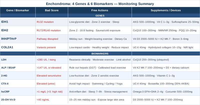
Moving Forward With Better Information
Enchondroma sits in an unusual clinical space — significant enough to require ongoing monitoring, yet benign enough that most conversations with physicians do not go deeper than imaging schedules. The genetic research of the past fifteen years has changed what is knowable and what is actionable. IDH1 and IDH2 mutations are identifiable, their downstream metabolic consequences are measurable, and there are targeted nutritional and lifestyle strategies that address the underlying biology rather than simply waiting for something to change on a scan.
Tracking six key biomarkers — LDH, alkaline phosphatase, 2-hydroxyglutarate, CTX-II, hsCRP, and 25-OH vitamin D — gives you a metabolic picture that imaging alone cannot provide. Understanding which of the four gene pathways is most relevant to your specific lesion transforms a passive posture into a focused and rational one.
The smart next step is not to start every supplement discussed here simultaneously. It is to bring these insights to a conversation with a knowledgeable orthopedic oncologist, integrative medicine physician, or functional medicine clinician, and ask which biomarkers make sense to start tracking in your specific situation. Better information leads to better decisions. That is the only promise worth making — and it is a meaningful one.
Cancer & Oncology Endocrine & Metabolic
Musculoskeletal: Bone Conditions
Autoimmune: Inflammatory Conditions Connective Tissue Conditions