This article contains AI generated content.
· UpdatedEnthesitis-Related Arthritis Genes and Biomarkers — 6 Genes and 7 Biomarkers to Track
Introduction
Enthesitis-related arthritis (ERA) is one of the most complex and often under-recognized subtypes of juvenile idiopathic arthritis. It targets the entheses — the anatomical points where tendons and ligaments attach to bone — and it can progress silently to involve the sacroiliac joints and spine long before imaging reveals it. If you or someone you care for has received this diagnosis, you already know the frustration: standard follow-up often cycles between CRP checks, NSAIDs, and watching for flares, without much insight into what is actually driving the disease at a biological level or how to intervene more precisely.
What makes ERA particularly layered is that it sits at the intersection of genetic susceptibility, immune dysregulation, and gut-joint communication. A single inflammatory marker can confirm that something is wrong without telling you which cytokine pathway is most active, how much structural risk exists, or whether the gut is acting as an upstream trigger. Generic advice — eat better, reduce stress, exercise — is not wrong, but it applies equally to every condition and answers none of the specific questions ERA raises.
This article does not offer a cure or an alternative to medical care. What it offers is a sharper lens: specific genetic variants that shape your ERA risk and behavior, and specific biomarkers that go well beyond standard CRP and ESR to reveal what is actually happening inside the tissue. Understanding these gives you better questions to bring to your rheumatologist and a more targeted framework for the lifestyle and nutritional decisions that sit alongside medical treatment.
Better information genuinely changes outcomes in chronic disease — not because it replaces medicine, but because it changes the quality of every decision made along the way. The sections below cover seven actionable biomarkers, six genetic variants with practical compensation strategies, a paradigm-shifting book that challenges conventional autoimmune thinking, and five complementary approaches with real clinical evidence. Each one adds a piece of information that can make the path forward less opaque.
Summary
Seven biomarkers beyond the standard panel — including IL-17A, MMP-3, serum calprotectin, and TNF-alpha — with cost ranges, optimal targets, and specific action plans with and without supplements. Six genetic variants — from the well-known HLA-B27 to ERAP1, IL23R, TNFA, PTPN22, and IL17A — each explained with what the risk allele does biologically and what can be done about it. Then a summary of the book that has probably changed more autoimmune trajectories than any single drug protocol, 10 specific insights from it that most rheumatologists never mention. Finally, five complementary modalities — including the Autoimmune Protocol, yoga, microbiome therapy, and photobiomodulation — selected for their actual clinical evidence in ERA-related inflammatory conditions, each with a specific protocol to apply. This article will not give you easy answers. It will give you better ones.
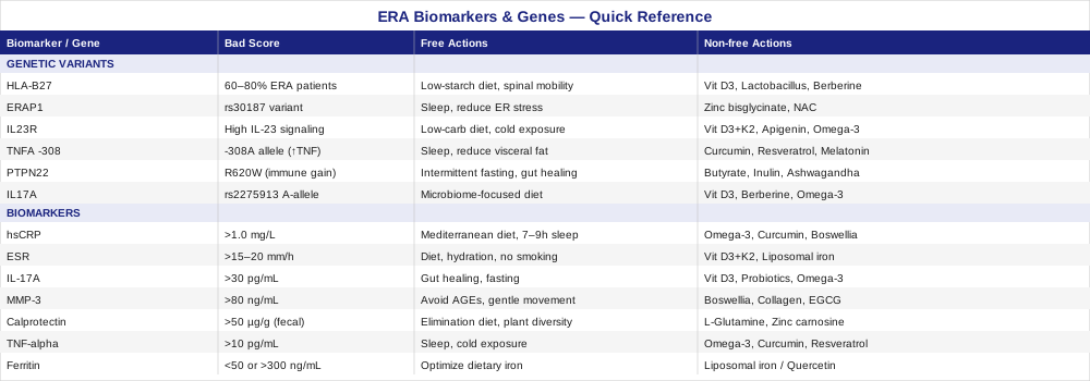
7 Biomarkers to Track in Enthesitis-Related Arthritis
Most ERA patients have CRP and ESR measured at every rheumatology visit. Those tests are useful, but they are blunt instruments. They confirm inflammation without identifying which pathways are active, how much structural risk exists, or whether gut dysfunction is amplifying joint disease. The seven biomarkers below offer a more granular picture — some are widely available, others require specialty labs, but all provide actionable information that standard panels miss.
1. High-Sensitivity CRP (hsCRP)
Why it matters and what it reveals
High-sensitivity CRP is the more precise version of standard CRP, capable of detecting low-grade systemic inflammation below the conventional test's threshold. In ERA, elevated hsCRP correlates with active enthesitis, synovitis, and axial involvement. More importantly, persistently elevated hsCRP between flares suggests ongoing subclinical inflammation driving gradual structural damage at entheseal insertion sites. Standard CRP only flags values above roughly 5 mg/L; hsCRP detects elevations from 0.3 mg/L. For ERA monitoring, this precision matters: some patients carry a normal-appearing standard CRP while hsCRP signals smoldering inflammation predictive of longer-term progression.
How to measure it
A standard blood test available at virtually any laboratory. Cost: $20–$50 depending on coverage and country. No special preparation required. Target: below 1.0 mg/L is optimal; 1.0–3.0 mg/L indicates moderate ongoing inflammation; above 3.0 mg/L signals active inflammatory burden requiring direct attention.
If the score is bad, the plan without supplements
The highest-yield lifestyle interventions to lower hsCRP: adopt a Mediterranean-style diet rich in oily fish, extra-virgin olive oil, vegetables, and legumes; eliminate refined sugars, seed oils (sunflower, corn, canola), and ultra-processed foods; achieve 7–9 hours of consistent sleep — poor sleep alone reliably raises CRP within days; and perform low-to-moderate aerobic exercise 3–5 times per week. Chronic psychological stress chronically elevates cortisol which paradoxically sustains low-grade inflammatory tone, so stress management is not optional. For ERA specifically, addressing gut permeability — through reduced alcohol intake, avoiding NSAIDs when possible (they damage the gut lining), and including fermented foods — can meaningfully reduce the upstream inflammatory load that drives hsCRP.
If the score is bad, the plan with supplements or equipment
- Omega-3 fatty acids (EPA+DHA): 2–4 g/day of combined EPA+DHA from high-quality fish oil. Multiple meta-analyses confirm reduction in systemic inflammatory markers. Take with food to improve absorption. Cycle: 6 months continuous, 4-week break to reassess baseline. Minor side effects: fish aftertaste, mild blood thinning at high doses. - Curcumin (BCM-95 or liposomal form): 500–1000 mg twice daily with meals. Targets NF-κB, the master switch upstream of TNF-alpha and IL-17 production. Cycle: 8 weeks on, 2 weeks off. Rare GI discomfort; avoid high doses in pregnancy. - Boswellia serrata (AKBA-standardized extract): 300–400 mg AKBA daily. Inhibits 5-LOX and complements omega-3 anti-inflammatory action via a distinct pathway. Side effects: occasional nausea; take with food.
2. Erythrocyte Sedimentation Rate (ESR)
Why it matters and what it reveals
ESR reflects the tendency of red blood cells to clump and settle — a proxy for acute-phase protein concentration. In ERA and other spondyloarthropathies, ESR rises during flares but can remain paradoxically normal in patients with primarily entheseal rather than synovial involvement. This makes ESR more useful as a longitudinal trend marker than a snapshot. A rising ESR over several months, even technically within range, carries clinical meaning. ESR is also influenced by anemia, age, and sex — factors to account for when interpreting results, since mild anemia of chronic disease is common in ERA.
How to measure it
Standard blood test, cost $10–$30. Targets: below 15 mm/h for adult men, below 20 mm/h for adult women; age- and sex-adjusted norms apply for pediatric ERA patients.
If the score is bad, the plan without supplements
Everything that reduces hsCRP also lowers ESR, since they track overlapping processes. Specific to ESR: optimize iron status — iron-deficiency anemia artificially elevates ESR, so investigate and address if present; ensure adequate hydration; and prioritize deep, consistent sleep. Eliminating tobacco exposure is non-negotiable for HLA-B27-positive spondyloarthropathy patients — smoking is a confirmed trigger for disease progression and reliably worsens ESR and structural outcomes.
If the score is bad, the plan with supplements or equipment
- Vitamin D3 + K2: target serum 25-OH-D of 60–80 ng/mL. Doses of 4000–6000 IU/day vitamin D3 paired with 100–200 mcg MK-7 K2. Vitamin D directly modulates TH17 activity, the central inflammatory arm in ERA. Retest every 3 months and adjust dose. Side effects: rare toxicity at very high doses — monitoring serum levels avoids this. - Anemia-driven high ESR: if iron-deficiency anemia is confirmed, liposomal iron 15–25 mg every other day (alternate-day dosing improves absorption by reducing hepcidin response). Pair with vitamin C 250 mg. Retest after 8–12 weeks. Constipation risk is lower with liposomal versus standard ferrous sulfate.
3. IL-17A
Why it matters and what it reveals
Interleukin-17A is arguably the most therapeutically significant biomarker in ERA. The IL-17 pathway is a central driver of entheseal inflammation: IL-17A is secreted by TH17 cells and innate lymphoid cells at the enthesis, where it recruits neutrophils, activates matrix metalloproteinases, and simultaneously promotes new bone formation and erosion. ERA patients consistently show elevated serum IL-17A compared to other JIA subtypes and healthy controls. This matters clinically because elevated IL-17A predicts who is most likely to benefit from anti-IL-17 biologics (secukinumab, ixekizumab), and persistently elevated values in the context of structural changes suggest an aggressive disease phenotype.
How to measure it
Measured by ELISA from serum or plasma; not universally included in standard panels but available through specialty and academic labs. Cost: $150–$400. Normal serum IL-17A is typically below 20–30 pg/mL in healthy individuals; ERA patients in active disease often show 40–200+ pg/mL.
If the score is bad, the plan without supplements
IL-17A is powerfully upregulated by gut dysbiosis, particularly gram-negative bacterial products crossing a compromised gut barrier. The highest-yield non-supplement strategy is an aggressive gut-healing protocol: remove inflammatory foods (gluten, dairy, refined sugars, seed oils), restore microbiome diversity through dietary fiber and fermented foods, and reduce intestinal permeability through time-restricted eating (12–16-hour overnight fast). Sleep is non-negotiable here: sleeping under 7 hours consistently dramatically elevates TH17 activity. Maintaining consistent circadian rhythm matters independently — clock gene disruption shifts T cell balance toward TH17 and away from regulatory T cells that control IL-17 overproduction.
If the score is bad, the plan with supplements or equipment
- Vitamin D3 (higher-dose, monitored): 5000–8000 IU/day with K2 100–200 mcg. Vitamin D directly suppresses IL-17A transcription via the vitamin D receptor on immune cells. Retest 25-OH-D every 3 months; adjust dose to hold 60–80 ng/mL. - EPA-dominant omega-3: 3–4 g/day of EPA specifically (rather than equal EPA+DHA). EPA competes with arachidonic acid, reducing the eicosanoid signaling environment that promotes TH17 differentiation. Take with largest meal for absorption. - Targeted probiotics (Lactobacillus reuteri + Bifidobacterium longum blend): these specific strains suppress TH17 activity by inducing regulatory T cells in gut-associated lymphoid tissue. 20–50 billion CFU daily with food. 3-month cycles; retest serum calprotectin afterward. Side effects: minor bloating in first week. - Far-infrared sauna: 20–30 minutes 3–4 times/week. Heat shock protein activation and improved lymphatic circulation support reduced cytokine expression. Begin with 10-minute sessions; avoid during acute flares with fever.
4. MMP-3 (Matrix Metalloproteinase-3)
Why it matters and what it reveals
MMP-3, also called stromelysin-1, is an enzyme produced by synoviocytes and fibroblasts in response to TNF-alpha and IL-1. In ERA and related spondyloarthropathies, MMP-3 is increasingly recognized as a superior biomarker to CRP for assessing structural damage risk: while CRP reflects current inflammation intensity, MMP-3 reflects connective tissue degradation activity — the process that directly drives joint destruction and entheseal scarring. Studies in adults with ankylosing spondylitis (the adult disease ERA can progress into) show that MMP-3 predicts radiographic progression and biologic treatment response better than ESR or CRP alone. Elevated MMP-3 in ERA, even alongside manageable CRP, warrants attention.
How to measure it
Available at specialty and academic medical labs; some commercial labs include it as a stand-alone order. Cost: $100–$250. Reference ranges vary by sex: roughly 17–60 ng/mL for males, 25–60 ng/mL for females. Values above 80–100 ng/mL in the context of ERA are clinically concerning regardless of CRP status.
If the score is bad, the plan without supplements
MMP-3 is directly stimulated by TNF-alpha and IL-1, so reducing those cytokines (strategies covered in the TNF-alpha section below) will lower MMP-3. Specific additional targets: eliminate dietary advanced glycation end products (AGEs) found in charred, fried, and ultra-processed foods — AGEs directly activate receptor-mediated MMP upregulation; reduce visceral adiposity (visceral fat is a major source of the TNF-alpha and IL-6 that drive MMP-3 expression); and avoid prolonged sedentary periods. Mechanical loading through gentle movement actually activates anti-inflammatory tissue repair programs at the connective tissue level.
If the score is bad, the plan with supplements or equipment
- Boswellia serrata (AKBA): directly inhibits MMP-3 and MMP-1 production via 5-LOX and NF-κB. 300–400 mg AKBA daily. 8-week cycles with 2-week breaks. - Hydrolyzed type II collagen peptides: 10–20 g/day. Provides substrate for joint matrix repair and may signal reduced MMP upregulation via feedback mechanisms. Low risk profile; sustained use (6+ months) is where the most consistent evidence lies. - Green tea extract (EGCG): 400–600 mg/day of standardized EGCG. Demonstrated inhibition of MMP-3 and MMP-13 in synoviocyte studies. Side effects: avoid high doses (above 800 mg/day) without food; very high doses carry hepatotoxicity risk with long-term use.
5. Serum and Fecal Calprotectin
Why it matters and what it reveals
Calprotectin is a calcium-binding protein released by neutrophils during activation. In ERA, elevated calprotectin serves as a dual signal: it reflects systemic neutrophil activity relevant to active enthesitis, and it reflects gut mucosal inflammation relevant to the gut-joint axis that drives ERA in many patients. Up to 30–60% of ERA patients have subclinical gut inflammation detectable through calprotectin measurement, even without obvious GI symptoms. This gut-joint connection is not incidental — microbiome dysbiosis and increased intestinal permeability are increasingly recognized as upstream triggers of spondyloarthropathy onset and flare cycles. Tracking calprotectin alongside joint biomarkers can reveal whether gut-directed interventions should be prioritized.
How to measure it
Fecal calprotectin: stool sample, widely available, $50–$150. Normal: below 50 µg/g; values above 200 µg/g signal significant mucosal inflammation. Serum calprotectin (S100A8/A9): specialty lab, $100–$200, more convenient but slightly less sensitive for gut-specific pathology. Both are useful; fecal is preferred when gut involvement is the primary question.
If the score is bad, the plan without supplements
A gut-directed elimination and restoration protocol is the most evidence-supported non-supplement intervention. Start with a 6-week strict elimination of gluten, dairy, refined sugars, seed oils, and alcohol. Simultaneously increase dietary plant diversity — aiming for 30+ different plant species per week supports microbiome richness in a way single-food interventions cannot. Polyphenol-rich foods (berries, dark chocolate above 85%, red onion, green tea) specifically feed Akkermansia muciniphila and Lactobacillus species that support gut barrier integrity. A 14–16 hour overnight fast reduces gut permeability markers measurably in clinical protocols.
If the score is bad, the plan with supplements or equipment
- L-glutamine: 5–10 g first thing in the morning on an empty stomach. Primary fuel for intestinal epithelial cells; supports tight junction protein expression (occludin, claudin-3). 3–6 month cycles. Minimal side effects; avoid very high doses in active cancer. - Zinc carnosine: 75–150 mg/day with food. One of the most clinically validated agents for gut barrier repair in human trials. 8-week cycles. - Saccharomyces boulardii: 250–500 mg/day. Yeast-based probiotic with specific evidence for reducing intestinal permeability and normalizing calprotectin in mucosal inflammatory conditions. Compatible with antibiotic use. - Multi-strain probiotics including soil-based organisms: Bacillus subtilis and Bacillus coagulans alongside Lactobacillus and Bifidobacterium species. 50–100 billion CFU daily. 12-week cycles. Reassess fecal calprotectin after the first cycle.
6. TNF-alpha (Serum)
Why it matters and what it reveals
Tumor necrosis factor-alpha (TNF-α) is one of the master regulators of ERA inflammation. It drives IL-6, IL-1, and MMP-3 production while activating NF-κB in entheseal fibroblasts and osteoclasts. The entire class of anti-TNF biologics used in ERA — etanercept, adalimumab, infliximab — was developed because TNF is so central to disease pathogenesis. Measuring serum TNF-alpha directly can help assess whether an anti-TNF biologic is targeting an active pathway, serve as a baseline before initiating or changing therapy, and identify TNF-driven disease activity between flares that may not manifest in standard markers.
How to measure it
Specialty ELISA or multiplex cytokine panel. Cost: $150–$400 as a stand-alone test; lower per-marker when bundled in a multiplex cytokine array. Normal serum TNF-alpha in healthy individuals: typically below 8–10 pg/mL; ERA patients in active phases often show 20–80+ pg/mL.
If the score is bad, the plan without supplements
Chronic sleep deprivation is one of the strongest non-infectious drivers of TNF-alpha elevation — addressing sleep quality alone produces measurable cytokine reduction within weeks. Visceral fat directly secretes TNF-alpha (adipose tissue is an active endocrine organ), so body composition management through resistance training and reduced refined carbohydrate intake is high-yield. Elimination of trans fats and seed oils reduces the omega-6 arachidonic acid load that feeds TNF-alpha production via eicosanoid cascades. Cold exposure (2–3 minutes cold shower ending, or brief ice bath) has preliminary evidence for reducing systemic cytokine burden via sympathetic activation and NF-κB inhibition.
If the score is bad, the plan with supplements or equipment
- EPA-dominant omega-3: 3–4 g/day. EPA specifically reduces TNF-alpha by outcompeting arachidonic acid at cyclooxygenase and lipoxygenase enzymes. Sustained use over 6+ months produces the most consistent results in inflammatory conditions. - Curcumin (liposomal or BCM-95): 1000–2000 mg/day. Among the best-characterized natural NF-κB inhibitors with actual human trial data. Combine with piperine (5–10 mg) if using standard curcumin to improve bioavailability roughly 20-fold. 8-week cycles. - Resveratrol: 250–500 mg/day with meals. Activates SIRT1 and inhibits NF-κB, reducing downstream TNF production. Cycle 8 weeks on, 2 weeks off. Can interact with blood thinners; inform your physician. - Melatonin (low-dose, 0.3–1 mg): regulates circadian immune rhythm and reduces overnight TNF-alpha spikes. Take 30 minutes before sleep. Low-dose is more physiologic and less likely to cause morning grogginess than standard 5–10 mg formulations.
7. Ferritin
Why it matters and what it reveals
Ferritin is primarily known as an iron storage protein, but it functions as a dual biomarker in ERA. It is both an acute-phase reactant (rising with inflammation) and a direct indicator of iron status. In ERA, elevated ferritin often reflects systemic inflammatory burden rather than iron excess — a crucial distinction, because treating high inflammation-driven ferritin with reduced iron intake will worsen the anemia of chronic disease that commonly accompanies ERA. Conversely, ferritin below 50–70 ng/mL in ERA patients signals true iron deficiency, which impairs immune regulation, worsens fatigue, and reduces exercise tolerance — all factors that feed the disease burden indirectly. The optimal window for ERA: ferritin 80–150 ng/mL, verified alongside transferrin saturation to distinguish inflammation from genuine iron overload.
How to measure it
Standard blood test, $20–$50, universally available. Always measure alongside transferrin saturation (TSAT) and full iron panel for correct interpretation. Ferritin above 300 ng/mL with TSAT below 20% signals inflammation-driven elevation; ferritin above 300 with TSAT above 45% warrants investigation for iron overload or macrophage activation syndrome, a rare but serious ERA complication.
If the score is bad, the plan without supplements
For inflammation-driven high ferritin: the goal is reducing the underlying inflammatory driver — the strategies covering hsCRP and IL-17A above apply directly. For low ferritin in ERA: optimize dietary heme iron (red meat, organ meats), pair plant-iron sources with vitamin C, and address gut absorption barriers — low stomach acid and intestinal permeability (both common in ERA patients with subclinical gut involvement) substantially impair iron absorption regardless of dietary intake.
If the score is bad, the plan with supplements or equipment
- Low ferritin: liposomal iron: 15–25 mg every other day. Alternate-day dosing reduces hepcidin response and improves net absorption by roughly 40% compared to daily dosing. Pair with vitamin C 250 mg. Retest after 8–12 weeks. Side effects: constipation is significantly mitigated with liposomal versus standard ferrous sulfate. - High ferritin (inflammation-driven): do not supplement iron. Support mild iron modulation with quercetin 500 mg/day (inhibits iron-induced oxidative stress and mildly chelates excess labile iron). IP6 (inositol hexaphosphate) 1–2 g/day on an empty stomach acts as a natural iron chelator; do not use long-term without lab monitoring.
6 Key Genes That Shape ERA Risk and Progression
Genetics does not determine destiny in ERA, but understanding your genetic architecture shifts how you can manage it. Several genes involved in antigen presentation, cytokine signaling, and T cell regulation have been shown to influence both susceptibility and disease severity. For each variant below, the focus is on what the risk allele does biologically and what practical interventions exist — with or without supplements. If you haven't had any genetic panel, most of these are available through clinical SNP testing or consumer genomic services.
1. HLA-B27
HLA-B27 is the most important genetic factor in ERA, present in 60–80% of ERA patients compared to roughly 8% of the general Western population. It encodes an MHC class I molecule that presents peptide fragments to CD8+ T cells. Three mechanisms are most supported by current research: abnormal peptide presentation activating autoreactive T cells; HLA-B27 heavy chain misfolding that triggers ER stress and IL-23 production; and direct molecular interaction between gut bacteria and HLA-B27-expressing cells at the gut mucosa. The subtype B*27:05 carries the highest spondyloarthropathy risk in Western populations.
Testing: a simple blood test for HLA-B27 antigen typing, $30–$100, routinely available and ordered by most rheumatologists evaluating ERA.
If the gene is bad, the plan without supplements
HLA-B27 increases ERA risk but only causes disease in combination with environmental triggers. The gut microbiome is the most modifiable factor: Klebsiella pneumoniae produces proteins with sequence homology to HLA-B27 and type VI collagen, and elevated gut Klebsiella loads correlate directly with flare activity in some research. A diet low in fermentable starches that feed Klebsiella — reducing refined bread, pasta, potatoes, and sugar — has been a consistent recommendation since Dr. Alan Ebringer's research in the 1990s and remains biologically plausible. Regular spinal mobility exercise is evidence-based for HLA-B27-positive patients and shown to slow structural progression in the adult spondyloarthropathy literature. Avoid antibiotic overuse, which disrupts the protective microbiome layer that keeps Klebsiella in check.
If the score is bad, the plan with supplements or equipment
- Vitamin D3 (5000–8000 IU/day monitored): modulates TH17 activation downstream of HLA-B27 antigen presentation. Monitor serum 25-OH-D quarterly; target 60–80 ng/mL. - Lactobacillus casei Shirota and Lactobacillus rhamnosus GG: strains with evidence for competitively excluding gram-negative bacteria including Klebsiella. 10–50 billion CFU/day. Long-term cycling: 3 months on, 1 month off. - Berberine: 500 mg twice daily with food. Antimicrobial against gram-negative bacteria and a microbiome modulator. 8-week courses with cycling to avoid receptor adaptation.
2. ERAP1 (Endoplasmic Reticulum Aminopeptidase 1)
ERAP1 trims peptides in the endoplasmic reticulum before they are loaded onto MHC class I molecules including HLA-B27. Risk variants — most importantly rs30187 (Lys528Arg) — alter the length and composition of the peptide repertoire presented by HLA-B27, effectively generating a more pro-inflammatory peptide landscape. Critically, ERAP1 risk alleles act epistatically with HLA-B27 — they only increase spondyloarthropathy risk in HLA-B27-positive individuals. Carrying ERAP1 risk variants without HLA-B27 has negligible effect on ERA risk.
If the gene is bad, the plan without supplements
ERAP1 affects protein processing in the ER, so supporting general ER homeostasis is the upstream intervention. Reducing chronic ER stress through consistent sleep, avoiding chronic caloric overload, and minimizing dietary lectins (particularly from nightshades and legumes, which some ERA patients react to) may reduce the misfolding and inflammatory signaling downstream of ERAP1 dysfunction. Both sauna heat stress and cold exposure activate heat shock proteins that support proper ER folding function — a physiologic lever with no drug interactions.
If the score is bad, the plan with supplements or equipment
- Zinc bisglycinate: ERAP1 is a zinc-dependent metalloprotease. 25–30 mg/day supports optimal enzyme function. Balance with copper (1–2 mg/day) to prevent copper depletion from prolonged zinc supplementation. Do not exceed 40 mg/day long-term without monitoring. - NAC (N-acetylcysteine): 600–1200 mg/day. Reduces ER oxidative stress and supports glutathione recycling, which is depleted during ER stress responses. Take on empty stomach or with light meal. 6–8 week cycles.
3. IL23R (Interleukin-23 Receptor)
The IL-23 receptor gene contains multiple variants associated with ERA and spondyloarthropathy broadly. The most well-studied variant (rs11209026, R381Q) is actually protective — it reduces TH17 differentiation. Conversely, the high-risk common alleles maintain strong IL-23 signaling that drives persistent TH17 activation and chronic IL-17A production. IL23R variant profiles partly explain why IL-23/IL-17 pathway biologics (ustekinumab, secukinumab) produce markedly different response rates between patients with otherwise similar clinical presentations.
If the gene is bad, the plan without supplements
The TH17 pathway is exquisitely sensitive to diet, light, and sleep. A low-carbohydrate, high-fat diet reduces IL-23-driven TH17 activity via ketone body signaling — beta-hydroxybutyrate directly inhibits the NLRP3 inflammasome upstream of IL-23 in a dose-dependent manner. Cold water immersion and consistent morning sunlight exposure to calibrate circadian immune rhythm are among the best-studied non-pharmaceutical interventions targeting TH17/IL-17 axis in individuals with high IL23R pathway activity.
If the score is bad, the plan with supplements or equipment
- Vitamin D3 + K2: target 60–80 ng/mL serum 25-OH-D. VDR signaling directly suppresses IL-23 production in dendritic cells. 4000–8000 IU/day with 100–200 mcg MK-7 K2; retest quarterly. - Apigenin (chamomile extract): 50–100 mg/day. Flavonoid with demonstrated TH17 suppression and regulatory T cell induction in early research. Very low risk profile; suitable for long-term use. - EPA-dominant omega-3: 3–4 g/day EPA specifically, as EPA reduces IL-23-induced TH17 differentiation more effectively than DHA in inflammatory models.
4. TNFA (-308 G>A Polymorphism)
The TNF-alpha gene promoter polymorphism at position -308 (rs1800629) — specifically the A allele — increases basal and stimulated TNF-alpha production. In ERA and juvenile spondyloarthropathies, the -308A allele correlates with greater inflammatory activity, more systemic features, and potentially better responsiveness to anti-TNF biologics (since TNF is the primary driver in A-allele carriers). Knowing whether you carry this allele can help predict treatment response and justify earlier aggressive management.
If the gene is bad, the plan without supplements
The -308A allele functions as a transcriptional volume control turned up for TNF. Lifestyle factors that most directly attenuate that amplification: visceral fat reduction through resistance training and dietary change (adipose tissue is the dominant non-immune source of TNF-alpha); consistent sleep to normalize circadian cytokine rhythms (TNF-alpha spikes measurably in sleep-deprived individuals overnight); and elimination of trans fats and seed oils to reduce the arachidonic acid cascade feeding TNF synthesis.
If the score is bad, the plan with supplements or equipment
- Curcumin (BCM-95): 500–1000 mg twice daily. The most studied natural NF-κB inhibitor with multiple human trials showing significant TNF-alpha reduction. 8-week cycles. - Resveratrol: 250–500 mg/day with meals. SIRT1 activation inhibits NF-κB transcriptional activity directly at the -308 promoter region. 8 weeks on, 2 weeks off. - Low-dose melatonin (0.3–1 mg): physiologic dose regulates circadian TNF-alpha peaks without the receptor downregulation associated with pharmacologic 5–10 mg doses.
5. PTPN22 (R620W Variant)
PTPN22 encodes a phosphatase that regulates T cell and B cell receptor signaling thresholds. The R620W variant (rs2476601, arginine to tryptophan at position 620) reduces the activation threshold for T cells, creating a more reactive immune baseline. It is associated with multiple autoimmune conditions — RA, type 1 diabetes, lupus, and ERA — suggesting it functions as a general "immune gain" switch rather than a disease-specific factor. In ERA, R620W carriers tend toward more aggressive disease onset and greater systemic involvement.
If the gene is bad, the plan without supplements
Supporting regulatory T cell (Treg) function is the primary target. Key strategies: intermittent fasting increases the Treg/TH17 ratio; butyrate-producing dietary fiber (resistant starch, inulin, cooked-and-cooled potato/rice, psyllium) is the most powerful dietary inducer of intestinal Foxp3+ Treg cells; and chronic psychological stress impairs Treg function via HPA axis dysregulation, making stress management unusually important in R620W carriers. Gut healing remains the highest-yield approach since gut-associated lymphoid tissue is where the Treg/TH17 balance is most plastic.
If the score is bad, the plan with supplements or equipment
- Sodium butyrate or tributyrin: 600–1200 mg/day with meals. Directly induces Foxp3+ Tregs in the intestine. 3-month cycles. - Prebiotic fiber (inulin or resistant starch): 5–10 g/day inulin powder, or 10–20 g resistant starch. Endogenously feeds butyrate-producing Clostridia species. Start low and increase gradually to avoid gas. - Ashwagandha KSM-66: 300–600 mg/day. Reduces cortisol-mediated immune dysregulation that impairs Treg function. 12-week cycles, 4-week break. Avoid in confirmed autoimmune thyroid conditions.
6. IL17A Gene Variants (rs2275913)
Several polymorphisms in the IL17A gene itself — distinct from the receptor genes that regulate its production — are associated with ERA susceptibility. The rs2275913 (-197 G>A) variant in the IL17A promoter region is linked to higher basal IL-17A transcription. A-allele carriers produce more IL-17A in response to the same inflammatory stimulus compared to G-allele homozygotes, effectively amplifying downstream entheseal inflammation for a given immune challenge.
If the gene is bad, the plan without supplements
A higher IL-17A transcriptional baseline means the gut microbiome optimization strategies described above become even more important. Reducing Prevotella copri and segmented filamentous bacteria — both strong TH17 inducers — through a low-inflammatory, high-diversity diet directly reduces the stimulation driving IL-17A overexpression in rs2275913-A carriers. Morning UVB sun exposure supports vitamin D-mediated IL-17A suppression; even 10–15 minutes of midday sunlight on forearms and face makes a measurable difference in serum vitamin D over weeks.
If the score is bad, the plan with supplements or equipment
- Apply the full supplement protocol from the IL-17A biomarker section above: vitamin D3 at therapeutic doses, EPA-dominant omega-3, and targeted Lactobacillus/Bifidobacterium probiotics are the highest-yield interventions for rs2275913-A carriers. - Berberine: 500 mg twice daily with food. Reduces Prevotella copri burden in the gut specifically. 8-week courses with cycling. - Physiologic melatonin (0.3–0.5 mg): melatonin receptors on immune cells modulate TH17/Treg balance, and IL17A promoter variants may particularly respond to melatonin-mediated circadian immune regulation.
What "The Autoimmune Fix" Gets Right About ERA
Tom O'Bryan's 2016 book The Autoimmune Fix is not specifically about ERA — but the framework it presents may be the single most practical paradigm for understanding what drives ERA onset and persistence that you will not hear in a standard rheumatology consultation. O'Bryan synthesizes decades of immunology research into a model that most practicing rheumatologists simply were not trained on: that autoimmunity is a spectrum, not an on/off switch, and that gut permeability is the required biological prerequisite for most autoimmune diseases to develop. Here are the 10 most impactful insights from the book in the context of ERA:
1. Autoimmunity Starts 3 to 10 Years Before Diagnosis
O'Bryan cites research showing that autoimmune biomarkers appear in blood years before clinical symptoms meet diagnostic criteria. For ERA, this means the biological process is underway long before enthesitis pain or morning stiffness begins — and that early detection through biomarkers and genetic panels can create a meaningful intervention window that most people never know exists.
2. The Three-Legged Stool: Genes, Gut Permeability, and Trigger
No autoimmune disease develops from genes alone. O'Bryan describes the essential interaction: a genetic predisposition (HLA-B27, PTPN22, etc.), a leaky gut that allows bacterial antigens into systemic circulation, and a triggering antigen (a specific microbe, food protein, or environmental toxin). Remove any one leg and the stool collapses. This is why two people with identical HLA-B27 status can have completely different ERA outcomes based on gut health and life exposures.
3. Leaky Gut Is the Non-Negotiable Upstream Step
Increased intestinal permeability — driven by gluten, NSAIDs, alcohol, dysbiosis, and chronic stress — allows bacterial lipopolysaccharide (LPS) and food protein fragments to reach systemic circulation, priming the immune system toward the exact TH17 overactivation that drives ERA. Closing the gut leak is described not as complementary medicine but as a prerequisite for durable immune regulation.
4. Gluten Activates the Same Molecular Mimicry as HLA-B27 Targets
O'Bryan presents research on molecular mimicry between gliadin peptides and human tissue proteins. For ERA patients with HLA-B27, this cross-reactivity between dietary antigens and joint tissues is a biologically plausible driver of entheseal inflammation — independent of celiac disease. This is why some ERA patients improve meaningfully on a strict gluten-free protocol even without a celiac diagnosis.
5. Stool Testing Reveals What Joint X-Rays Never Will
O'Bryan strongly advocates for comprehensive stool analysis as a window into the gut-immune axis. In ERA, fecal calprotectin, zonulin (gut permeability marker), and microbiome profiling can identify the specific gut dysfunction driving systemic inflammation — information that allows targeted rather than trial-and-error interventions.
6. Food Sensitivities Are Not Allergies and Are Easily Missed
IgG-mediated food sensitivities (as distinct from IgE-mediated allergies) cause delayed inflammatory reactions that rarely appear on standard allergy panels. O'Bryan presents evidence that unidentified food sensitivities maintain a chronic low-grade immune activation that sustains autoimmune processes — including the TH17-dominant pattern seen in ERA. An elimination-rechallenge protocol is the most accessible way to identify these.
7. Infections Are Molecular Mimics, Not Just Coincidences
Specific bacterial and viral infections share protein sequence homology with human joint and connective tissue antigens. For ERA, Klebsiella pneumoniae is the most studied example: its nitrogenase and pullulanase sequences mimic HLA-B27 and type VI collagen. O'Bryan frames chronic low-level gut infections not as background noise but as active inflammatory drivers requiring identification and targeted treatment.
8. The Brain-Gut-Immune Axis Means Stress Literally Opens Your Gut
Psychological stress increases cortisol, which directly impairs tight junction proteins in the gut epithelium — measurably increasing permeability within hours of an acute stress event. For ERA patients who notice that major life stressors reliably precede flares, this is the mechanism: stress physically opens the gut, increases LPS exposure, and amplifies the TH17 response. This is not a metaphor; it is a documented physiological cascade.
9. Total Toxic Load Compounds Genetic Risk Nonlinearly
O'Bryan argues that environmental toxins — heavy metals, pesticide residues, phthalates, BPA — do not simply add to genetic risk, they compound it. They act as gut-disrupting and immune-activating agents that lower the threshold at which genetic susceptibility becomes clinical disease. For ERA patients, reducing environmental toxic exposure (filtered water, organic food, non-plastic food storage) is not a luxury intervention but a load-reduction strategy.
10. You Can Test for Autoimmunity Before It Has a Name
One of the book's most actionable messages: advanced antibody panels can detect tissue-specific autoimmunity years before symptoms qualify for a diagnosis. For individuals with HLA-B27 positivity and a family history of spondyloarthropathy, running periodic anti-nuclear antibody, anti-CCP, and tissue transglutaminase panels alongside the biomarkers described in this article can catch an emerging process before structural damage accumulates.
Complementary and Integrative Approaches
The following five modalities have meaningful human clinical evidence for conditions on the ERA-spondyloarthropathy spectrum. They are presented as additions to — not replacements for — medical treatment, and each comes with a specific protocol and a note on evidence quality.
The Autoimmune Protocol (AIP) — Sarah Ballantyne
The Autoimmune Protocol, developed by Dr. Sarah Ballantyne in her book The Paleo Approach and extensively referenced in her research-focused work, is a structured elimination diet designed to reduce gut permeability, modulate immune activation, and identify food-based inflammatory triggers. It removes all grains, legumes, dairy, nightshades, eggs, nuts and seeds, NSAIDs, and processed foods during a 4–6 week elimination phase, then systematically reintroduces foods to identify personal triggers. ERA falls squarely within its target category as an autoimmune-spectrum inflammatory arthritis with known gut-immune interaction.
The evidence base includes a prospective study by Konijeti et al. (2017) in Inflammatory Bowel Diseases demonstrating significant reductions in inflammatory markers including CRP and fecal calprotectin in IBD patients following AIP — directly relevant to ERA's gut-joint axis. While ERA-specific RCTs are lacking, the biological mechanisms and the IBD overlap (ERA and IBD share the same TH17-dominant inflammatory pathway and frequent clinical co-occurrence) make this evidence directly applicable.
Protocol: begin the strict elimination phase for 6 weeks, then reintroduce one food group every 5–7 days while tracking joint symptoms, energy, and gut function. Maintain the full elimination phase during active flares. Work with a nutritionist familiar with AIP to avoid deficiency during the elimination phase, particularly for calcium and vitamin E.
Yoga
Yoga is one of the few physical movement interventions with documented evidence specifically in spondyloarthropathy. For ERA patients, particularly those with axial involvement or progression toward ankylosing spondylitis features, yoga addresses the core deficits of the disease: spinal mobility loss, entheseal tightness around the sacroiliac joints, and the postural compensation patterns that develop around chronic pain. It also exerts measurable effects on systemic inflammation through vagal nerve activation and cortisol modulation.
A systematic review by Lee et al. examining yoga in ankylosing spondylitis — the adult form ERA can progress to — documented significant improvements in Bath Ankylosing Spondylitis Disease Activity Index (BASDAI) scores and functional measures after 8–12 weeks of structured practice. The evidence is sufficient to recommend yoga as an adjunct in ERA-spectrum disease, with the caveat that poses involving prolonged hip flexion or asymmetric spinal loading should be modified during active flares.
Protocol: aim for 3 sessions per week of 45–60 minutes, focusing on hip-opening sequences, thoracic extension, and gentle spinal rotation. Iyengar yoga, which emphasizes precision and uses props extensively, is particularly appropriate for ERA patients as it allows modification without compromising the therapeutic intention of each pose. Avoid hot yoga during active disease; begin with an instructor familiar with inflammatory arthritis.
Mindfulness-Based Stress Reduction (MBSR)
MBSR is an 8-week structured program developed by Jon Kabat-Zinn that combines mindfulness meditation, body scanning, and gentle movement. Its relevance to ERA is twofold: it directly addresses the chronic stress-gut permeability cascade described in the O'Bryan framework above, and it provides neurologically validated pain modulation that reduces the central sensitization component of entheseal pain. In chronic inflammatory arthritis broadly, MBSR has been shown to reduce perceived pain severity, disease-related distress, and several inflammatory markers including CRP.
A study by Zangi et al. (2012) in Annals of the Rheumatic Diseases examining a mindfulness-based group intervention in inflammatory arthritis patients showed significant improvements in fatigue, coping, and self-efficacy — dimensions of disease burden that standard pharmacotherapy does not address. The stress-reducing effects of MBSR have a plausible and direct mechanism for reducing ERA flare frequency via the cortisol-gut-immune axis.
Protocol: commit to the full 8-week MBSR course (available through university medical centers and online via programs such as Palouse Mindfulness). The critical component is the daily 30–45 minute home practice — weekend-only meditation produces insufficient cortisol modulation to affect inflammatory biology meaningfully. After completing the initial course, maintain a daily 20-minute practice.
Microbiome-Directed Therapies
Emerging research has identified specific gut microbiome signatures in spondyloarthropathy patients — lower Faecalibacterium prausnitzii and Akkermansia muciniphila (both gut-protective species), and higher Prevotella copri and Ruminococcus gnavus (both pro-inflammatory). Microbiome-directed therapy in ERA means using targeted dietary and probiotic interventions to specifically shift these ratios toward a less inflammatory baseline, directly addressing the upstream gut-joint axis trigger. This is no longer fringe science; the gut microbiome-spondyloarthritis connection has been investigated in multiple controlled studies.
Research published in journals including Arthritis & Rheumatology and Gut Microbes has documented these specific dysbiosis patterns in ERA and ankylosing spondylitis patients and shown correlations between microbiome composition and disease activity scores. While therapeutic manipulation trials are still maturing, the mechanistic evidence is strong enough to justify structured intervention now.
Protocol: Phase 1 (weeks 1–4): strict elimination of refined sugars and alcohol, which preferentially feed Prevotella copri. Phase 2 (weeks 5–12): introduce fermented vegetables (sauerkraut, kimchi) daily, add a high-diversity prebiotic fiber protocol (15–20 g/day mixed fibers), and start a pharmaceutical-grade multi-strain probiotic including Lactobacillus acidophilus, Bifidobacterium infantis, and Faecalibacterium prausnitzii precursors. Reassess fecal calprotectin and serum calprotectin at 12 weeks. Phase 3: maintain long-term with periodic fecal microbiome analysis to track shifts.
Low-Level Laser Therapy (LLLT) / Photobiomodulation
Low-level laser therapy, also called photobiomodulation, uses specific wavelengths of red and near-infrared light (typically 630–1000 nm) to stimulate mitochondrial function in target tissues, reduce local inflammation, and accelerate tissue repair. Its relevance to ERA is specifically at the enthesis — the target tissue of this disease — where photobiomodulation has been shown to reduce inflammatory cytokine expression in connective tissue, improve collagen organization, and reduce pain in enthesopathies and tendinopathies.
A Cochrane-aligned meta-analysis on LLLT in musculoskeletal pain documented significant short-term pain reduction and functional improvement in tendinopathy and joint conditions with LLLT at doses of 3–9 J/cm². Evidence specific to ERA-type enthesitis is limited to small trials and case series, but the mechanism is directly applicable to entheseal tissue biology.
Protocol: use a Class 3B or Class 4 device delivering 630–850 nm wavelengths. Apply to active entheseal sites (Achilles insertion, plantar fascia, sacroiliac joints) for 60–120 seconds per site, 3–5 times per week. Allow 48 hours between sessions on the same site. Home devices are available in the 50–100 mW range; clinical devices are more powerful but require professional administration. Do not apply directly over active inflammatory swelling in acute flares without professional guidance. Side effects at correct dosing are minimal; avoid over injected joints.
Conclusion
Enthesitis-related arthritis is a condition where precision matters more than volume of effort. The seven biomarkers covered here go meaningfully beyond CRP and ESR to reveal which inflammatory pathways are most active, whether structural damage is underway, and whether the gut is contributing upstream. The six genetic variants provide a biological context that makes those biomarkers more interpretable and the interventions more targeted. Together they support a more individualized conversation with your rheumatologist — one grounded in actual data rather than symptom severity alone.
None of these strategies work in isolation, and none of them replace evidence-based medical treatment. But the combination of knowing your biomarker baseline, understanding your genetic risk architecture, addressing gut health systematically, and applying the best-evidenced complementary modalities gives you a substantially more complete toolkit than standard care alone provides. The next smart step is to pick the two or three highest-priority items from this article — whether that is testing IL-17A, discussing your HLA-B27 status in detail with your rheumatologist, starting an AIP elimination protocol, or ordering a fecal calprotectin — and act on them deliberately. Better data leads to better decisions. That is where meaningful change begins.
Musculoskeletal: Joint Conditions Tendon & Ligament Conditions Spine Conditions
Autoimmune: Inflammatory Conditions