This article contains AI generated content.
· UpdatedEosinophilic Cellulitis — 5 Genes and 6 Biomarkers to Track
Introduction
Living with eosinophilic cellulitis means navigating a condition that most physicians see only a handful of times in their careers. The swollen, erythematous, sometimes blistering patches that appear without warning — often misdiagnosed as bacterial cellulitis before biopsy reveals something quite different — are alarming enough on their own. But the deeper frustration for many people is the uncertainty: why did this happen, why does it keep coming back, and is there anything beyond corticosteroids that can actually make a lasting difference?
Standard management of Wells syndrome tends to be reactive. Flare up, treat with prednisone, wait for remission, repeat. What this approach misses is that the condition is driven by a specific immune dysregulation pattern — eosinophil overactivation rooted in Th2-dominant signaling — that varies considerably from person to person. Two people with identical histology on skin biopsy can have very different upstream drivers: different cytokine profiles, different genetic predispositions, different environmental triggers. Generic treatment protocols, by necessity, ignore those differences.
This article takes a more targeted approach. It focuses on the biomarkers that give you a real-time window into your eosinophilic disease activity — what to measure, what the numbers mean, and what to do differently when they are off. It also covers the genetic layer: the five genes most relevant to eosinophilic skin inflammation, what it means to carry an unfavorable variant, and what practical steps can compensate. Neither framework promises remission, but together they offer a level of biological self-knowledge that changes how you manage this condition.
The evidence base specific to Wells syndrome is limited by its rarity. Where direct evidence exists, it is cited. Where the science is extrapolated from related eosinophilic conditions — eosinophilic esophagitis, hypereosinophilic syndrome, atopic dermatitis — that context is stated clearly. Better information does not guarantee better outcomes, but it reliably improves the quality of decisions made along the way.
Summary
This article covers 6 actionable biomarkers for eosinophilic cellulitis — from the Absolute Eosinophil Count and IgE to more specialized signals like Eosinophil Cationic Protein, IL-5, and Periostin — each with a specific plan for improving your numbers with and without supplements. It also reviews 5 key genes (IL5, IL5RA, CCR3, STAT6, and FLG) that may wire certain individuals toward eosinophilic overactivation, with gene-aware lifestyle protocols that go beyond generic anti-inflammatory advice. Beyond the lab data, you will find a summary of The Allergy Solution by Dr. Leo Galland — a physician-authored guide challenging the standard allergy management paradigm — plus evidence-based complementary approaches including the Autoimmune Protocol, microbiome strategies, breathing protocols, and photobiomodulation. If you have been managing flares reactively, this article gives you a framework for getting ahead of them.
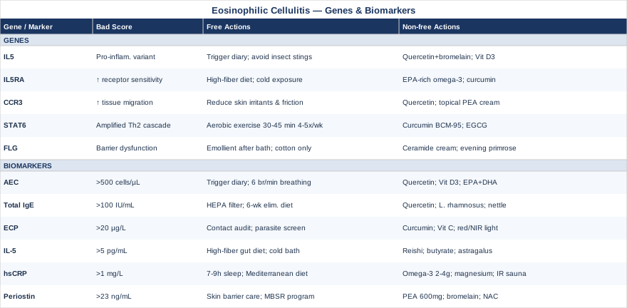
6 Biomarkers to Track for Eosinophilic Cellulitis
Biomarker tracking gives you something clinical observation alone cannot: objective, quantifiable data about what your immune system is doing right now, and whether it is trending better or worse over time. For a condition driven by eosinophil dysfunction, certain markers are particularly informative — some capture the number and activation state of eosinophils directly, others measure the upstream cytokines driving their production, and still others reflect the downstream tissue damage they cause. Selecting the right set and knowing what to do when one is off is what turns passive monitoring into active disease management.
Biomarker 1 — Absolute Eosinophil Count (AEC)
Why it matters and what it reveals. The Absolute Eosinophil Count is the most accessible and direct measure of eosinophil burden in the bloodstream. In eosinophilic cellulitis, blood eosinophilia is documented in roughly 50–75% of patients during active disease across published case series. This makes AEC both a diagnostic flag and an ongoing monitoring reference point. It does not capture what eosinophils are doing inside tissue — that requires more specific markers — but it provides a rapid, affordable window into systemic eosinophilic load and responds to treatment within days to weeks.
Normal AEC is 100–500 cells/µL. Mild eosinophilia is 500–1500 cells/µL; moderate eosinophilia is 1500–5000 cells/µL; hypereosinophilia exceeds 5000 cells/µL. In Wells syndrome, levels during flares most commonly fall in the mild-to-moderate range and may normalize during remission, though normalization of blood counts does not guarantee tissue remission.
How to Measure It
AEC is calculated from a complete blood count (CBC) with differential, one of the most commonly ordered blood tests in clinical medicine. Cost ranges from $20 to $80 depending on location and insurance coverage. Available through any physician and through most direct-to-consumer lab platforms. Measure at baseline, during any active flare, and monthly when tracking a treatment response. Ensure the count is reported as an absolute number (cells/µL), not just a percentage.
If the AEC Is Elevated — the Plan Without Supplements
Begin with a structured trigger identification protocol. Eosinophilic flares in Wells syndrome have been linked to insect stings and bites, parasitic infections, specific medications (NSAIDs, penicillins, carbamazepine, certain anticonvulsants), and in some cases foods high in histamine. A daily exposure-and-symptom diary maintained for 4–8 weeks can reveal personal patterns that would otherwise remain invisible.
Beyond avoidance, vagal tone improvement is a legitimate non-pharmacological lever on eosinophil levels. Slow nasal diaphragmatic breathing at 4–6 breaths per minute (physiological coherence breathing), practiced for 15–20 minutes daily, has measurable autonomic effects. Pair this with moderate aerobic exercise at 150+ minutes per week, which suppresses Th2 cytokine production via anti-inflammatory myokines. Both approaches reduce the sympathetic tone that amplifies mast cell and eosinophil reactivity.
If the AEC Is Elevated — the Plan With Supplements or Equipment
- Quercetin: 500–1000 mg/day in divided doses with food. A bioflavonoid with mast cell-stabilizing properties and documented effects on eosinophil survival in vitro. Cycle 8 weeks on, 2 weeks off. Minimal side effects; high doses may interfere with thyroid hormone assays. Human eosinophil-specific data are still emerging. - Vitamin D3 + K2: 2000–5000 IU D3/day paired with 100–200 mcg K2. Vitamin D has direct immunomodulatory effects on Th2 responses and correlates inversely with eosinophilia severity in atopic populations. Target serum 25-OH-D at 50–70 ng/mL and retest after 8–12 weeks. No cycling needed; year-round supplementation is appropriate in most latitudes. - EPA+DHA fish oil: 2–4 g combined EPA+DHA daily with food. Omega-3 fatty acids shift eicosanoid production away from arachidonic acid-derived pro-eosinophilic mediators. Effects accumulate over 8–12 weeks of consistent use. Monitor for additive blood-thinning effects if on anticoagulants.
Biomarker 2 — Total IgE
Why it matters and what it reveals. Total serum IgE is the signature marker of Th2 immune activation — the immune phenotype that underlies most eosinophilic diseases. Elevated IgE signals that your immune system is running a chronically allergic program driven by IL-4 and IL-13, which in turn creates the cytokine environment that sustains eosinophil recruitment and tissue infiltration. In Wells syndrome, elevated total IgE is documented in a meaningful subset of patients, particularly those with concurrent atopic disease such as eczema, asthma, or allergic rhinitis. It also serves as a proxy for how allergic the overall immune background is — which informs both trigger identification and treatment choices.
Normal total IgE is generally below 100 IU/mL, though laboratory reference ranges vary. Levels above 150–200 IU/mL suggest significant Th2 activation; levels above 1000 IU/mL warrant investigation for hyperallergic states or occult parasitic infection.
How to Measure It
Total IgE is a standard blood test available through any physician and most direct-to-consumer lab services. Cost: $30 to $100. Specific allergen IgE panels can be added to identify which allergens are maintaining the Th2 response, adding $100–300 depending on panel breadth. Retest at 3–6 months when tracking a treatment response. Note that IgE has a half-life of approximately 2 weeks, so it responds more slowly to intervention than AEC.
If Total IgE Is Elevated — the Plan Without Supplements
The most foundational non-supplement approach is systematic allergen load reduction. Dust mite encasements on all bedding, a HEPA air purifier in the bedroom (running continuously overnight — your longest sustained allergen exposure window), and laundering bedding at 60°C weekly address the most common indoor allergen sources. For individuals with food-associated flares, a structured 6-week elimination of the most common allergenic foods (gluten, dairy, eggs, shellfish, soy) followed by methodical reintroduction identifies dietary drivers.
A Mediterranean-style anti-inflammatory diet maintained consistently over 12 weeks has been shown to reduce total IgE and associated inflammatory markers in atopic populations. Core elements: abundant extra-virgin olive oil, oily fish (salmon, sardines, mackerel) 3 times/week, diverse colorful vegetables, legumes, and strict reduction of ultra-processed foods, refined sugars, and high-histamine foods during active periods.
If Total IgE Is Elevated — the Plan With Supplements or Equipment
- Quercetin: 500–1000 mg/day. IgE reduction is a relevant secondary effect of quercetin's Th2 suppression mechanism. Same cycling as above. - Lactobacillus rhamnosus GG + Bifidobacterium probiotic combination: 10–20 billion CFU/day consistently. IgE production is regulated by gut microbiome composition; probiotic use shows IgE-reducing effects in atopic populations in multiple randomized trials, particularly when begun early. Long-term use is safe. Choose a product with guaranteed CFU counts through expiration, kept refrigerated. - HEPA air purification equipment: A quality HEPA unit ($150–400) in the bedroom is an investment in chronic allergen reduction that compounds over months of use. HEPA filtration to 0.3 microns removes the most biologically active airborne allergen particle sizes. - Stinging nettle leaf extract: 600 mg freeze-dried/day. Pilot human data supports antihistamine and modest IgE-modulating effects. Cycle 4–6 weeks. Pause if taking blood pressure medications, as additive hypotensive effects are possible.
Biomarker 3 — Eosinophil Cationic Protein (ECP)
Why it matters and what it reveals. The Absolute Eosinophil Count tells you how many eosinophils are circulating. Eosinophil Cationic Protein tells you how activated they are — specifically, how much toxic granule content they are releasing into surrounding tissue. This distinction is critical in eosinophilic cellulitis, because the characteristic "flame figures" seen on skin biopsy — eosinophil granule deposits coating and destroying dermal collagen fibers — are caused directly by ECP and related granule proteins (major basic protein, eosinophil peroxidase). A patient can have a modestly elevated AEC and still have aggressively degranulating tissue eosinophils that blood count alone would miss.
ECP is especially useful for evaluating whether an intervention is actually reducing eosinophil activation at the tissue level, not just shifting peripheral numbers. ECP elevation in apparent remission indicates ongoing subclinical tissue disease. Normal ECP is typically below 13–20 µg/L depending on the assay used.
How to Measure It
ECP is measured by ELISA from serum, less commonly ordered than AEC and typically requiring a specialist referral or a direct-to-consumer specialty lab. Cost: $50 to $150. A critical pre-analytical note: blood must be processed correctly for ECP to be accurate. Eosinophils release ECP ex vivo if the sample sits at room temperature too long. Most validated protocols specify collection in a gold-top tube, held at room temperature for exactly 60 minutes before centrifugation. Confirm your lab follows this protocol before trusting the result.
If ECP Is Elevated — the Plan Without Supplements
High ECP with mild or absent blood eosinophilia suggests active tissue degranulation is occurring beneath the clinical surface. The first priority is ruling out occult parasitic infection — a stool ova-and-parasite examination plus serologies for strongyloides and toxocara is worthwhile, as helminth-driven eosinophilic degranulation can be silent systemically.
Beyond infectious causes, skin contact triggers deserve close attention. Fragrance-containing products, synthetic fabrics in direct skin contact, latex, harsh cleansers, and nickel-containing metal in jewelry against the skin can sustain low-grade dermal eosinophil activation independently of systemic flares. A contact allergen audit — switching to certified fragrance-free products, cotton or bamboo fabrics, and avoiding known sensitizers for 4–6 weeks — is a practical first step.
If ECP Is Elevated — the Plan With Supplements or Equipment
- Curcumin (high-bioavailability formulation): 500–1000 mg/day of BCM-95, Meriva, or piperine-enhanced curcumin. Curcumin suppresses NF-κB and STAT6, both of which drive eosinophil granule release. 12-week cycles with a 2–4 week pause. Avoid high doses if on anticoagulants. - Vitamin C: 1000–2000 mg/day in divided doses. Ascorbic acid supports tissue repair mechanisms and modulates eosinophil degranulation in in vitro models. Gastrointestinal tolerance is the primary dose-limiting factor; reduce if loose stools occur. No cycling needed. - Photobiomodulation (red/near-infrared light therapy): Low-level light at 630–850 nm applied to affected skin areas for 10–20 minutes, 3–5 times/week. Photobiomodulation reduces pro-inflammatory cytokine expression in dermal tissue and has been used for inflammatory skin conditions including eosinophilic-driven presentations. Home devices range from $150 to $600; clinical panels are available at dermatology and integrative medicine practices. Avoid use over active blistered lesions.
Biomarker 4 — Interleukin-5 (IL-5)
Why it matters and what it reveals. IL-5 is the cytokine most specifically responsible for eosinophil production in bone marrow, eosinophil survival in circulation, and activation upon tissue entry. It is the upstream master regulator of eosinophilic disease — which is precisely why biologics targeting the IL-5 pathway (mepolizumab, reslizumab, benralizumab) have transformed management of severe eosinophilic asthma and hypereosinophilic syndrome, and are increasingly being explored for Wells syndrome in case reports and small series.
Measuring serum IL-5 answers an important question: is the eosinophilia being driven by ongoing, active IL-5 signaling, or is it more of a structural feature? If IL-5 is clearly elevated, interventions targeting the Th2 cytokine environment become the priority rather than focusing only on eosinophils themselves. Normal serum IL-5 is generally very low (below 2–5 pg/mL); any detectable elevation warrants attention.
How to Measure It
IL-5 requires a cytokine panel or targeted ELISA ordered through a specialty or research laboratory. It is not part of standard clinical blood panels and typically needs to be requested through an allergist, immunologist, or functional medicine physician. Cost: $100 to $300 depending on whether it is ordered individually or as part of a broader cytokine inflammatory panel. Pre-analytical handling is important — IL-5 degrades rapidly; samples should be processed and frozen promptly.
If IL-5 Is Elevated — the Plan Without Supplements
Elevated IL-5 points upstream to persistent Th2 skewing. The most evidence-supported non-pharmacological strategy for reducing Th2 dominance is gut microbiome optimization. Th2/Th1 immune balance is strongly influenced by microbial diversity and short-chain fatty acid production. A high-fiber diet (targeting 35–40 g/day from diverse plant sources), regular consumption of fermented foods (plain yogurt, kefir, kimchi, sauerkraut), and strict avoidance of gut-disrupting factors (unnecessary antibiotics, artificial sweeteners, ultra-processed foods) consistently shifts immune phenotype away from Th2 dominance over 8–16 weeks of adherence.
Cold water immersion (1–4 minutes at 10–15°C, 3–4 times/week) increases norepinephrine, activates Th1-promoting immune pathways, and provides a counterbalance to Th2 dominance. Begin with 30–60 seconds at the end of a warm shower and progress gradually over 2–3 weeks. Contraindicated with Raynaud's phenomenon, certain cardiovascular conditions, and in some pregnancy situations.
If IL-5 Is Elevated — the Plan With Supplements or Equipment
- Reishi mushroom extract (Ganoderma lucidum): 1–3 g/day standardized extract. Reishi contains beta-glucans and triterpenes with documented Th1/Th2 rebalancing effects in human trials on atopic conditions. Cycle 8–12 weeks on, 2–4 weeks off. Avoid concurrent use with immunosuppressant medications; monitor liver enzymes with extended use. - Sodium butyrate or tributyrin: 300–600 mg/day. Short-chain fatty acid that reduces intestinal permeability, restores epithelial integrity, and shifts mucosal immune phenotype away from Th2. Take with meals. May cause transient gas or bloating in the first 1–2 weeks. - Astragalus root extract: 500–1000 mg/day standardized to polysaccharide content. Evidence for Th1 promotion and IL-5 modulation in allergic contexts. Cycle with reishi rather than stacking simultaneously. Avoid in patients on immunosuppressive therapy.
Biomarker 5 — High-Sensitivity C-Reactive Protein (hsCRP)
Why it matters and what it reveals. High-sensitivity CRP is the benchmark marker of systemic low-grade inflammation, and one of the core biomarkers prioritized by physicians like Peter Attia in longevity-focused medicine. In the context of eosinophilic cellulitis, hsCRP serves a particular function: it tracks systemic inflammatory burden over time and responds to lifestyle interventions in a measurable way. Because Wells syndrome involves tissue inflammation that can extend well beyond visible lesions, and because many patients carry a background of low-grade systemic inflammation that amplifies flares, hsCRP gives a broad readout of the overall inflammatory environment — independent of eosinophil-specific counts.
Normal hsCRP is below 1 mg/L. Values between 1–3 mg/L indicate moderate inflammatory risk; above 3 mg/L indicates significant systemic inflammation. Trends over months matter as much as single absolute values.
How to Measure It
hsCRP is a widely available blood test, distinct from standard CRP in its sensitivity at low levels. Cost: $20 to $50. Available through virtually all clinical labs and direct-to-consumer services. Measure every 3 months when tracking a treatment response; annually for baseline maintenance. Avoid measuring within 72 hours of any infection, injury, or intense exercise, as hsCRP spikes acutely with any inflammatory insult and the result will not reflect baseline.
If hsCRP Is Elevated — the Plan Without Supplements
Sleep optimization is among the highest-leverage non-supplement tools for reducing hsCRP. Controlled human sleep deprivation studies consistently show that even one week of 6-hour nights significantly elevates CRP and other inflammatory markers. Targeting 7.5–9 hours with consistent sleep and wake times, a room temperature of 65–68°F (18–20°C), and a strict no-screens-before-bed rule for 60–90 minutes addresses the single most underestimated driver of chronic inflammation.
Mediterranean diet adherence is the most evidence-dense dietary intervention for CRP reduction across multiple populations and conditions. The key elements — abundant olive oil, oily fish 2–3 times/week, diverse vegetables, legumes, and elimination of ultra-processed foods — reliably lower hsCRP by 20–40% over 12 weeks in clinical trials.
If hsCRP Is Elevated — the Plan With Supplements or Equipment
- EPA+DHA omega-3s: 2–4 g combined EPA+DHA/day has the strongest meta-analytic evidence for hsCRP reduction of any supplement. Consistent use is key; effects accumulate over 8–12 weeks. - Magnesium glycinate or threonate: 300–400 mg elemental magnesium/day. Magnesium deficiency is independently associated with elevated CRP; repletion lowers inflammatory markers in deficient individuals. Take in the evening. Good long-term safety profile; no cycling required. - Infrared sauna (if accessible): 15–20 minute sessions 3–4 times/week show hsCRP reduction in human studies of cardiovascular and inflammatory conditions. Home far-infrared units range from $1,500 to $4,000; sessions at wellness centers run $20–50. Not suitable during active skin flares with open lesions.
Biomarker 6 — Periostin
Why it matters and what it reveals. Periostin is an extracellular matrix protein secreted by epithelial and stromal cells in direct response to IL-4 and IL-13 — the two defining cytokines of Th2 tissue inflammation. It is a downstream functional readout of how much IL-4/IL-13-driven tissue remodeling is occurring, and has been validated clinically to predict biologic response in severe asthma and to track active eosinophilic esophagitis. In eosinophilic skin conditions, elevated periostin reflects that the Th2 inflammatory cascade is actively restructuring dermal collagen and extracellular matrix — not merely that eosinophils are circulating.
What makes periostin particularly useful is that it can remain elevated even when blood eosinophil counts have normalized, revealing ongoing subclinical tissue inflammation in patients who appear to be in remission. For people who feel they are doing better but whose skin keeps reacting or whose lesions keep recurring, periostin can confirm that the underlying Th2 program is still active. Normal serum periostin is typically below 23 ng/mL in adults; levels above 25–30 ng/mL outside of active flares warrant investigation.
How to Measure It
Periostin is measured by ELISA from serum. It is not a standard clinical order in most settings but availability is expanding as its utility in eosinophilic diseases is increasingly recognized. Request through an allergist or immunologist; some functional medicine and precision medicine labs offer it as a standalone panel. Cost: $100 to $250.
If Periostin Is Elevated — the Plan Without Supplements
High periostin signals that IL-4 and IL-13 are actively driving tissue remodeling beneath the visible surface. The non-supplement priority is reducing the epithelial stress signals that trigger IL-4/IL-13 release. On the skin: switch to certified fragrance-free personal care products, pure cotton or bamboo clothing against the skin, and gentle emollient application immediately after bathing. On the gut side: food sensitizers (particularly dairy and gluten in atopic individuals) drive IL-13 production at the epithelial level — a structured elimination trial is worthwhile if gut symptoms coexist.
A structured stress-reduction program has direct mechanistic relevance here. Psychological stress upregulates corticotropin-releasing hormone on mast cells, which amplifies IL-4 and IL-13 production. Mindfulness-Based Stress Reduction (MBSR) — an 8-week program of 30–45 minutes of daily practice — has shown measurable reduction in Th2 cytokine output in atopic populations in randomized trials. The investment is time, not money.
If Periostin Is Elevated — the Plan With Supplements or Equipment
- Palmitoylethanolamide (PEA): 600–1200 mg/day. An endogenous lipid mediator with documented mast cell-stabilizing and anti-inflammatory effects in multiple human skin condition trials. Well tolerated; no significant drug interactions identified. Long-term use appears safe; no cycling required. - Bromelain: 500–1000 GDU/day on an empty stomach between meals. Pineapple-derived proteolytic enzyme with documented Th2-modulating properties, reducing IL-4 and IL-13 in animal and emerging human data. Cycle 8 weeks on, 2 weeks off. Avoid if allergic to pineapple; potential interaction with anticoagulants at high doses. - N-acetylcysteine (NAC): 600–1200 mg/day with food. Antioxidant that reduces oxidative stress driving IL-4/IL-13 transcription and supports epithelial tissue repair. Take with food to minimize GI effects. Avoid if on nitroglycerin.
Understanding what your biomarkers reveal in real time is only part of the picture. Knowing the genetic architecture that may be making your immune system prone to these patterns adds another layer of practical insight.
The Genetic Side of Eosinophilic Cellulitis — 5 Genes Worth Knowing
An important caveat: genetic research specific to Wells syndrome is very limited. Most evidence referenced here comes from related eosinophilic and atopic conditions — eosinophilic esophagitis, hypereosinophilic syndrome, atopic dermatitis, eosinophilic asthma — with overlapping immune pathways. This information should be understood as directional, not definitive, for eosinophilic cellulitis specifically.
The genetics of eosinophilic inflammation tell a story about immune architecture — individuals whose Th2 immune systems are structurally wired to respond more strongly and more persistently to inflammatory stimuli than average. Understanding your genetic tendencies in this area doesn't explain everything, but it can shift management thinking from pure symptom suppression toward addressing biological predispositions that your care team may never have considered.
Gene 1 — IL5 (Interleukin-5)
What it does. The IL5 gene encodes interleukin-5, the primary cytokine driving eosinophil production from bone marrow progenitors, their survival in circulation, and their activation in tissue. Variants in the IL5 promoter region (particularly the -703 C/T polymorphism, studied extensively in asthma and eosinophilic esophagitis populations) are associated with higher IL-5 transcription and greater eosinophilic responses to the same inflammatory stimulus. If you carry pro-inflammatory IL5 variants, your immune system may generate disproportionate eosinophil surges in response to triggers that produce only modest responses in others. This is one plausible explanation for why Wells syndrome-like flares are triggered in some individuals by insect stings or medications but not in most exposed people.
If the Gene Is Unfavorable — the Plan Without Supplements
Prioritize trigger identification and elimination above all else. With a high-IL-5 genetic background, each inflammatory stimulus produces an amplified eosinophilic response. A 6–8-week elimination and reintroduction protocol for potential dietary and environmental triggers, maintained with a detailed daily log, is the highest-leverage non-pharmacological step. Protect yourself against insect stings with appropriate repellents and long sleeves; carry an antihistamine during outdoor activities.
If the Gene Is Unfavorable — the Plan With Supplements or Equipment
- Quercetin + bromelain combination: These have complementary effects on mast cell activation and downstream IL-5 release. Quercetin 500 mg twice daily with bromelain 500 GDU between meals. 8-week cycles with 2 weeks off. - Vitamin D3 optimization to 60–70 ng/mL: Vitamin D directly modulates IL-5 gene transcription. Achieving optimal serum levels (60–70 ng/mL 25-OH-D) may require 4000–6000 IU/day; recheck after 10–12 weeks. Pair with vitamin K2 (200 mcg/day).
Gene 2 — IL5RA (IL-5 Receptor Alpha)
What it does. IL5RA encodes the alpha subunit of the IL-5 receptor expressed on eosinophils. This protein determines how sensitively eosinophils respond to circulating IL-5. Variants that increase receptor expression or signaling sensitivity result in stronger eosinophilic activation at the same IL-5 concentration — meaning eosinophils are effectively more reactive even when IL-5 levels appear normal. This is the protein targeted by benralizumab, a biologic that depletes eosinophils by binding IL5RA directly. Variants in this gene have been associated with differential biologic response in eosinophilic asthma, suggesting their relevance to treatment personalization across eosinophilic conditions.
If the Gene Is Unfavorable — the Plan Without Supplements
Since IL5RA variants increase eosinophil sensitivity to IL-5 signaling, the strategy is to minimize IL-5 exposure — keeping upstream Th2 production as low as possible so that even sensitized receptors have less to respond to. This means all the Th2-reducing upstream strategies: high-fiber diet for microbiome diversity, cold exposure protocols for Th1/Th2 rebalancing, allergen load reduction. Think of it as keeping the signal (IL-5) low because the antenna (IL5RA) is amplified.
If the Gene Is Unfavorable — the Plan With Supplements or Equipment
- High-EPA omega-3 formulation: Formulations with at least 2 g EPA/day (prioritizing EPA over DHA ratio) show some evidence for reducing IL-5 receptor sensitivity on eosinophils. Consistent use 8–12+ weeks required to see effect. - Curcumin: Downregulates STAT5, the downstream transcription factor activated by IL-5 receptor signaling. 500–1000 mg/day of a high-bioavailability formulation. 12-week cycles.
Gene 3 — CCR3 (C-C Chemokine Receptor 3)
What it does. CCR3 encodes the receptor through which eotaxins (CCL11, CCL24, CCL26) attract eosinophils from blood into tissue. When CCL11 (eotaxin-1) is produced at a site of skin inflammation, it binds CCR3 on circulating eosinophils and pulls them into the dermis — a process central to the histological picture of Wells syndrome. Variants that increase CCR3 surface expression or signaling efficiency mean eosinophils migrate into tissue more aggressively in response to the same eotaxin signal. This could explain the pronounced dermal eosinophil infiltration seen in some Wells syndrome patients even when peripheral blood counts are only mildly elevated.
If the Gene Is Unfavorable — the Plan Without Supplements
Reduce local skin eotaxin production by minimizing epithelial stressors. Physical irritation to skin produces local CCL11 upregulation independently of systemic immune status. Protect skin from friction, mechanical trauma, insect bites, and chemical irritants. Identify areas most prone to recurrent lesions and be particularly vigilant about allergen and irritant contact at those sites.
If the Gene Is Unfavorable — the Plan With Supplements or Equipment
- Quercetin (oral): Reduces CCL11/eotaxin production from human keratinocytes in ex vivo studies. 500–1000 mg/day as part of an ongoing cycle. - Topical palmitoylethanolamide (PEA) cream: Applied 1–2 times daily to areas prone to lesion formation. PEA modulates local mast cell activity — the primary cellular source of cutaneous eotaxin release. Combine with oral PEA 600 mg/day for systemic and local coverage.
Gene 4 — STAT6
What it does. STAT6 is the transcription factor activated by both IL-4 and IL-13 — the two defining cytokines of Th2 tissue inflammation. When IL-4 or IL-13 binds to their receptor, STAT6 is phosphorylated and translocates to the nucleus, activating a cascade of Th2-promoting genes including IgE production, eosinophil chemoattractants, and periostin. Gain-of-function STAT6 variants amplify the entire Th2 program in response to the same cytokine stimulus. These variants have been identified in both sporadic and familial eosinophilic gastrointestinal disease and atopic conditions, and are increasingly recognized across the spectrum of eosinophilic inflammatory disorders.
If the Gene Is Unfavorable — the Plan Without Supplements
Consistent moderate-intensity aerobic exercise is the most evidence-supported non-supplement strategy for STAT6 modulation. Exercise suppresses STAT6 phosphorylation downstream of IL-4/IL-13 receptor activation in human studies — partly through catecholamine effects on immune cells and partly through the anti-inflammatory myokine IL-6 and irisin release. Target 30–45 minutes of brisk walking, cycling, or swimming 4–5 days/week. This is not a metaphor for "stay active"; the specific dose-response matters.
If the Gene Is Unfavorable — the Plan With Supplements or Equipment
- Curcumin: Directly inhibits STAT6 phosphorylation in T cells — one of the most mechanism-specific supplement applications in this article. 500–1000 mg/day of BCM-95 or Meriva formulation. 12-week cycles with 2–4 weeks off. - EGCG (green tea extract): 400–800 mg standardized EGCG/day. EGCG inhibits both STAT6 and STAT3 activation in T cells and has human trial data in atopic conditions. Take with food. Cycle 8–10 weeks on, 2 weeks off. Choose decaffeinated extract if caffeine-sensitive.
Gene 5 — FLG (Filaggrin)
What it does. Filaggrin is the structural protein responsible for organizing the keratin cytoskeleton in the outer epidermis and producing natural moisturizing factors that maintain skin barrier integrity. Loss-of-function FLG mutations are the strongest single genetic risk factor for atopic dermatitis and significantly predispose carriers to sensitization, allergen penetration, and eosinophilic skin inflammation. When the filaggrin-dependent skin barrier is impaired, environmental allergens, microbes, and irritants penetrate more easily, driving the Th2 immune response — including eosinophil recruitment — that can initiate and sustain eosinophilic cellulitis.
Approximately 10% of people of European ancestry carry at least one loss-of-function FLG mutation (R501X and 2282del4 are the most studied). This is not a rare polymorphism. For Wells syndrome patients with concurrent atopic features or a history of childhood eczema, FLG status is worth investigating, as it has direct implications for long-term skin management strategy. The landmark 2006 Nature Genetics study by Palmer et al. established FLG mutations as the primary genetic determinant of atopic skin barrier dysfunction.
If the Gene Is Unfavorable — the Plan Without Supplements
Aggressive, consistent skin barrier repair is the cornerstone. Apply a thick, fragrance-free emollient — petroleum-based (plain Vaseline), or a ceramide-containing medical moisturizer (CeraVe, La Roche-Posay Cicaplast) — immediately after bathing while the skin is still slightly damp, every single day without exception. Use lukewarm water only. Wash all clothing in fragrance-free detergent. Wear only cotton or bamboo fabric against the skin. This is zero-cost except for the products and the highest-leverage intervention available for FLG-impaired barrier function.
If the Gene Is Unfavorable — the Plan With Supplements or Equipment
- Ceramide-dominant medical moisturizer (applied topically): Products containing ceramides 1, 3, and 6-II — the specific lamellar lipids depleted in FLG-deficient skin — replenish barrier integrity topically. Apply twice daily to all skin surfaces, not only affected areas. This is a foundational long-term strategy, not a cycling product. - Evening primrose oil (oral): 1–2 g GLA/day. Gamma-linolenic acid supports skin barrier lipid composition and is incorporated into the ceramide and sphingolipid pools that filaggrin normally organizes. 12-week trial minimum; assess skin dryness and flare frequency. Cycle as needed. - High-linoleic sunflower seed oil (dietary): 1–2 tablespoons/day in food. Linoleic acid is a key substrate for the lamellar body lipids that maintain epidermal water barrier function. No cycling required.
The genetics and biomarker layers together give you a map of your own immune predispositions. The next section brings in a framework from a physician who has spent his career connecting those predispositions to practical solutions.
The Allergy Solution by Dr. Leo Galland — 10 Ideas That Challenge Standard Allergy Management
The Allergy Solution (2016) by Dr. Leo Galland and Jonathan Galland is one of the most practically useful books on immune dysregulation for people navigating eosinophilic and allergic conditions. Dr. Galland, a pioneer of integrative medicine who trained at NYU and has published extensively on functional immunology, argues that the global surge in allergic disease is driven not just by allergens, but by a breakdown in the biological systems that regulate immune tolerance. The book draws on decades of clinical experience and peer-reviewed research to challenge the conventional allergy management paradigm. Here are ten of its most impactful ideas.
1. Allergy Is a Loss of Immune Tolerance, Not Just Allergen Exposure
Galland's central thesis is that allergic and eosinophilic diseases are fundamentally disorders of immune regulation — the loss of the body's ability to maintain appropriate tolerance to harmless substances. Most conventional allergy care focuses on allergen avoidance and symptom suppression. Galland argues this misses the upstream dysregulation that allows the immune system to overreact in the first place. Restoring regulatory immune function — not just blocking the response — is the actual therapeutic target.
2. The Gut Is the Headquarters of Immune Regulation
Seventy percent of the immune system resides in the gut-associated lymphoid tissue. The gut microbiome teaches the immune system what to tolerate and what to attack. A dysbiotic gut — lacking microbial diversity, depleted in short-chain fatty acid-producing bacteria — produces a Th2-skewed, low-tolerance immune state that underlies eosinophilic and allergic conditions. Galland prescribes specific dietary and probiotic interventions to restore microbial diversity as a primary treatment approach, not an afterthought.
3. Vitamin D Deficiency Is the Most Underappreciated Driver of Allergic Immune Dysregulation
Vitamin D acts as an immune hormone, not just a calcium regulator. Regulatory T cells — the immune cells responsible for suppressing inappropriate Th2 responses — require vitamin D for their development and function. Galland documents the consistent inverse relationship between vitamin D status and allergic disease severity across populations, and argues for aggressive repletion to levels most physicians consider unnecessary (targeting 60–70 ng/mL rather than the standard "adequate" threshold of 30 ng/mL).
4. Environmental Chemicals Shift Immunity Toward the Allergic Mode
Certain pesticides, plasticizers (particularly BPA and phthalates), flame retardants, and heavy metals have documented Th2-promoting and Th1-suppressing effects in human studies. Galland calls these "immune disruptors" and argues they represent a systematically underappreciated environmental driver of eosinophilic and allergic disease. Reducing exposure — through organic food prioritization, filtered water, fragrance-free products, and avoidance of plastic food storage — is not wellness theater but a measurable immunological intervention.
5. The Threshold Effect — Small Loads Add Up
Galland introduces a practical model: the allergic threshold. No single exposure may trigger a reaction, but when multiple burdens stack simultaneously — emotional stress, a particular food, higher pollen count, poor sleep, a new chemical exposure — the combined load exceeds the threshold and a flare results. This explains the seemingly random pattern of flares in Wells syndrome. The intervention is not to identify one trigger but to systematically lower total load across all categories so that even combined stressors stay below threshold.
6. Sugar and Ultra-Processed Foods Feed Immune Dysregulation
Refined sugars and ultra-processed foods are not merely unhealthy in a general sense — they specifically promote the gut dysbiosis, systemic inflammation, and Th2 skewing that underlie eosinophilic conditions. Galland documents the mechanisms: fructose-driven hepatic inflammation, gut permeability from emulsifiers and artificial additives, and the depletion of microbiome species that produce butyrate and maintain immune tolerance. Removing these from the diet is one of the highest-leverage dietary steps available.
7. Food Sensitivity Is a Spectrum, Not a Binary
Galland distinguishes between IgE-mediated food allergy (immediate, potentially severe) and food sensitivity (delayed, inflammatory, immune-mediated but non-IgE). Many people with eosinophilic conditions have significant food sensitivities that do not appear on standard allergy testing. Elimination diets lasting 3–6 weeks, followed by careful reintroduction with symptom monitoring, are Galland's clinical tool for identifying these invisible drivers.
8. Sleep Deprivation Is an Immune Dysregulator With Direct Allergic Consequences
Galland dedicates significant attention to sleep as a direct immunological variable. During sleep, the immune system performs regulatory housekeeping — producing IL-10, TGF-beta, and other tolerance-promoting cytokines, and reducing Th2 cytokine production. Cutting sleep short disrupts this process, elevates IgE, raises eosinophil counts, and lowers the flare threshold. Seven to nine hours is not a lifestyle recommendation; in Galland's framework, it is an immune intervention.
9. Stress Activates Eosinophil-Promoting Immune Pathways
Galland documents the direct pathways by which psychological stress activates corticotropin-releasing hormone on mast cells, triggering histamine, eotaxin, and IL-5 release. Stress is not merely a trigger for psychological symptoms — it is a measurable activator of the same immune pathways driving eosinophilic skin inflammation. Stress reduction techniques are not soft add-ons; they are immunologically active interventions.
10. The 4R Protocol for Gut Restoration
Galland's clinical framework for restoring immune regulation centers on four sequential steps: Remove (eliminate gut pathogens, food sensitivities, and harmful exposures), Replace (restore digestive enzymes and stomach acid if depleted), Reinoculate (replenish beneficial microbiome species through probiotics and prebiotic fiber), and Repair (support gut epithelial integrity with glutamine, zinc carnosine, and butyrate). This is a structured, sequenced approach that addresses the gut-immune axis systematically rather than haphazardly.
Complementary Approaches With Relevant Evidence
The following modalities have meaningful evidence in related inflammatory and immune conditions. None replaces medical management of eosinophilic cellulitis, but each may serve as a useful adjunct — particularly for reducing flare frequency, managing stress-driven immune activation, or supporting skin and gut barrier function.
Mindfulness Meditation and MBSR
Mindfulness-Based Stress Reduction (MBSR) is an 8-week structured program originally developed by Jon Kabat-Zinn involving 30–45 minutes of daily practice combining body scan, seated meditation, and mindful movement. Its relevance to eosinophilic cellulitis lies in the direct pathway between psychological stress and Th2 cytokine production — particularly IL-4, IL-13, and the mast cell-activating corticotropin-releasing hormone that triggers eotaxin and IL-5 release.
A 2016 meta-analysis published in Brain, Behavior, and Immunity found that mindfulness-based interventions produced significant reductions in inflammatory biomarkers including CRP and pro-inflammatory cytokines across multiple clinical populations. Studies in atopic dermatitis specifically have shown reduced flare frequency and severity with mindfulness training. While direct Wells syndrome data are absent (given the condition's rarity), the immune mechanisms are shared.
The practical implementation is straightforward. Use a structured MBSR program — either in-person through a certified instructor (available at most major hospital systems) or via validated digital programs (the Sounds True MBSR program mirrors the original clinical protocol). Commit to daily practice for 8 weeks before evaluating effect. Supplementary tools such as slow breathing devices (Resperate, Moonbird) can add coherence breathing training for an additional autonomic regulation benefit.
Sarah Ballantyne's Autoimmune Protocol (AIP)
The Autoimmune Protocol, developed by Dr. Sarah Ballantyne (a PhD research scientist and author of The Paleo Approach), is a structured dietary and lifestyle elimination protocol specifically designed to reduce immune dysregulation in autoimmune and inflammatory conditions. It removes all common dietary immune triggers — grains, legumes, dairy, eggs, nightshades, nuts, seeds, alcohol, and processed foods — for a minimum of 30 days, then reintroduces them systematically to identify individual drivers. The protocol also addresses sleep, stress, movement, and gut barrier support as integrated components.
Eosinophilic cellulitis sits in a gray zone between autoimmune and allergic conditions, with features of both. The AIP's mechanism — reducing dietary antigen load, improving gut barrier integrity, and modulating mucosal immune activation — directly addresses the gut-immune axis implicated in eosinophilic disease. A 2017 pilot study in Inflammatory Bowel Diseases found the AIP produced significant symptom reduction and decreased inflammatory markers in Crohn's disease and ulcerative colitis — both eosinophil-involved conditions. While no Wells syndrome-specific AIP trial exists, the mechanistic rationale is strong.
Ballantyne's protocol is best followed with her book The Paleo Approach or her online resource platform for full reintroduction guidance. The elimination phase is highly restrictive and requires meal planning — starting with a 30-day commitment is more feasible than attempting it indefinitely. Work with a dietitian experienced in elimination protocols to ensure nutritional adequacy during the elimination phase.
Microbiome-Directed Therapies
The gut-eosinophil axis is well-established: microbiome composition directly influences Th2/Th1 balance, IgE production, and eosinophil survival signals. Dysbiosis — characterized by reduced microbial diversity, depletion of butyrate-producing species, and overgrowth of pro-inflammatory bacteria — creates the immune environment that sustains eosinophilic disease. Microbiome-directed therapies attempt to correct this upstream driver.
Evidence for microbiome intervention in eosinophilic conditions comes primarily from eosinophilic esophagitis (EoE) and atopic dermatitis research. A 2020 study in Gut identified specific microbial signatures associated with EoE and demonstrated that dietary modification could shift microbiome composition in parallel with symptom improvement. Probiotic strains Lactobacillus rhamnosus GG and Bifidobacterium longum have the strongest evidence for atopic immune modulation in randomized trials.
Practically, microbiome-directed therapy for eosinophilic cellulitis involves three concurrent strategies: a high-fiber, diverse plant-rich diet to feed commensal microbiome species (targeting 35–40 g fiber/day from at least 30 different plant sources weekly); daily consumption of fermented foods (plain yogurt, kefir, kimchi, sauerkraut — one to two servings); and targeted probiotic supplementation with clinically validated strains (Lactobacillus rhamnosus GG 10–20 billion CFU/day, taken consistently with food). Allow 12–16 weeks for meaningful microbiome shifts to occur.
Breathing-Based Therapies
Breathing-based interventions — specifically slow diaphragmatic breathing at 4–6 breaths per minute (physiological coherence breathing) — activate the parasympathetic nervous system via the vagus nerve, reduce sympathetic tone, and produce measurable anti-inflammatory effects through cholinergic anti-inflammatory pathways. The vagus nerve directly suppresses cytokine release from macrophages and mast cells via the nicotinic acetylcholine receptor — a pathway discovered in the 2000s by Kevin Tracey and colleagues. For a condition like eosinophilic cellulitis, where stress and sympathetic activation are documented flare triggers, vagal tone improvement is a biologically grounded intervention.
A 2018 meta-analysis in Frontiers in Human Neuroscience confirmed that slow-paced breathing at 6 breaths/minute significantly increases heart rate variability (HRV) — the primary index of vagal tone — across multiple populations. Studies in atopic and allergic conditions specifically show that HRV-biofeedback breathing reduces symptom severity and inflammatory markers. While direct Wells syndrome data are absent, the inflammatory pathway is directly relevant.
The practical implementation requires 15–20 minutes of intentional slow breathing daily, ideally at the same time (morning or evening). Inhale for 5 seconds, exhale for 5 seconds — this produces exactly 6 breaths per minute. A biofeedback device (HeartMath Inner Balance sensor, $130–180) provides real-time HRV feedback that significantly improves learning speed and adherence. Alternatively, free smartphone apps (Breathwrk, Paced Breathing) guide the rhythm without hardware.
Low-Level Laser Therapy and Photobiomodulation
Low-level laser therapy (LLLT) and broader photobiomodulation (PBM) use non-thermal light at specific wavelengths (typically 630–850 nm in the red to near-infrared range) to stimulate mitochondrial energy production, reduce local inflammatory cytokine expression, and accelerate tissue repair in treated skin areas. In eosinophilic skin inflammation, PBM is relevant because it acts on the dermal tissue directly affected by eosinophil degranulation — reducing the local inflammatory environment that sustains eosinophil recruitment and granule release.
A 2017 systematic review in Photomedicine and Laser Surgery documented anti-inflammatory effects of PBM across multiple inflammatory dermatological conditions, with consistent reductions in TNF-alpha, IL-6, and local edema. Specific Wells syndrome data are absent, but the anti-inflammatory and tissue-repair mechanisms are directly applicable to dermal eosinophilic inflammation. Evidence in eosinophilic esophagitis animal models shows PBM reduces eosinophil counts at treated tissue sites.
For home use, panel-based near-infrared devices (Joovv Go, Mito Red Light panels) applied to affected skin areas for 10–20 minutes at 3–5 sessions per week represent a reasonable entry point. Avoid applying over active blistering lesions or open skin. Clinical-grade devices at dermatology offices and integrative medicine practices deliver higher irradiance with more precise wavelength targeting. Results in inflammatory skin conditions typically require 6–12 weeks of consistent use before meaningful assessment.
Conclusion
Eosinophilic cellulitis is a rare, complex, and genuinely understudied condition. What current medicine offers is real but limited — and there is substantial space between "your eosinophils are high" and a fully personalized management plan that accounts for why they are high and what specifically is driving them in your case. Biomarker tracking and genetic awareness don't fill that space entirely, but they fill more of it than most patients currently have access to.
The six biomarkers covered here — AEC, total IgE, ECP, IL-5, hsCRP, and periostin — give you a layered view of your eosinophilic disease activity at different points in the inflammatory cascade. The five genes — IL5, IL5RA, CCR3, STAT6, and FLG — give you a map of the structural predispositions that may be amplifying that cascade. Together, they provide the foundation for more targeted interventions, better-informed conversations with your care team, and a more active role in your own management.
The smart next step is not to implement everything at once. Start with what is most accessible: get your AEC, IgE, and hsCRP measured, review your sleep and diet fundamentals, and work with a physician — ideally an allergist, immunologist, or dermatologist familiar with eosinophilic conditions — to interpret the results in the context of your full history. If you want to go deeper, add ECP, IL-5, and periostin, and consider genetic testing through a panel that includes the variants discussed here. Each piece of data narrows the gap between "eosinophilic cellulitis" as a label and your specific version of it as a biological reality you can actually work with.