This article contains AI generated content.
· UpdatedEosinophilic Fasciitis Genes And Biomarkers: 5 Genes And 7 Biomarkers To Track
Introduction
Eosinophilic fasciitis is one of those conditions that can leave you feeling like you are fighting in the dark. The diagnosis itself often takes months or years, the list of available treatments is short, and most of what you find online stops at "corticosteroids and wait." If that is where you are, this article is meant to give you something more useful than a shrug.
What makes this condition particularly frustrating is how uneven the standard conversation around it tends to be. The inflammation is real, the joint stiffening is real, the fatigue is real, yet the monitoring tools offered to most patients rarely go beyond a basic blood count and a physician's clinical impression. There is more to track, more to understand, and more to work with than that.
The deeper you look into the biology of eosinophilic fasciitis, the more useful patterns emerge. Specific measurable biomarkers correlate with disease activity, tissue fibrosis, and immune dysregulation, giving you an actual dashboard rather than guesswork. Underneath those markers, a handful of genetic variants appear to influence who develops this condition and how severely it progresses.
This article takes both angles seriously. The core section is a practical walkthrough of seven biomarkers you can actually measure, with specific action plans for each. After that, a shorter section covers five genes that appear relevant to EF and what to do if your variants are not in your favor. Further along, you will find a book summary that challenges the standard treatment narrative, and a set of complementary approaches with real clinical context. None of this replaces your physician, but better information tends to lead to better conversations and better decisions.
Summary
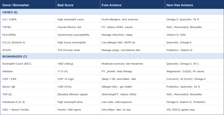
This article covers seven trackable biomarkers for eosinophilic fasciitis, including eosinophil count, aldolase, CRP, IgE, TGF-β1, IL-5, and ferritin, each with a concrete action plan whether or not you use supplements. It then reviews five genes most relevant to EF, explains what each one may affect, and lays out practical compensation strategies. A book summary challenges the idea that EF is purely a drug-management problem. Finally, complementary approaches are evaluated with clinical evidence, including the Autoimmune Protocol, photobiomodulation, mindfulness, and physical therapies with documented anti-fibrotic or immune-modulatory effects. If you have been told to simply wait and manage, what follows offers a more active framework for understanding and influencing your condition.
7 Biomarkers That Can Help You Track and Influence Eosinophilic Fasciitis
Most people with eosinophilic fasciitis have their eosinophil count checked once and then receive a prescription. That is a starting point, not a monitoring strategy. Tracking biomarkers over time lets you see whether the disease is progressing, stabilizing, or responding to interventions, and it gives you something concrete to discuss with your physician. The seven markers below were selected for three reasons: they are biologically meaningful for EF specifically, they are measurable through standard or specialty labs, and acting on them is realistic.
Biomarker 1: Absolute Eosinophil Count (AEC)
Why it matters: The absolute eosinophil count is the defining biomarker of eosinophilic fasciitis. Peripheral blood eosinophilia is present in the vast majority of EF cases at diagnosis, often exceeding 500 to 3,000 cells per microliter when the disease is active. It is both a diagnostic signal and a useful marker of disease activity over time, though it can normalize even while tissue inflammation continues.
What it may reveal: A persistently elevated AEC suggests ongoing immune activation and eosinophil recruitment to the fascia. A sharp rise after apparent remission can signal relapse before symptoms intensify. A count that remains elevated despite corticosteroids may indicate an inadequate treatment response or a secondary driver such as a parasitic infection or hematologic disorder.
How to measure it: The AEC is derived from a complete blood count with differential, one of the least expensive standard blood tests available. Cost ranges from $10 to $50 depending on the laboratory and insurance status. Ideally, retest every 6 to 12 weeks while the condition is active, and every 3 to 6 months once it stabilizes.
If the AEC is high, the plan without supplements
The first step is identifying any modifiable trigger. Heavy strenuous exercise is a well-documented precipitant of EF; reducing intensity to moderate aerobic activity (walking, swimming at a relaxed pace) removes a known eosinophil activator. Review all current medications for eosinophilia as a side effect, including certain antibiotics, NSAIDs, and some cholesterol medications. A low-histamine dietary trial for 4 weeks (removing fermented foods, alcohol, cured meats, aged cheeses) can reduce overall mast cell and eosinophil activity. Prioritize 7 to 9 hours of sleep nightly; sleep restriction measurably increases circulating eosinophil activation markers.
If the AEC is high, the plan with supplements or equipment
Quercetin: 500 to 1,000 mg per day with meals. Quercetin stabilizes mast cells, reduces eosinophil degranulation, and inhibits IL-5-driven eosinophil survival. Cycle 12 weeks on, 4 weeks off to maintain sensitivity. Side effects are mild: occasional headache or digestive discomfort at higher doses.
Omega-3 fatty acids (EPA + DHA): 2 to 4 grams per day from fish oil or algae-based sources. EPA and DHA shift eicosanoid production toward less pro-inflammatory prostaglandins and reduce IL-5 expression. Take with the largest meal to minimize GI upset. Ongoing use is appropriate; no cycling needed.
Vitamin C: 1 to 3 grams per day in divided doses (sodium ascorbate or liposomal form to improve tolerability). Supports eosinophil apoptosis and general antioxidant defense. Bowel tolerance is the dose ceiling for plain ascorbic acid.
Biomarker 2: Aldolase
Why it matters: Aldolase is a glycolytic enzyme found in skeletal muscle. Its elevation in EF reflects muscle and fascial inflammation, and it is one of the more EF-specific findings since it helps distinguish EF from conditions like systemic sclerosis where fascial thickening occurs without significant muscle involvement.
What it may reveal: Elevated aldolase confirms active myofascial inflammation even when imaging is inconclusive. It tends to track with disease activity: values often fall as corticosteroids take effect, and they rise again with relapse. It is also useful for detecting subclinical flares before joint symptoms worsen.
How to measure it: Aldolase is ordered as a standalone serum test. Cost ranges from $20 to $60. Normal reference range is approximately 1.0 to 7.5 U/L for adults; EF patients in active phases often present significantly above this range.
If aldolase is high, the plan without supplements
Gentle physical therapy focused on range-of-motion and fascial stretching is both therapeutic and a useful monitoring tool: if mobility improves alongside falling aldolase, the intervention is working. Gentle movement outperforms strict rest in inflammatory myofascial conditions. Dietary focus should emphasize protein adequacy (1.2 to 1.6g per kg of body weight daily) for tissue repair, and elimination of processed foods and seed oils known to exacerbate oxidative stress in muscle tissue. Heat therapy via a warm bath or heating pad applied to affected areas for 15 to 20 minutes after gentle exercise may reduce local inflammation and aid fascial mobility.
If aldolase is high, the plan with supplements or equipment
Magnesium glycinate: 300 to 400 mg at bedtime. Magnesium is a cofactor in over 300 enzymatic reactions including those involved in muscle energy metabolism. Deficiency worsens muscle inflammation. Start at 200 mg and increase gradually to avoid loose stools.
Coenzyme Q10: 100 to 300 mg per day with fat-containing meals (ubiquinol form is preferred for those over 40). Supports mitochondrial function in muscle cells and reduces oxidative muscle injury. Ongoing use appropriate; no cycling needed.
Far-infrared sauna: If accessible, 15- to 20-minute sessions 3 to 4 times per week. Far-infrared penetrates deeper into fascial and muscle tissue than standard steam sauna and may support metabolic waste clearance and fascial compliance. Begin with shorter sessions (8 to 10 minutes) and ensure good hydration.
Biomarker 3: CRP and ESR (Inflammation Composite)
Why it matters: C-reactive protein (CRP) and erythrocyte sedimentation rate (ESR) are general systemic inflammation markers. They are not specific to EF, but they are reliable indicators of overall inflammatory burden. Both are elevated in active EF and both tend to improve with effective treatment. Tracking them together gives a broader picture than either alone.
What it may reveal: High-sensitivity CRP above 3 mg/L is clinically meaningful and indicates elevated cardiovascular and systemic inflammatory risk independent of EF. ESR is a slower-moving marker, making it more useful for tracking trends over weeks rather than days. A discordance between the two (one elevated, one normal) sometimes occurs and warrants repeat testing to clarify the pattern.
How to measure it: Standard CRP: $10 to $30. High-sensitivity CRP (hs-CRP): $15 to $40. ESR: $10 to $25. All are available through any standard laboratory.
If CRP/ESR are elevated, the plan without supplements
Sleep is the single most powerful lever here: one night of inadequate sleep (less than 6 hours) measurably spikes CRP the following morning. Aim for 7 to 9 hours with consistent sleep and wake times. An anti-inflammatory dietary approach, specifically eliminating refined sugars, refined carbohydrates, and seed oils (canola, soybean, corn) while increasing vegetables, oily fish, and olive oil, has documented CRP-lowering effects within 4 to 8 weeks. Time-restricted eating (eating within a 10- to 12-hour window) reduces fasting insulin and inflammatory cytokine production. Low-intensity daily movement (walking 30 minutes/day) lowers CRP more reliably than sporadic intense exercise.
If CRP/ESR are elevated, the plan with supplements or equipment
Curcumin (as BCM-95 or phytosome form): 500 to 1,500 mg per day. Standard curcumin has poor bioavailability; formulations like BCM-95 or Meriva show significantly better absorption in clinical trials. Multiple randomized controlled trials demonstrate CRP reduction with curcumin supplementation. Cycle 12 weeks on, 4 weeks off; avoid at anticoagulant doses. Take with meals.
Vitamin D3 with K2: 2,000 to 5,000 IU per day, targeting blood levels of 40 to 60 ng/mL. Vitamin D deficiency is independently associated with elevated inflammatory markers and worse autoimmune outcomes. K2 (100 to 200 mcg MK-7 form) directs calcium appropriately. Test 25-OH vitamin D every 6 months while adjusting dose.
Omega-3 (EPA + DHA): As noted under AEC; the CRP-lowering effect of omega-3s is well-documented across multiple meta-analyses.
Biomarker 4: Serum IgE
Why it matters: Immunoglobulin E (IgE) is the antibody class most associated with allergic responses and eosinophilic activation. IgE is not universally elevated in EF, but when it is, it points to a Th2-dominant immune pattern — the same immunological skew that drives eosinophil recruitment and activation. Elevated IgE also suggests that allergen burden or gut permeability may be contributing factors.
What it may reveal: Very high total IgE (above 200 IU/mL) may suggest an atopic or allergic component that is worth investigating separately. It can also flag the possibility of a parasitic infection, which should be ruled out before settling on an idiopathic EF diagnosis. In the context of EF management, tracking IgE over time helps assess whether immune dysregulation is improving.
How to measure it: Total serum IgE is a standard immunology test. Cost: $30 to $80. Some physicians also order specific allergen IgE panels; these are more expensive ($100 to $300+) but can identify specific triggers.
If IgE is elevated, the plan without supplements
A structured allergen elimination trial (removing the top six food allergens: gluten, dairy, eggs, soy, corn, and tree nuts for 30 days, then reintroducing one at a time) is the most practical first step. Reduce environmental allergen load: HEPA air filtration in the bedroom, mite-proof mattress covers, and minimizing exposure to mold. Improving gut barrier function is central: remove alcohol, highly processed foods, and chronic NSAID use, which all directly compromise intestinal permeability.
If IgE is elevated, the plan with supplements or equipment
Probiotics (specifically Lactobacillus rhamnosus GG and L. acidophilus): A 60- to 90-day course has the most evidence for shifting Th2-dominant immune responses toward better Th1/Th2 balance. After the initial course, maintenance dosing every other day may sustain the benefit. GI discomfort in the first week is common and usually resolves.
Quercetin: As noted above; quercetin stabilizes IgE-mediated mast cell activation.
Vitamin D3: Optimizing vitamin D levels (40 to 60 ng/mL) specifically supports immune regulation and can modulate IgE-mediated responses. Dose as described under Biomarker 3.
Biomarker 5: TGF-β1 (Transforming Growth Factor Beta 1)
Why it matters: TGF-β1 is the master regulator of fibrosis in connective tissue. In eosinophilic fasciitis, eosinophils degranulate and release profibrotic mediators including TGF-β1, which then drives fibroblast activation and collagen deposition in the fascia. This is the mechanism behind the skin hardening and joint restriction characteristic of EF. Tracking TGF-β1 gives you a window directly into fibrotic activity.
What it may reveal: Elevated serum TGF-β1 confirms ongoing fibrotic signaling even when surface symptoms seem stable. It is particularly useful for assessing whether a treatment is actually halting fibrosis or merely masking symptoms. Some research suggests that corticosteroids, while effective at reducing eosinophilia, do not fully suppress TGF-β1-mediated fibrosis.
How to measure it: TGF-β1 is measured via ELISA-based serum testing, typically available through specialty or functional medicine labs. Cost: $100 to $200. This is not a standard panel item, so you may need to specifically request it or order through a direct-to-consumer lab.
If TGF-β1 is elevated, the plan without supplements
Physical therapy with a focus on myofascial release and active stretching is the most evidence-supported mechanical approach to limiting fascial fibrosis. Sustained gentle stretching signals fibroblasts through mechanotransduction to remodel collagen rather than lay down excessive scar tissue. Advanced glycation end-products (AGEs), found in high-temperature cooked processed foods, stimulate TGF-β1 directly; reducing dietary AGEs (by preferring poached, steamed, and raw preparations over charred or fried foods) is a meaningful and underused intervention. Far-infrared sauna (as noted above) may also reduce TGF-β1 activity through thermal stress pathways.
If TGF-β1 is elevated, the plan with supplements or equipment
NAC (N-acetyl cysteine): 600 to 1,800 mg per day in divided doses with meals. NAC has documented anti-fibrotic effects in pulmonary and hepatic fibrosis models, operating through glutathione replenishment and direct inhibition of TGF-β1 signaling. Take with food to reduce nausea. Ongoing use is appropriate; no cycling required. Do not combine with nitroglycerin.
Resveratrol (as pterostilbene or trans-resveratrol): 250 to 500 mg per day. Resveratrol suppresses SMAD2/3 phosphorylation downstream of TGF-β1 in multiple cell types, directly dampening fibrotic gene expression. Cycle 12 weeks on, 4 weeks off. Avoid at high doses in pregnancy and with anticoagulants.
Boswellia serrata (standardized to AKBA): 300 to 500 mg three times per day. Boswellic acids inhibit 5-lipoxygenase and have anti-fibrotic properties supported by human studies in inflammatory conditions. 12-week cycles are a reasonable approach; GI side effects are uncommon.
Biomarker 6: Interleukin-5 (IL-5)
Why it matters: IL-5 is the cytokine that drives eosinophil production in bone marrow, prolongs eosinophil survival in circulation, and recruits them to tissues. It is the central upstream signal in eosinophilic inflammation. Monoclonal antibodies targeting IL-5 (mepolizumab, reslizumab) have become major treatments in severe eosinophilic asthma and are being explored in EF. Knowing your circulating IL-5 level offers direct insight into the intensity of eosinophil-driving activity.
What it may reveal: A persistently elevated IL-5 level signals that eosinophilic drive is high regardless of whether peripheral AEC is currently controlled by treatment. It may also help identify patients most likely to respond to anti-IL-5 biologics, though this clinical application is still being studied for EF specifically.
How to measure it: Serum IL-5 is measured via multiplex cytokine panel or standalone ELISA at specialty or research-affiliated laboratories. Cost: $100 to $300. This is a more advanced marker; discuss with your physician whether it is appropriate to add to your monitoring panel.
If IL-5 is elevated, the plan without supplements
Reducing Th2-polarizing stimuli is the primary goal. Key practical approaches include: limiting refined carbohydrate intake (high glycemic load promotes Th2 polarization), reducing aflatoxin exposure (inspect stored grains and nuts for mold), and addressing any undiagnosed parasitic infections via stool testing. Short cold exposure (cold showers ending with 30 to 60 seconds of cold water) may shift the Th1/Th2 balance over time; this is early evidence, but the mechanism is plausible and the practice carries no meaningful risk.
If IL-5 is elevated, the plan with supplements or equipment
Omega-3 fatty acids: 2 to 4 grams EPA + DHA per day. DHA specifically downregulates IL-5 production and reduces Th2 skewing. This is one of the better-supported nutritional interventions for IL-5 modulation. Take with meals.
Vitamin D3: Vitamin D directly suppresses IL-5 gene expression through VDR binding at the IL5 promoter region. Optimizing to 40 to 60 ng/mL is essential.
Probiotics: Lactobacillus strains improve Th1/Th2 cytokine balance, reducing IL-5 production. A 90-day course with ongoing maintenance dosing is a reasonable protocol. Choose multi-strain products with documented refrigeration chain integrity.
Biomarker 7: Complete Blood Count and Serum Ferritin
Why it matters: This final biomarker slot is actually two closely related tests that serve a critical and often overlooked monitoring purpose in EF. Between 8 and 20 percent of EF cases are associated with hematologic disorders, most notably aplastic anemia, myelodysplastic syndrome, T-cell lymphoma, and multiple myeloma. A CBC with differential and periodic bone marrow biopsy evaluation are the primary tools for identifying this overlap. Serum ferritin, beyond reflecting iron stores, is a reliable acute-phase reactant and a surrogate marker of systemic inflammatory burden.
What it may reveal: Unexplained anemia, thrombocytopenia, or abnormal white cell morphology on a CBC are red flags for hematologic comorbidity in EF and should prompt specialist referral. Persistently elevated ferritin (above 300 ng/mL in men or 150 ng/mL in women) indicates ongoing systemic inflammation even when other markers normalize. Very high ferritin (above 1,000 ng/mL) may indicate macrophage activation syndrome or hemophagocytic lymphohistiocytosis, rare but serious complications.
How to measure it: CBC with differential: $10 to $50. Serum ferritin: $15 to $40. Both are widely available. Recommended monitoring: every 3 to 6 months in stable EF, more frequently during active disease.
If CBC or ferritin is abnormal, the plan without supplements
Any unexplained abnormality in the CBC warrants prompt physician evaluation, not self-management. For elevated ferritin without hematologic pathology, an anti-inflammatory dietary approach (as described under Biomarker 3) is the primary lifestyle lever. Ferritin tracks broadly with inflammation: addressing upstream biomarkers (CRP, AEC) will typically pull ferritin down over months. Avoid supplemental iron unless documented deficiency is confirmed.
If ferritin is elevated, the plan with supplements or equipment
IP6 (inositol hexaphosphate): 1 to 4 grams per day on an empty stomach can moderately reduce excess ferritin. Do not combine with iron supplementation. Cycle 8 to 12 weeks at a time with reassessment.
Green tea extract (EGCG): 400 to 800 mg per day has mild iron-chelating properties and general anti-inflammatory effects. Take between meals. Avoid in significant liver disease.
Address root inflammation first; ferritin will follow.
With a clear picture of seven actionable biomarkers, the next layer worth examining is what your genetics may be contributing to this pattern, and what, if anything, can be done about it.
The Genetics Behind Eosinophilic Fasciitis: What Your DNA May Be Telling You
Genetic research on eosinophilic fasciitis specifically is limited — the condition is too rare for large genome-wide association studies. What exists is a mosaic of insights drawn from related eosinophilic conditions, fibrotic diseases, and autoimmune genetics research. The following five genes represent the most biologically plausible and relevant genetic contributors based on current understanding. Think of this section as a framework for interpreting your genetic data rather than a definitive diagnostic map.
Gene 1: IL5 and IL5RA — The Eosinophil Production Axis
The IL5 gene encodes interleukin-5, and IL5RA encodes its receptor. Variants in both have been associated with elevated eosinophil counts in eosinophilic esophagitis and other eosinophilic conditions. Higher baseline IL-5 signaling genetically predisposes to exaggerated eosinophil responses when immune challenges occur.
If the gene variant is unfavorable, the plan without supplements
Minimize the environmental inputs that activate IL-5 production: avoid known personal allergens, reduce exposure to inhaled mold, and maintain low-allergen dietary patterns. Manage physical exertion carefully, since strenuous exercise is a documented EF trigger and may interact with heightened IL-5 responsiveness. Prioritize gut barrier integrity through diet (fermented foods, fiber diversity).
If the gene variant is unfavorable, the plan with supplements or equipment
Omega-3 fatty acids and Quercetin (as described under Biomarker 1 and 6) directly dampen IL-5 production and receptor signaling respectively. Vitamin D optimization is particularly important here: low vitamin D allows IL-5-driven eosinophilic activity to amplify unchecked. For those with confirmed unfavorable IL5/IL5RA variants and persistent eosinophilia, discuss with your physician whether anti-IL-5 biologic therapy (mepolizumab) has been studied in your specific presentation.
Gene 2: TGFB1 — The Fibrosis Risk Gene
The TGFB1 gene has a well-characterized functional polymorphism (rs1800470, also known as the codon 10 L/P variant) that affects TGF-β1 production levels. The high-producer variant leads to greater collagen deposition in response to inflammatory stimuli. In the context of EF, where TGF-β1-driven fibrosis is central to morbidity, carrying the high-producer variant may worsen fascial thickening and prolong recovery.
If the gene variant is unfavorable, the plan without supplements
Aggressive early intervention with physical therapy is particularly important for TGFB1 high-producer variants. Fascial fibrosis driven by genetics responds to mechanical loading signals: sustained stretching, myofascial release, and progressive joint mobilization help counterbalance fibroblast activation. Dietary reduction of advanced glycation end-products (charred and processed foods) is a meaningful lifestyle-level intervention for high TGF-β1 expressors. Regular sauna use (3 to 4 sessions per week) may also partially counteract TGFB1 pro-fibrotic signaling.
If the gene variant is unfavorable, the plan with supplements or equipment
NAC, Resveratrol, and Boswellia (described under Biomarker 5) are the strongest candidates for TGFB1 high-producers. For those with significant fascial involvement, consider discussing pentoxifylline with your physician: this prescription medication has anti-fibrotic properties and has been used adjunctively in fibrotic conditions. For equipment, consider a percussive massage device (Theragun-style) for regular fascial stimulation.
Gene 3: HLA-DRB1 — The Immune Presentation Gate
HLA (Human Leukocyte Antigen) genes control how the immune system presents antigens to T cells. Certain HLA-DRB1 alleles are consistently associated with autoimmune susceptibility across multiple conditions. While no large studies have confirmed specific HLA-DRB1 alleles in EF, the pattern in related fibroinflammatory conditions (like systemic sclerosis) suggests that immune presentation genes shape who develops eosinophilic and fibrotic autoimmune responses.
If the gene variant is unfavorable, the plan without supplements
HLA-DRB1 variants cannot be changed, but the environmental triggers that activate aberrant antigen presentation can be managed. Reduce molecular mimicry triggers by avoiding chronic low-grade infections and considering gluten elimination if HLA-DRB1*07 or similar alleles are present (this allele increases celiac susceptibility and cross-reactive autoimmune risk). Managing oxidative stress through diet and sleep is the most practical general lever.
If the gene variant is unfavorable, the plan with supplements or equipment
Vitamin D optimization (40 to 60 ng/mL) is arguably the highest-impact supplement for HLA-associated autoimmune risk, as VDR signaling directly modulates antigen presentation. N-acetyl cysteine supports glutathione production and reduces oxidative activation of antigen-presenting cells. These are safe ongoing interventions with broad benefit.
Gene 4: CCL11 (Eotaxin-1) — Eosinophil Recruitment to Tissue
CCL11 encodes eotaxin-1, the chemokine that physically recruits eosinophils into tissues from circulation. Even with a normal eosinophil count, upregulated eotaxin-1 activity can drive disproportionate tissue eosinophil accumulation. Variants that increase CCL11 expression may explain why some patients develop severe fascial eosinophil infiltration while others with similar peripheral counts do not.
If the gene variant is unfavorable, the plan without supplements
Environmental allergen reduction directly lowers eotaxin-1 production in mucosal and connective tissues. A low-allergen diet (eliminating identified personal food triggers) and improving indoor air quality (HEPA filtration, mold remediation) are the highest-impact practical steps. Endurance exercise at low intensity (as opposed to high-intensity training) does not significantly upregulate eotaxin-1 and preserves fitness without added tissue recruitment risk.
If the gene variant is unfavorable, the plan with supplements or equipment
Quercetin specifically blocks CCR3, the receptor through which eotaxin-1 recruits eosinophils. This makes Quercetin particularly relevant for CCL11 high-expressors. Dose as previously described. Omega-3 fatty acids also suppress eotaxin-1 production through PPAR-γ pathway modulation.
Gene 5: STAT6 — The Th2 Amplifier
STAT6 is the intracellular signal transducer for IL-4 and IL-13, two cytokines that powerfully drive Th2 immune polarization and eosinophilic activity. Gain-of-function STAT6 mutations cause severe eosinophilic multi-organ disease in rare cases; more common variants that increase STAT6 signaling efficiency are associated with atopic conditions and eosinophilia. In EF, STAT6 amplification may sustain eosinophilic activity beyond the initial trigger.
If the gene variant is unfavorable, the plan without supplements
Reducing Th2-promoting environmental exposures is again central: manage atopic conditions (asthma, eczema, allergic rhinitis) aggressively, since these conditions mutually amplify STAT6 signaling. Saline nasal irrigation reduces the allergen load that triggers STAT6-driven mucosal immune responses. Avoid prolonged antibiotic use when possible, as microbiome disruption strongly promotes Th2 skewing through STAT6-dependent pathways.
If the gene variant is unfavorable, the plan with supplements or equipment
Probiotics (specifically strains documented to improve Th1/Th2 balance, such as L. rhamnosus and L. acidophilus) can partially compensate for STAT6 pro-Th2 variants. Vitamin D optimization suppresses STAT6-mediated IL-13 signaling. For STAT6 high-activity variants combined with persistently elevated eosinophilia, discussion of dupilumab (an IL-4/IL-13 receptor antagonist) with your physician is worth exploring.
Understanding your genetic predispositions can sharpen which biomarkers to prioritize and which interventions are most likely to move the needle. The following section offers a framework that ties many of these threads together.
Rethinking the Root: Key Insights From "The Autoimmune Fix" by Tom O'Bryan
Tom O'Bryan is a functional medicine clinician and researcher whose 2016 book The Autoimmune Fix draws on hundreds of peer-reviewed studies to challenge a core assumption of conventional autoimmune care: that autoimmune conditions are defined by the organ they affect rather than by shared upstream triggers that can be addressed. The book does not mention EF specifically, but its framework applies precisely to the biological terrain that allows EF to develop and persist.
1. Autoimmune disease develops in stages — and most people miss the window
O'Bryan describes a three-stage model: silent autoimmunity, autoimmune reactivity, and full autoimmune disease. Most people are diagnosed at stage three. Stage one and two can last years and are often detectable through specific antibody testing. For EF, this means the inflammatory and fibrotic cascade likely began long before the first visible skin change or lab abnormality.
2. Leaky gut is not a fringe concept — it is mechanistically upstream
Intestinal permeability allows partially digested food proteins and microbial fragments to enter systemic circulation, where they activate immune responses. O'Bryan cites extensive evidence linking increased gut permeability to systemic autoimmune activation. In eosinophilic conditions specifically, gut dysbiosis and permeability are closely linked to mucosal eosinophilia and broader immune dysregulation.
3. Gluten is a uniquely potent trigger for intestinal permeability
Gliadin, a component of gluten, directly triggers zonulin release, which opens tight junctions in the intestinal lining even in people without celiac disease. O'Bryan references multiple studies showing transient intestinal permeability in non-celiac individuals following gluten exposure. Whether gluten is a trigger for a specific individual requires testing and elimination trial, not assumption, but for those with HLA-DRB1 variants associated with gluten sensitivity, this is a high-priority investigation.
4. Your immune system responds to your environment, not just your genes
The book presents compelling evidence that epigenetic modifications, driven by diet, stress, environmental toxins, and infections, can activate genetic susceptibilities that would otherwise remain silent. This is particularly relevant for the five genes discussed in the previous section: variants are not destiny; expression is shaped by environment.
5. Molecular mimicry is underappreciated as an ongoing trigger
Certain food proteins and microbial antigens share amino acid sequences with human tissue proteins. When the immune system attacks these look-alikes, it can inadvertently attack self. For eosinophilic and fibrotic conditions, proteins in certain grains and dairy have documented molecular mimicry with connective tissue components.
6. Systemic inflammation is measured poorly in conventional care
O'Bryan argues that standard inflammatory panels (CRP, ESR) are too insensitive to catch early-stage immune dysfunction. He recommends a broader panel that includes homocysteine, Lp-PLA2, and specific autoantibody tests. This aligns with the biomarker approach in this article: measuring more gives you more to work with.
7. The microbiome directly programs immune identity
The gut microbiome shapes the development and calibration of the immune system throughout life. O'Bryan reviews evidence that specific bacterial species (particularly Akkermansia muciniphila and Bifidobacterium) act as natural Th1/Th2 balancers. Dysbiosis tilts the balance toward Th2 and eosinophilic activity.
8. Environmental toxins accumulate and create long-term immune noise
Organophosphate pesticides, phthalates, and heavy metals have all been shown to alter immune gene expression. For eosinophilic conditions, pesticide exposure in particular correlates with altered eosinophil behavior. O'Bryan advocates for testing metal burden and reducing exposure through filtered water, organic food, and toxin-reducing sauna protocols.
9. Stress is not separate from immune function — it is immune function
Chronic psychological stress activates HPA axis dysregulation, which suppresses Th1 activity and permits Th2 and eosinophilic dominance. O'Bryan ties cortisol dysregulation directly to autoimmune flares and recommends measuring DHEA-to-cortisol ratio as a proxy for HPA balance.
10. The elimination diet is not a punishing restriction — it is a diagnostic instrument
O'Bryan's practical centerpiece is a structured elimination protocol, very similar to what the Autoimmune Protocol formalizes (discussed next). He frames it not as a treatment but as a diagnostic: removing the most immunogenic foods tells you what is actually driving your immune activation. For EF patients, this reframe makes the difficult early weeks more tolerable.
The Autoimmune Fix points toward a direction that several integrative medicine practitioners have codified in structured protocols — including one specifically designed for autoimmune conditions.
Complementary Approaches With Meaningful Evidence
The Autoimmune Protocol (Sarah Ballantyne, PhD)
The Autoimmune Protocol (AIP) is a structured dietary and lifestyle framework developed by Sarah Ballantyne, a researcher with a PhD in medical biophysics who published the framework in her book The Paleo Approach. It is specifically designed for autoimmune and immune-dysregulated conditions. Its relevance to EF is direct: EF involves immune dysregulation, eosinophilic activation, and pro-inflammatory cytokine production, all of which the AIP addresses through dietary elimination and gut microbiome restoration.
A pilot study published in Inflammatory Bowel Diseases (Konijeti et al., 2017) demonstrated that AIP led to clinical remission in 73 percent of patients with inflammatory bowel disease after 6 weeks, with significant reductions in inflammatory markers. While no EF-specific trial exists, the AIP's mechanisms (gut barrier restoration, reduction of immune-activating foods, microbiome support) are directly relevant to eosinophilic and fibrotic autoimmune presentations. The study is referenced at PubMed (PMID 29202018).
The AIP involves a 30- to 90-day elimination of grains, legumes, dairy, eggs, nightshades, nuts, seeds, alcohol, and all processed foods, followed by a structured reintroduction phase. For EF patients, this can be adopted cautiously alongside standard treatment. Begin with the dietary phase; the lifestyle additions (sleep, stress, gentle movement) can be layered in progressively. The protocol is demanding, but it has a well-developed patient community and documented support resources at ThePaleoMom.com.
Mindfulness-Based Stress Reduction (MBSR)
MBSR is an 8-week structured program combining mindfulness meditation, body scan, and gentle yoga, originally developed by Jon Kabat-Zinn at the University of Massachusetts. It is relevant to EF because chronic psychological stress is a known driver of Th2 immune polarization, cortisol dysregulation, and inflammatory cytokine production — all of which amplify eosinophilic disease activity.
A meta-analysis published in JAMA Internal Medicine (Goyal et al., 2014) reviewing 47 trials found that mindfulness meditation programs produced moderate reductions in anxiety, depression, and pain, and small but significant reductions in inflammatory biomarkers including CRP. The study is available at PubMed (PMID 24395196). A subsequent immunological study demonstrated that MBSR practice reduced circulating IL-6 and modulated Th1/Th2 cytokine balance in participants with elevated baseline inflammation.
For EF patients, an 8-week MBSR course (available in-person or online through certified programs) is a realistic and low-risk addition to standard care. Daily practice of 30 to 45 minutes, including a 10-minute body scan and 20 minutes of seated meditation, is the standard protocol. Maintaining this practice beyond the 8-week course is associated with sustained benefit.
Low-Level Laser Therapy (LLLT) / Photobiomodulation
Low-level laser therapy uses specific wavelengths of light (typically 630 to 980 nm) to stimulate mitochondrial function, reduce oxidative stress, and modulate inflammatory signaling in target tissues. In EF, where fascial fibrosis and localized inflammation are the primary tissue-level problems, LLLT's documented anti-fibrotic and anti-inflammatory effects in connective tissue are mechanistically relevant.
Clinical evidence is most robust in musculoskeletal and fibrotic applications. A systematic review published in Lasers in Medical Science (Tumilty et al., 2010) found that LLLT significantly reduced pain and improved functional outcomes in musculoskeletal soft tissue conditions. More relevant to fibrosis, a 2014 study by Silveira et al. in Photomedicine and Laser Surgery demonstrated that LLLT reduced TGF-β1 expression and collagen deposition in an experimental fibrosis model.
For EF patients, LLLT can be applied by a physiotherapist or sports medicine physician trained in photobiomodulation, targeting affected fascial areas (typically the limbs and trunk). Parameters of 4 to 8 Joules per cm² at 808 to 850 nm, applied 2 to 3 times per week over 8 to 12 weeks, represent a reasonable starting protocol. Home devices with lower-power output are available but are likely less effective than professional-grade equipment; they remain a reasonable maintenance option. Evidence for EF specifically is limited to mechanistic plausibility, so set expectations accordingly.
Massage Therapy (Myofascial Release Technique)
Massage therapy, specifically myofascial release (MFR), is a hands-on technique targeting the connective tissue sheaths (fascia) surrounding muscles and organs. For eosinophilic fasciitis, where fascial thickening, fibrosis, and restricted mobility are hallmark features, MFR offers a direct mechanical intervention for tissue compliance and range of motion.
A systematic review of myofascial release for musculoskeletal conditions (Ajimsha et al., 2015, Complementary Therapies in Clinical Practice) found significant improvements in pain, mobility, and function across multiple conditions involving fascial restriction. No EF-specific randomized trial exists, but the fascial remodeling mechanisms are directly applicable. A single case report in the occupational therapy literature describes meaningful functional improvement in an EF patient following a structured manual therapy program.
Sessions should be conducted by a licensed massage therapist or physical therapist with specific myofascial release training. For EF patients, direct deep tissue work on significantly thickened or fibrotic areas should be gentle and progressive, avoiding excessive pressure that can exacerbate inflammation. Starting with weekly 60-minute sessions and transitioning to biweekly maintenance once mobility improves is a practical approach. Self-myofascial release using a foam roller on less severely affected areas is a useful daily adjunct.
Breathing-Based Therapies
Diaphragmatic breathing and controlled breathing protocols (including slow-paced breathing at 5 to 6 breaths per minute, resonance frequency breathing, and box breathing) stimulate the vagus nerve and activate the parasympathetic nervous system. This directly counteracts the sympathetic-dominant state that promotes Th2 immune polarization and pro-inflammatory cytokine production — a state that many patients with chronic inflammatory conditions spend too much time in.
A randomized controlled trial published in BMC Complementary and Alternative Medicine (Bhattacharya et al., 2014) demonstrated that slow-paced breathing (6 breaths per minute for 15 minutes) significantly reduced salivary cortisol and pro-inflammatory cytokine levels compared to uncontrolled breathing. Vagal activation through breathing is also documented to reduce TNF-α and IL-1β production through the cholinergic anti-inflammatory pathway. While EF-specific studies do not exist, the inflammatory pathways modulated are directly relevant.
Practical application: practice slow-paced breathing (inhale for 5 seconds, exhale for 5 seconds) for 15 to 20 minutes twice daily. Apps such as Breathwrk, Insight Timer, or a simple metronome facilitate this. Box breathing (4 seconds in, 4 hold, 4 out, 4 hold) is an alternative that some find easier to sustain. Consistent daily practice over 8 to 12 weeks is required for meaningful autonomic adaptation. No side effects; easily combined with other interventions.
Conclusion
Eosinophilic fasciitis is rare, under-researched, and often managed with a single tool. This article has outlined a more comprehensive framework: seven biomarkers that track disease activity and fibrotic burden, five genetic variants that help explain individual susceptibility and severity, a root-cause perspective informed by autoimmune research, and complementary approaches with evidence sufficient to warrant serious consideration.
None of this replaces the guidance of a physician with experience in rare inflammatory diseases, and none of it promises remission. What it does offer is a clearer view of the terrain and more options for engaging with it actively. The most useful next step depends on where you are: if you have never had a full biomarker panel, start there. If your current panel is being tracked but nothing is improving, review the genetic and complementary angles. If you have the biology addressed but your lifestyle is not aligned, the dietary and stress protocols deserve a genuine trial. Take the next smart step — whatever that is for you — with a clear head and a conversation with a physician who will engage with the details.
Musculoskeletal: Muscle Conditions
Skin: Inflammatory Skin Conditions
Autoimmune: Inflammatory Conditions Connective Tissue Conditions