This article contains AI generated content.
· UpdatedErosive Inflammatory Osteoarthritis Genes Biomarkers — 6 Genes and 7 Biomarkers to Track
Introduction
Erosive inflammatory osteoarthritis (EIOA) sits in uncomfortable territory between two well-mapped diseases. It shares the mechanical wear of classical osteoarthritis but also carries the synovial inflammation more typical of rheumatoid disease. For those living with it — swollen, painful finger joints, central erosions on X-ray, and flares that do not always follow a predictable pattern — that combination means both physical discomfort and diagnostic uncertainty. Standard advice to protect your joints and manage inflammation is not wrong, but it treats every EIOA patient as a single type.
The reality is more specific. Underlying inflammatory drivers, cartilage degradation rates, genetic susceptibilities, and metabolic contributions vary widely from person to person. A protocol designed for average inflammation will be too mild for one person and unnecessary for another. What looks like the same diagnosis on paper can involve very different active biology beneath the surface.
A smarter approach starts with measurement. Tracking the right biomarkers gives you a real-time view of which processes — inflammatory signaling, cartilage breakdown, metabolic dysfunction — are actually active in your joints right now. Understanding which genetic variants you carry takes this one step further: it identifies the biological pathways that are working against you by design, and opens the door to targeted compensating strategies.
This article maps both dimensions. The first and primary section covers seven measurable biomarkers — including what they reveal, what levels to aim for, cost estimates, and step-by-step plans with and without supplements. The second section examines six genes implicated in EIOA risk and erosive progression, with practical steps to partially offset less favorable variants. A third section draws on Peter Attia's evidence-based longevity framework for its most relevant insights on inflammation and joint health. Finally, five complementary modalities with meaningful clinical evidence round out the picture. Better information rarely cures a condition, but it consistently leads to better decisions.
Summary
This article covers erosive inflammatory osteoarthritis from two angles that most clinical appointments never reach: the biomarkers you can track today (seven, with cost ranges and specific action plans), and the six genes that shape your underlying joint biology. You will find concrete protocols — both with and without supplements — for every biomarker and gene discussed, along with specific cycling schedules and side effects. Beyond the biology, the article includes ten of the most impactful insights from Peter Attia's longevity work as they apply to inflammatory joint disease, and five complementary approaches — including tai chi, low-level laser therapy, the Autoimmune Protocol, and microbiome-directed strategies — each backed by human clinical evidence. If you have been managing EIOA with generic advice, the layers you are about to read may change how you approach it.
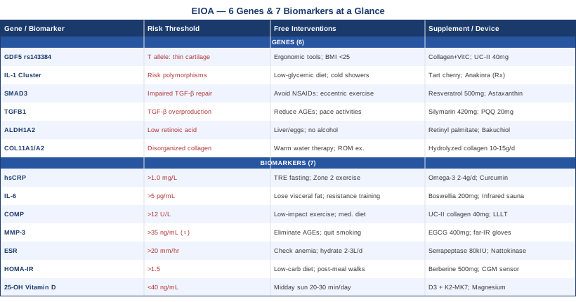
7 Biomarkers to Track for Erosive Inflammatory Osteoarthritis
Tracking biomarkers in EIOA is not about chasing numbers for their own sake. Each of the following seven markers tells you something specific: whether active inflammation is driving your disease, whether cartilage is degrading faster than it should be, and whether metabolic factors are contributing to the inflammatory environment. Together, they offer considerably more resolution than clinical examination or X-ray alone.
Biomarker 1: High-Sensitivity C-Reactive Protein (hsCRP)
Why it matters: CRP is produced by the liver in response to inflammatory signals, particularly interleukin-6 (IL-6). In EIOA, circulating hsCRP levels are consistently and significantly higher than in non-erosive hand osteoarthritis, directly reflecting the degree of active synovial inflammation. Studies comparing erosive and non-erosive hand OA cohorts find that hsCRP correlates with both disease activity and radiographic erosion progression. Peter Attia, who advocates for aggressive inflammation tracking in the context of chronic disease prevention, treats hsCRP not as a binary flag but as a continuous variable where lower is almost always better — and where even values labeled "normal" by standard labs may represent meaningful chronic inflammatory activity in EIOA.
Optimal range: Under 0.5 mg/L (Attia's preferred target for chronic disease management). Most labs flag anything below 1.0 mg/L as normal, but in EIOA, values in the 1.0–3.0 mg/L range indicate ongoing low-grade inflammation worth actively addressing.
How to Measure It
Standard blood draw ordered by a GP or rheumatologist. Cost: $20–50 USD. Ensure the lab runs the high-sensitivity version (hsCRP), not standard CRP, which lacks precision at lower levels. Draw fasting, and avoid testing within a week of any acute illness or injection-site inflammation, which will artificially elevate results. Retest every 3–6 months while making changes.
If hsCRP Is High: Plan Without Supplements
- Time-restricted eating (14–16 hour overnight fast): Reduces fasting insulin and downstream inflammatory signaling. Begin with 12 hours and extend gradually over 2–3 weeks. - Zone 2 aerobic exercise, 150–180 minutes per week: At a pace where conversation remains comfortable. Zone 2 training upregulates mitochondrial function and measurably reduces IL-6 output from adipose tissue within 6–8 weeks of consistent effort. - Remove ultra-processed foods and seed oils: Trans fats and refined carbohydrates are among the most reliable dietary drivers of CRP elevation. A whole-food diet with adequate fiber is the structural foundation — not an optional add-on. - Prioritize sleep (7–9 hours): Sleep restriction below 7 hours reliably elevates CRP within days. Address sleep before adding any other intervention.
If hsCRP Is High: Plan With Supplements or Equipment
- Omega-3 fatty acids (EPA+DHA), 2–4 g/day: Evidence for CRP reduction in inflammatory conditions is robust across multiple RCTs. Use a high-quality fish oil or algae-based supplement. Take with the largest meal to reduce GI side effects. Cycling: Can be used long-term. Side effects: Fish burps, mild blood-thinning at higher doses — consult a physician if taking anticoagulants. - Bioavailable curcumin, 500–1000 mg/day: Choose BCM-95, Meriva, or CurcuWIN formulations. Multiple RCTs demonstrate CRP reduction in inflammatory arthritis. Cycling: 5 days on, 2 days off is a common approach; no established clinical reason to cycle. Side effects: Can inhibit CYP3A4 enzymes — potential drug interactions with certain medications. - Red light / photobiomodulation panel (630–850 nm): 10–20 minutes per session, 3–5 times/week over affected joints. Emerging evidence suggests modulation of NF-κB pathway, one of the primary CRP-inducing pathways in synovial tissue. Cost of device: $150–600 USD.
Biomarker 2: Interleukin-6 (IL-6)
Why it matters: IL-6 sits upstream of hsCRP — it is the primary cytokine signal that triggers CRP production. But its importance in EIOA goes beyond being a CRP precursor. IL-6 directly activates osteoclasts, the bone-resorbing cells responsible for the central erosions that characterize EIOA radiographically. Elevated IL-6 in both serum and synovial fluid is a consistent finding in EIOA versus non-erosive OA, and the fact that IL-6 receptor blockers (like tocilizumab) have shown benefit in erosive OA in early trials confirms its mechanistic centrality. Measuring IL-6 directly gives you an earlier and more specific inflammatory signal than CRP alone.
Optimal range: Under 3.1 pg/mL (standard lab reference). In EIOA management, target the lower third of the reference range. Values above 5 pg/mL in the absence of acute illness indicate sustained inflammatory activity.
How to Measure It
Serum IL-6 assay via blood draw. Cost: $60–150 USD. Less routinely ordered than CRP — request it specifically from a rheumatologist or access it through a direct-to-consumer lab. Draw in the morning, fasting, for most reliable results. Sensitive to acute stress, so avoid testing during or immediately after high-intensity exercise.
If IL-6 Is High: Plan Without Supplements
- Reduce visceral adiposity: Visceral fat is a primary IL-6 secretion site. A 5–10% reduction in body weight in overweight individuals measurably reduces circulating IL-6 within 8–12 weeks. - Resistance training, 3 sessions/week: Despite acutely increasing IL-6 during exercise, regular resistance training reduces resting IL-6 through durable anti-inflammatory adaptation — particularly by increasing skeletal muscle mass, which competes with adipose tissue as a metabolic driver. - Contrast hydrotherapy on affected joints: 3 minutes warm water, 1 minute cold, 3–4 cycles. Some evidence for reducing local inflammatory cytokine levels in inflammatory arthritis when applied consistently.
If IL-6 Is High: Plan With Supplements or Equipment
- Boswellia serrata (standardized AKBA extract), 200–400 mg/day: Inhibits 5-LOX enzyme, which reduces leukotriene production upstream of IL-6 signaling. RCT evidence exists for knee OA. Cycling: 8 weeks on, 2 weeks off. Side effects: Mild GI symptoms, rare skin rash. - Magnesium glycinate or malate, 300–400 mg/day: Low magnesium is independently associated with elevated IL-6. Cycling: Can be used continuously. Side effects: Loose stools at higher doses. - Infrared sauna, 20–30 minutes at 45–55°C, 3–4 times/week: Several controlled studies in rheumatoid arthritis populations show reductions in IL-6 and pain scores after 4 consecutive weeks. Caution: Start with shorter sessions at lower temperatures; avoid during acute flares. Cost of home unit: $1,200–3,500 USD.
Biomarker 3: Cartilage Oligomeric Matrix Protein (COMP)
Why it matters: COMP is a structural protein released from cartilage when it undergoes mechanical stress or enzymatic degradation. Elevated serum COMP does not merely reflect existing damage — it reflects ongoing, active cartilage breakdown. In EIOA specifically, COMP levels are higher than in non-erosive OA and correlate with radiographic erosion scores over time, making it both a diagnostic and prognostic marker. OARSI (Osteoarthritis Research Society International) has endorsed COMP as one of the most clinically informative OA cartilage biomarkers in multiple systematic reviews. It tells you not just that joint damage is present but whether it is accelerating.
Optimal range: Reference ranges vary by assay. In erosive OA populations, serum COMP above 12 U/L (Anamar ELISA) is associated with active cartilage turnover. Trends over time matter more than any single reading — establish a baseline and monitor every 6–12 months.
How to Measure It
Serum COMP via specialized laboratory; not routine in all clinical settings. Cost: $100–300 USD. Specialty labs and rheumatology centers offer this test. Establish a baseline reading before making interventions so you have a reference point.
If COMP Is Elevated: Plan Without Supplements
- Replace high-impact with low-impact activity during active periods: Swimming, cycling, and elliptical training load the cardiovascular system without the repetitive joint impact that accelerates COMP release. Reserve hand-loading exercises for remission phases. - Joint mechanics assessment: An occupational therapist can identify daily grip patterns and tool-use habits that accumulate cartilage stress in the DIP and PIP joints most affected in EIOA. Simple modifications — lighter tools, adapted jar openers, ergonomic keyboard supports — measurably reduce cumulative loading. - Mediterranean dietary pattern: Cohort studies link Mediterranean diet adherence to lower cartilage degradation markers, including COMP, likely through combined antioxidant and omega-3 anti-inflammatory effects.
If COMP Is Elevated: Plan With Supplements or Equipment
- Undenatured type II collagen (UC-II), 40 mg/day: A double-blind RCT by Lugo et al. (2016, Journal of the International Society of Sports Nutrition) demonstrated significant joint pain reduction and functional improvement; the proposed mechanism is oral tolerance, reducing immune activity directed at cartilage collagen. Cycling: Assess at 3 months. Side effects: Generally well tolerated. - Glucosamine sulfate 1,500 mg + chondroitin 1,200 mg/day: The GAIT trial and European meta-analyses show modest but significant structural benefit in OA, with some evidence of COMP reduction at 6 months. Cycling: Long-term use appears safe and may be more effective over time. Side effects: Possible blood sugar elevation in diabetics; shellfish allergy concern for crustacean-sourced glucosamine. - Low-level laser therapy (LLLT), 630–850 nm, 4–8 J/cm², 3x/week: Evidence from Cochrane reviews on hand OA supports pain reduction and functional improvement; the mechanism likely involves reduced proteolytic enzyme activity that drives COMP release from cartilage matrix.
Biomarker 4: Matrix Metalloproteinase-3 (MMP-3)
Why it matters: MMP-3 (stromelysin-1) is an enzyme that cleaves collagen and aggrecan — the structural scaffolding of cartilage. In EIOA, synovial fibroblasts and macrophages produce excess MMP-3 in response to inflammatory stimuli, and serum MMP-3 levels correlate with both structural joint damage and disease activity in published erosive OA studies. Where COMP reflects the downstream result of cartilage breakdown, MMP-3 sits closer to the upstream machinery — it is part of the enzymatic process doing the damage. Controlling MMP-3 levels is therefore one of the clearest mechanistic targets in EIOA.
Optimal range: Under 28–35 ng/mL in women; under 59 ng/mL in men (standard reference ranges). EIOA patients often present 2–3 times above these ranges. Japanese clinical rheumatology guidelines have already incorporated MMP-3 as a standard monitoring marker in erosive arthritis.
How to Measure It
Serum MMP-3 via specialty laboratory. Cost: $100–200 USD. Not universally available through primary care — rheumatology referral or direct-access labs required. Monitor every 6 months when actively managing EIOA.
If MMP-3 Is Elevated: Plan Without Supplements
- Eliminate refined sugars and minimize AGEs: Advanced glycation end-products from high-temperature dry cooking (grilling, frying) activate RAGE receptors and upregulate MMP expression directly. Wet cooking methods — steaming, poaching — substantially reduce dietary AGE load. - Quit smoking if applicable: Cigarette smoke is a direct inducer of MMP-3 in synovial tissue, with dose-dependent evidence. This is one of the clearest environmental modifiers of erosive OA progression. - Reduce alcohol to under 5 units per week: Heavy alcohol consumption elevates systemic MMP activity. The benefit of reduction is measurable within 4–8 weeks.
If MMP-3 Is Elevated: Plan With Supplements or Equipment
- EGCG (green tea extract), 400–600 mg/day of standardized extract: EGCG inhibits NF-κB, which drives MMP-3 transcription, with supporting in vitro and early human data. Take with food. Cycling: 8–12 weeks on, 4 weeks off. Side effects: Liver toxicity at very high doses (above 800 mg EGCG/day); do not use on an empty stomach. - Far-infrared compression gloves, 30–60 minutes/day: Small RCTs on hand OA demonstrate reductions in local inflammatory markers and MMP activity with consistent daily use. Cost: $40–150 USD. Practical to use while reading or watching screens. - Low-dose doxycycline (20–50 mg/day): Has documented MMP inhibition properties independent of its antibiotic activity; studied specifically in OA contexts. This is prescription-only — discuss with your physician. Side effects: GI upset, photosensitivity, microbiome disruption with prolonged use.
Biomarker 5: Erythrocyte Sedimentation Rate (ESR)
Why it matters: ESR is one of the oldest and most accessible inflammation markers, and while it is less specific than hsCRP or IL-6, this accessibility makes it practically valuable. In EIOA, ESR is significantly elevated compared to non-erosive OA and tracks well with disease activity over months. It is the marker most likely already in your chart if you have ever seen a rheumatologist. Its primary value for EIOA management is as a longitudinal trend indicator: watching whether ESR rises or falls over 3–6 months tells you whether your overall inflammatory burden is moving in the right direction without requiring expensive specialty testing every cycle.
Optimal range: Under 20 mm/hr (women under 50); under 30 mm/hr (women over 50). ESR rises modestly with age, so age-adjusted interpretation is necessary to avoid over-treating benign age-related changes.
How to Measure It
Standard blood test, widely available. Cost: $10–30 USD. Often ordered alongside CRP as a basic inflammatory panel. Retest every 3–6 months to track trends rather than relying on any single measurement. Do not retest during acute illness or after a recent injury.
If ESR Is High: Plan Without Supplements
- Check and correct anemia: ESR is elevated by anemia independently of inflammation — iron deficiency in particular. Always request a simultaneous CBC and ferritin. Correcting iron deficiency can normalize ESR partially before any anti-inflammatory intervention. - Adequate hydration (2–3 liters of water daily): Dehydration increases blood viscosity and ESR. This is the simplest first step and often overlooked. - All foundational anti-inflammatory interventions apply: The Zone 2 exercise, sleep, and dietary changes described for hsCRP and IL-6 all contribute to ESR normalization — the markers share common upstream drivers.
If ESR Is High: Plan With Supplements or Equipment
- Serrapeptase, 40,000–80,000 IU/day on an empty stomach: A proteolytic enzyme with modest evidence for reducing fibrinogen and acute-phase proteins that drive ESR elevation. Cycling: 4 weeks on, 2 weeks off. Side effects: Rare GI discomfort; avoid in active bleeding. - Nattokinase, 2,000–4,000 FU/day alongside serrapeptase: Both enzymes reduce plasma protein aggregation that contributes to ESR. Cycling: As above. Side effects: Blood-thinning effect — caution with anticoagulants.
Biomarker 6: Fasting Insulin / HOMA-IR
Why it matters: The connection between metabolic dysfunction and inflammatory joint disease is now well-established. Insulin resistance — measurable as fasting insulin or as HOMA-IR (fasting insulin × fasting glucose ÷ 22.5) — drives elevated IL-6, IL-1β, and TNF-α through multiple mechanisms: adipokine dysregulation, NLRP3 inflammasome activation, and impaired glucose-dependent immune regulation. Peter Attia describes fasting insulin as one of the most undertracked biomarkers in standard care — a compound driver of chronic inflammatory disease that almost never appears on a routine panel. A fasting insulin above 10 µIU/mL in the setting of EIOA should be treated as an active, modifiable disease driver rather than an incidental metabolic finding.
Optimal range: Fasting insulin under 5 µIU/mL (Attia's preferred target); HOMA-IR under 1.5. Standard labs consider fasting insulin up to 25 µIU/mL "normal" — too broad a range for chronic inflammatory disease management, where values between 8–15 µIU/mL represent meaningful metabolic stress.
How to Measure It
Fasting blood draw for insulin and glucose together, then calculate HOMA-IR. Cost: $30–80 USD. Draw after 10–12 hours of fasting in the morning. Available through primary care — request both insulin and glucose simultaneously. Retest every 6 months while making dietary and exercise interventions.
If HOMA-IR Is High: Plan Without Supplements
- Low-carbohydrate dietary trial (50g net carbs/day or fewer, 3–6 months): This single dietary change consistently reduces fasting insulin by 30–60% in insulin-resistant individuals. Not required permanently — only until insulin sensitivity is restored. - Post-meal walks, 10–15 minutes after each meal: Reduces postprandial glucose spikes significantly without requiring a gym. The glucose disposal occurs primarily in working muscle — any movement helps. - Progressive resistance training, 3 sessions/week: Skeletal muscle is the body's primary site of glucose disposal. Adding muscle mass via compound resistance training is the single most durable intervention for HOMA-IR improvement over 6–12 months.
If HOMA-IR Is High: Plan With Supplements or Equipment
- Berberine, 500 mg 2–3 times/day with meals: Head-to-head comparisons with metformin in T2D studies show comparable glucose-lowering effects via AMPK activation. Cycling: 8–12 weeks on, 4 weeks off (tolerance may develop). Side effects: GI upset; potential interactions with CYP3A4 substrates. - Continuous glucose monitor (CGM), worn for 14 days: Devices such as Freestyle Libre or Dexterity provide real-time feedback on how specific foods, sleep quality, and exercise affect glucose dynamics — the most powerful behavioral tool available for improving insulin sensitivity. Cost: $80–150 USD for a 14-day sensor. - Myo-inositol + D-chiro-inositol (40:1 ratio), 2–4 g/day: Primarily studied in PCOS but with broader insulin-sensitizing effects via insulin receptor signaling. Cycling: Can be used continuously. Side effects: Very well tolerated; mild nausea at higher doses.
Biomarker 7: 25-Hydroxyvitamin D (25-OH Vitamin D)
Why it matters: Vitamin D functions as a pleiotropic hormone — not merely a bone nutrient — with extensive immunomodulatory activity at the cellular level. Vitamin D receptors are expressed on synovial fibroblasts and immune cells in the inflamed joint, and deficiency removes a key restraint on NF-κB-driven inflammatory signaling. Epidemiological data suggest that EIOA patients have systematically lower 25-OH D levels than age-matched non-erosive OA patients. Low vitamin D is independently associated with higher inflammatory cytokine levels and faster cartilage loss in OA populations. It is also one of the most modifiable biomarkers on this list.
Optimal range: 40–60 ng/mL (100–150 nmol/L) — Peter Attia's preferred range for chronic disease management. Standard lab ranges flag anything above 20 ng/mL as "sufficient," which is far too conservative for inflammatory joint disease.
How to Measure It
Serum 25-OH Vitamin D3 via standard blood draw. Cost: $30–60 USD. Test in late summer (when levels are highest) and late winter (when lowest) to understand annual variation. Always retest 3 months after changing your supplementation dose to calibrate appropriately.
If Vitamin D Is Low: Plan Without Supplements
- Midday sun exposure, 20–30 minutes on arms and legs (10am–2pm): Direct skin exposure to midday sun produces 10,000–20,000 IU of vitamin D3 in fair skin — far more effective than any oral dose. Consistency across seasons matters far more than occasional long exposures. - Medical-grade UVB lamp (Sperti or equivalent): For northern latitudes or office-bound individuals unable to access midday sun, a UVB lamp used 5 minutes/day provides meaningful vitamin D synthesis. Cost: $250–450 USD.
If Vitamin D Is Low: Plan With Supplements or Equipment
- Vitamin D3 (cholecalciferol) + K2-MK7 co-administration: For deficiency below 30 ng/mL: 5,000–8,000 IU/day of D3 with 100–200 mcg K2-MK7 to direct calcium properly. Retest after 3 months and adjust. Maintenance at 40–60 ng/mL typically requires 2,000–4,000 IU/day. Side effects: Hypercalcemia at very high doses (above 10,000 IU/day long-term) — always retest before dose escalation. - Magnesium glycinate co-supplementation, 300–400 mg/day: Vitamin D hydroxylation is magnesium-dependent. Supplementing D3 alone in individuals who are also magnesium-deficient often produces a partial or absent response. These two should be taken together.
6 Key Genes That Shape Erosive Inflammatory Osteoarthritis Risk
Biomarkers tell you what is happening now. Genetics tell you what you are working with. Knowing which biological pathways are under genetic pressure gives you a framework for where standard interventions may be insufficient and where targeted compensatory strategies are worth prioritizing. Gene variants are probabilities, not destinies — but when multiple risk variants cluster together with an inflammatory lifestyle, the combination is rarely favorable.
The following six genes have meaningful evidence linking them specifically to erosive or inflammatory OA risk, progression, or susceptibility.
Gene 1: GDF5 (Growth Differentiation Factor 5) — rs143384
What it does: GDF5 encodes a growth factor essential for joint development and ongoing cartilage homeostasis. The rs143384 T allele reduces GDF5 expression in joint tissues, leading to smaller articular cartilage surfaces, reduced cartilage thickness, and faster thinning under mechanical load. This is the most replicated osteoarthritis susceptibility variant in the literature, with significant associations confirmed in multiple genome-wide association studies across European and Asian populations. Carriers of the risk allele have a structural disadvantage in cartilage maintenance that does not produce symptoms early in life but compounds with age and inflammatory burden.
If the Variant Is Present: Plan Without Supplements
- Minimize repetitive hand impact: High-volume grip work, power tool use, and repetitive pinching accelerate cartilage wear in GDF5 risk carriers. Ergonomic tools, lighter grips, and typing modifications are meaningful load-management strategies. - Intrinsic hand muscle strengthening: Stronger periarticular muscles reduce cartilage contact pressure. Therapeutic putty exercises, towel wringing (low resistance, high reps), and wrist extensor work 3–4 times/week provides durable protection. - Maintain BMI under 25: GDF5 reduction amplifies the mechanical loading effect on joint surfaces. Every additional kilogram of body weight increases joint reaction forces in small joints — weight normalization removes a compounding risk.
If the Variant Is Present: Plan With Supplements or Equipment
- Collagen synthesis support stack: Undenatured type II collagen (40 mg/day) + vitamin C (1,000 mg/day) + hydrolyzed collagen peptides (10–15 g/day). GDF5 reduction impairs cartilage matrix production — supporting precursor availability through multiple pathways is the practical compensatory approach. Cycling: Can be used continuously. Side effects: Generally safe; vitamin C above 2,000 mg/day may cause GI discomfort. - BPC-157 (Body Protection Compound): A peptide with anabolic signaling in tendon and cartilage tissue in animal models; human evidence is still early-stage. Consult a physician before use — not yet FDA-approved. Typical dose in studies: 250–500 mcg/day. Side effects: Not well characterized in human trials.
Gene 2: IL-1 Gene Cluster (IL1A, IL1B, IL1RN)
What it does: The interleukin-1 cluster on chromosome 2q encodes IL-1 alpha, IL-1 beta, and the IL-1 receptor antagonist (IL-1Ra). Polymorphisms in IL1B and IL1RN shift the balance toward excess IL-1β production and reduced natural inhibition. IL-1β drives osteoclast activation — the central mechanism of bone erosion in EIOA. People with these variants produce more IL-1β per unit of inflammatory stimulus, making them disproportionately susceptible to joint erosion in the presence of any triggering inflammation, as documented in cohort studies of inflammatory OA.
If the Variant Is Present: Plan Without Supplements
- Strict low-glycemic diet: Glucose spikes activate the NLRP3 inflammasome, the primary upstream activator of IL-1β cleavage. A low-glycemic diet (glycemic load under 80/day) is one of the highest-leverage dietary strategies for IL-1 cluster risk carriers. - Regular cold exposure (cold showers or brief cold immersion, 2–5 minutes, 4–5 days/week): Activates cold shock proteins and suppresses NLRP3 inflammasome activity with documented downstream reduction in IL-1β. Cost: zero.
If the Variant Is Present: Plan With Supplements or Equipment
- Tart cherry extract, 500–1,000 mg/day of standardized powder: Contains anthocyanins that inhibit NLRP3 and reduce IL-1β in preliminary human studies. Cycling: Can be used continuously. Side effects: Excellent safety profile; may mildly lower blood pressure. - Soy isoflavones (daidzein/genistein), 40–80 mg/day: Phytoestrogens have documented IL-1β suppression in joint tissue. Particularly relevant for post-menopausal women, who represent the majority of EIOA cases. Cycling: Can be used continuously. Side effects: Monitor in women with estrogen-sensitive cancers — discuss with physician. - Biological IL-1 inhibitors (Anakinra/Canakinumab): Prescription therapies that specifically block IL-1β or its receptor; used in clinical EIOA management when conservative measures fail. Consult a rheumatologist — these are not OTC options.
Gene 3: SMAD3
What it does: SMAD3 is a transcription factor that mediates TGF-β signaling — the pathway responsible for cartilage repair, anti-inflammatory macrophage polarization in joints, and chondrocyte survival under stress. Variants in SMAD3 (identified in multiple OA genome-wide association studies) reduce the efficiency of this protective signaling. When TGF-β/SMAD3 signaling is impaired, chondrocytes are more susceptible to apoptosis under inflammatory conditions, and cartilage repair after injury or inflammation is significantly reduced. SMAD3 variants connect directly to both structural degradation and the perpetuation of the inflammatory cycle in EIOA.
If the Variant Is Present: Plan Without Supplements
- Avoid long-term NSAID use: Chronic NSAID use inhibits prostaglandin E2, which — in SMAD3-reduced individuals — is paradoxically needed for TGF-β-mediated cartilage repair signaling. Chronic NSAIDs may accelerate cartilage loss in SMAD3 variant carriers despite reducing symptoms. Discuss this specifically with your rheumatologist. - Eccentric exercise protocols: Low-load eccentric muscle contractions (reverse wrist curls, finger extensor exercises with resistance band, 2 sets of 15 reps/day) upregulate TGF-β production in periarticular tissues via mechanotransduction — a direct compensatory signal for SMAD3 insufficiency.
If the Variant Is Present: Plan With Supplements or Equipment
- Trans-resveratrol, 500 mg/day: Upregulates SIRT1, which indirectly supports SMAD3 downstream activity in chondrocytes. Use Japanese knotweed-sourced trans-resveratrol for highest bioavailability. Cycling: 3 months on, 1 month off. Side effects: Blood-thinning at higher doses; CYP3A4 interactions. - Astaxanthin, 8–12 mg/day: Activates the Nrf2 pathway, reducing oxidative stress in chondrocytes — stress that is disproportionately damaging when SMAD3 repair signaling is compromised. Cycling: Can be used continuously. Side effects: May turn stool slightly orange at higher doses; excellent overall safety profile.
Gene 4: TGFB1 (Transforming Growth Factor Beta 1)
What it does: TGFB1 encodes TGF-β1, which sits at the hub of the inflammatory-repair balance in joint tissue. At appropriate levels, TGF-β1 promotes cartilage matrix synthesis and anti-inflammatory macrophage polarization. However, certain TGFB1 polymorphisms lead to overproduction of TGF-β1 in synovial tissue, paradoxically driving fibrosis, osteophyte formation, and subchondral bone changes — features prominent in erosive OA. This dual role means TGFB1 risk variants can worsen EIOA through the opposite mechanism from SMAD3 deficiency: too much TGF-β1 signaling in the wrong tissue compartment, rather than too little.
If the Variant Is Present: Plan Without Supplements
- Load management to reduce mechanical TGF-β1 induction: Excess TGF-β1 expression is amplified by mechanical overuse. Pacing repetitive activities, using adaptive tools, and avoiding sustained gripping reduce the mechanical stimulus that triggers overexpression. - Reduce dietary AGEs (anti-fibrotic diet): Advanced glycation end-products from high-temperature dry cooking activate RAGE receptors and amplify TGF-β1-mediated fibrosis. Switching to wet cooking methods (steaming, poaching, braising) is the most practical dietary intervention.
If the Variant Is Present: Plan With Supplements or Equipment
- Silymarin (milk thistle), 420 mg/day of standardized extract: Documented anti-fibrotic effects via TGF-β1 pathway inhibition in multiple organ systems, including hepatic and renal fibrosis studies. Cycling: Can be used continuously. Side effects: Very rare; may mildly lower blood sugar. - Pyrroloquinoline quinone (PQQ), 20 mg/day: Mitochondrial antioxidant that reduces fibrogenic signaling in connective tissue. Emerging evidence — largely preclinical — but with a favorable safety profile. Cycling: Can be used continuously. Side effects: Generally well tolerated.
Gene 5: ALDH1A2 (Aldehyde Dehydrogenase 1A2)
What it does: ALDH1A2 converts retinaldehyde to retinoic acid, a metabolite of vitamin A critical for cartilage development and joint homeostasis. Variants in ALDH1A2 were identified in genome-wide association studies of hand and finger OA (including erosive forms) as associated with increased susceptibility. Reduced ALDH1A2 activity lowers local retinoic acid signaling in joint tissues, impairing chondrocyte differentiation, cartilage matrix maintenance, and the regulation of inflammatory cytokines via retinoic acid receptors. This gene represents a nutritional genomics target — the pathway is directly diet-modifiable.
If the Variant Is Present: Plan Without Supplements
- Increase dietary preformed vitamin A and carotenoids: Liver (pâté, liverwurst), egg yolk, and high-fat dairy provide retinol that partially bypasses the impaired ALDH1A2 conversion step. Orange, yellow, and dark leafy vegetables provide provitamin A carotenoids as an additional substrate. - Reduce alcohol: Alcohol competes with ALDH1A2 for aldehyde metabolism. Even moderate drinking further reduces available ALDH1A2 capacity in individuals who already carry lower-function variants — a compounding effect worth removing.
If the Variant Is Present: Plan With Supplements or Equipment
- Retinyl palmitate (preformed vitamin A), 5,000–10,000 IU/day: Does not require ALDH1A2 activity for absorption and utilization — bypasses the impaired enzymatic step entirely. Continuous use at this dose is safe for most adults. Caution: Do not exceed 25,000 IU/day long-term; do not use in pregnancy without medical supervision. - Bakuchiol, 0.5% topical or 250 mg oral: A natural retinol-like compound that activates retinoic acid receptors through an alternative pathway not dependent on ALDH1A2 activity. Cycling: Can be used continuously topically. Side effects: Very low — a safer retinol alternative for those sensitive to conventional retinoids.
Gene 6: COL11A1 / COL11A2 (Type XI Collagen)
What it does: Type XI collagen regulates the diameter and spacing of type II collagen fibrils in cartilage — governing the structural organization of the primary load-bearing protein in articular cartilage. Variants in COL11A1 and COL11A2 produce disorganized cartilage matrix architecture from early life, making articular cartilage disproportionately vulnerable to mechanical damage and enzymatic erosion. These variants are associated specifically with generalized OA including hand involvement, and interact with inflammatory factors (particularly MMP-3 and IL-1β) to accelerate the structural damage that defines EIOA.
If the Variant Is Present: Plan Without Supplements
- Daily warm water therapy for affected finger joints (38–42°C, 10–15 minutes): Improves synovial fluid distribution, maintains range of motion, and reduces mechanical joint stress during daily activities — protecting structurally vulnerable cartilage from cumulative damage. - Finger-specific range-of-motion exercises in warm water: Reduce contact stresses on affected DIP and PIP joints while maintaining movement and preventing further stiffness-related loading patterns.
If the Variant Is Present: Plan With Supplements or Equipment
- Hydrolyzed collagen peptides (type I + II), 10–15 g/day: Provides proline and glycine substrates for type XI collagen synthesis. Evidence from RCTs by Zdzieblik et al. demonstrates that collagen peptides accumulate in cartilage tissue after 3 months of daily supplementation. Cycling: Long-term use appears safe. Side effects: Generally well tolerated; rare allergic reactions with beef or marine collagen. - Vitamin C (ascorbic acid), 500–1,000 mg/day: Required for hydroxylation of proline and lysine in collagen synthesis — a rate-limiting step in collagen fiber cross-linking that becomes critical when the structural template is already genetically compromised. Cycling: Can be continuous. Side effects: Kidney oxalate risk at doses above 2,000 mg/day long-term in susceptible individuals.
What Peter Attia's "Outlive" Reveals About Inflammatory Joint Disease
Outlive: The Science and Art of Longevity (2023) by Peter Attia, MD, does not address erosive osteoarthritis as a named condition. What it does, more valuably, is dismantle the reactive medical model — treating inflammation after it has already caused damage — and replace it with a proactive, biomarker-driven framework that applies directly to EIOA. Attia spent years as a surgical oncologist before pivoting to longevity medicine and built one of the most rigorously science-based arguments for early intervention in chronic inflammatory disease. Here are the ten insights from Outlive most directly relevant to managing EIOA.
1. Fasting Insulin Is the Most Undertracked Biomarker in Standard Care
Routine blood panels almost never include fasting insulin, even though insulin resistance is among the most prevalent silent drivers of systemic inflammation. For EIOA patients, a fasting insulin above 10 µIU/mL represents a modifiable inflammatory load that typically goes undetected and untreated through standard rheumatology follow-up.
2. Zone 2 Training Is the Most Powerful Anti-Inflammatory Tool Most People Are Not Using
Zone 2 aerobic exercise — low intensity, 150–180 minutes per week — upregulates mitochondrial biogenesis via PGC-1α. The downstream effects include lower resting IL-6, reduced adipose tissue inflammation, and improved glucose disposal. Attia considers this the single most evidence-backed lifestyle intervention for chronic inflammatory disease available to most people.
3. Sleep Deprivation Is an Active Inflammatory Intervention — in the Wrong Direction
Below 7 hours per night, consistently, the body elevates CRP, IL-6, and cortisol. Attia cites data showing that even mild restriction (6.5 versus 8 hours) measurably increases inflammatory markers within days. For EIOA, sleep is not a lifestyle nicety — it is a first-order clinical variable that should be assessed before any supplement is added.
4. Muscle Mass Is a Longevity Organ — and a Joint Protector
Skeletal muscle is the body's primary glucose sink and a key regulator of adipokine balance. Attia argues that building and maintaining muscle has a more durable anti-inflammatory effect than any drug long-term. For EIOA specifically, intrinsic hand and forearm muscle strength also directly reduces cartilage loading in affected finger joints — a dual benefit beyond the systemic anti-inflammatory effect.
5. Visceral Fat Is the Inflammatory Driver — Not Subcutaneous Fat
Visceral adipose tissue is metabolically active — it secretes IL-6, TNF-α, and leptin at high rates. Attia describes it as "an endocrine gland working against you." BMI misses visceral adiposity entirely. Waist-to-hip ratio or DEXA body composition scanning is necessary to identify and monitor this specific inflammatory compartment.
6. Omega-3 to Omega-6 Ratio Matters More Than Total Fat Intake
Western diets average a 15:1 to 20:1 omega-6 to omega-3 ratio. Attia cites evidence that a ratio closer to 4:1 significantly reduces inflammatory prostaglandin production. For EIOA, this means two practical simultaneous changes: increase fatty fish or omega-3 supplementation, and reduce seed oils (sunflower, corn, soybean) that are ubiquitous in processed foods.
7. Chronic Inflammation Is a Disease of Mismatch
Attia frames chronic inflammatory disease as a mismatch between evolutionary biology and modern environmental conditions: excess calories, sedentary behavior, disrupted sleep, and chronic psychological stress. This framework points toward root causes rather than symptoms. Treating EIOA with anti-inflammatory medication while maintaining an inflammatory lifestyle is, as Attia frames it, mopping the floor while the tap is running.
8. Glucose Variability Drives Inflammation Independent of Average HbA1c
Using continuous glucose monitor data, Attia shows that glucose spikes — independent of average HbA1c — activate inflammatory cascades including NLRP3 and IL-1β. A person with a "normal" HbA1c of 5.4% may still spike repeatedly to 180 mg/dL after meals. A 2–4 week CGM wear reveals this pattern and enables targeted dietary correction that a standard A1c result completely misses.
9. Protein Intake Is Systematically Underestimated — Especially in Older Women
Attia recommends 1.6–2.2 g of protein per kilogram of body weight per day — substantially above the 0.8 g/kg RDA. Adequate protein is required for collagen synthesis, muscle preservation, and immune function. Most older women, who represent the majority of EIOA patients, consume well under 1 g/kg/day — creating a silent structural deficit in cartilage and muscle maintenance.
10. Psychological Stress Is Hard Biology, Not Soft Psychology
Attia dedicates significant space in Outlive to the HPA axis: chronic psychological stress chronically elevates cortisol, which dysregulates immune signaling and amplifies inflammatory responses in joint tissue. He advocates for structured stress management — therapy, breathwork, sleep — with the same discipline applied to exercise and nutrition, because the downstream inflammatory effects are equally measurable.
Complementary Approaches With Evidence in Inflammatory Joint Disease
Standard EIOA treatment addresses the condition from one direction. These five approaches access distinct biological mechanisms — mechanical, photonic, immunological, microbial, and dietary — that conventional treatment cannot reach. Each has meaningful human clinical evidence in hand osteoarthritis, inflammatory arthritis, or a closely related condition.
Tai Chi
Tai chi is a slow, low-impact movement practice combining coordinated flow with mindful breathing. For EIOA, its relevance operates on multiple levels simultaneously: it reduces systemic CRP and IL-6 in controlled trials, improves hand joint range of motion, addresses the stress-inflammation connection via HPA axis regulation, and accomplishes all of this without any compressive loading on vulnerable finger joints. It is one of very few exercise modalities where EIOA patients can participate without risk of exacerbating erosive joints.
A randomized controlled trial by Wang et al., published in Arthritis Care and Research, compared tai chi to physical therapy in OA and found equivalent pain reduction while also improving psychological well-being and balance outcomes. A broader body of RCT evidence confirms consistent CRP reduction after 8–12 weeks of regular practice in inflammatory arthritis populations.
For EIOA, Sun style or Yang style (24-form) practiced 2–3 times/week for a minimum of 8 weeks produces the documented biological effects. Beginner classes are widely available in person and online. The critical parameter is consistency over 8+ weeks — shorter courses do not show the same anti-inflammatory adaptation. Given the finger joint vulnerability in EIOA, avoid styles with weighted props or intensive grip work; classical empty-hand forms are ideal.
Low-Level Laser Therapy (Photobiomodulation)
Low-level laser therapy (LLLT) applies non-thermal photonic energy at 630–1000 nm wavelength to biological tissue, stimulating cytochrome c oxidase in mitochondria, reducing local reactive oxygen species, and downregulating NF-κB activity in synovial tissue. In EIOA, this mechanism directly modulates the inflammatory cascade at the joint level — a topical, device-based anti-inflammatory intervention that complements systemic approaches without systemic side effects.
A Cochrane systematic review on LLLT for OA and subsequent meta-analyses confirm significant pain relief and functional improvement versus sham in hand OA, with effects persisting 4–8 weeks after a treatment course. Studies using 4–8 J/cm² per session, applied 3 times/week for 4–6 weeks, show the most consistent outcomes. See the PubMed evidence base for trial details. Evidence is stronger for pain and function than for structural modification.
For home use, a handheld LLLT device (808 nm, 200–500 mW, CE-marked or FDA-cleared) applied 10–20 minutes per session directly to each affected finger joint is practical and accessible. Clinical devices are more powerful, but consistent home application daily or every other day may outperform less frequent clinical sessions. Cost of home device: $150–600 USD. Start with 3 sessions/week and assess pain response at 4 weeks before extending. Avoid application over areas of active infection, malignancy, or open wounds.
Mindfulness-Based Stress Reduction (MBSR)
MBSR is an 8-week structured program combining mindfulness meditation, body scan, and gentle movement developed by Jon Kabat-Zinn at UMass Medical School. Its value in EIOA extends well beyond pain perception: MBSR measurably reduces cortisol, IL-6, and CRP in chronic inflammatory condition populations by downregulating HPA axis reactivity and reducing sympathetic nervous system tone — directly modulating the hormonal environment that drives joint inflammation. This makes it a biological intervention with psychological mechanics, not the reverse.
A trial by Zautra et al. in rheumatoid arthritis — a closely related inflammatory joint disease — demonstrated significant reductions in both self-reported pain and circulating inflammatory markers in the MBSR arm compared to education-only controls. The intervention's effectiveness appears most pronounced in patients with high baseline stress load, which describes most people managing a chronic pain condition.
Commit to the full 8-week curriculum: two formal sits of 20–45 minutes daily for best results. Apps such as Insight Timer and Palouse Mindfulness (free online MBSR course) provide accessible entry points. Hospital-based MBSR programs certified by the Center for Mindfulness are the gold standard. The key biological requirement is 8 weeks of consistent daily practice — shorter trials show inconsistent anti-inflammatory effects.
Microbiome-Directed Therapies
The gut-joint axis is an emerging frontier with increasingly solid mechanistic grounding. Dysbiosis — reduced microbial diversity and increased gut permeability — allows bacterial endotoxins (LPS) to enter circulation, where they activate TLR4 receptors on immune cells, triggering systemic IL-1β and TNF-α production. This mechanism links gut health directly to synovial inflammation. Several cohort studies have documented microbiome composition differences between erosive and non-erosive OA patients, pointing to gut ecology as a modifiable factor in EIOA inflammatory activity.
Specific microbiome-directed interventions with emerging evidence in inflammatory joint disease include high-fiber prebiotic diets targeting Akkermansia muciniphila and Faecalibacterium prausnitzii (both anti-inflammatory species) and probiotic supplementation with Lactobacillus casei Shirota and Lactobacillus acidophilus, which have demonstrated reductions in inflammatory markers in OA-related pilot RCTs. Evidence quality is still early-stage for EIOA specifically; effects in adjacent inflammatory arthritis populations are more established.
Practical protocol: increase dietary fiber to 30–40 g/day from diverse plant sources (targeting 30+ different plant foods per week), include a daily fermented food (kefir, kimchi, sauerkraut), and add a broad-spectrum probiotic (at least 10 billion CFU mixed Lactobacillus/Bifidobacterium strains) for 8–12 weeks. Stool microbiome testing (Genova GI Effects, Viome) can identify specific deficiencies to target more precisely. Use antibiotics only when medically necessary — they disrupt the anti-inflammatory microbial ecology that suppresses systemic joint inflammation.
The Autoimmune Protocol (AIP) — Sarah Ballantyne
EIOA occupies an ambiguous position in the autoimmune landscape. It has features of autoimmunity — synovial lymphocytic infiltration, elevated IL-17, anti-collagen antibodies in some patients — without meeting formal autoimmune classification. This overlap makes the Autoimmune Protocol (AIP), developed by Sarah Ballantyne, PhD, a directly relevant consideration. The AIP is a structured dietary elimination framework removing common immunogenic foods (grains, legumes, dairy, eggs, nightshades, nuts, seeds, vegetable oils) for 30–90 days, followed by systematic reintroduction while monitoring inflammatory response.
Ballantyne's protocol was tested in a 2017 pilot study by Konijeti et al. in Inflammatory Bowel Diseases, showing significant reduction in inflammatory markers over a 6-week elimination phase in Crohn's and ulcerative colitis patients. No published RCT exists specifically in EIOA, but the shared inflammatory mechanisms — intestinal permeability, food-driven antigen presentation, and gut-joint axis signaling — make the principle directly applicable. Ballantyne's detailed review of lectins, gut permeability, and systemic joint inflammation is available in her book The Paleo Approach.
For EIOA, the most practical implementation is a 4–6 week elimination phase structured around concurrent symptom and biomarker tracking: log joint symptoms daily, and measure hsCRP before and after. Reintroduce one food category every 5–7 days. Foods most commonly implicated in inflammatory flares in EIOA-adjacent populations include nightshades (tomatoes, peppers), high-lectin grains, and industrial seed oils. Individual responses vary significantly — systematic reintroduction, not permanent restriction, is the goal.
Conclusion
Erosive inflammatory osteoarthritis is a condition with multiple active biological layers, and most standard management addresses only one or two of them. The encouraging reality is that most of these layers are measurable and many are meaningfully modifiable.
Tracking the seven biomarkers covered here — hsCRP, IL-6, COMP, MMP-3, ESR, fasting insulin, and vitamin D — gives you a real-time view of which processes are most active in your specific case. Understanding variants in GDF5, IL-1 cluster, SMAD3, TGFB1, ALDH1A2, and COL11A1/2 tells you which biological weaknesses to work around before they compound. The complementary approaches — tai chi, photobiomodulation, MBSR, microbiome support, and the Autoimmune Protocol — each address distinct mechanisms that pharmaceuticals alone often cannot reach. And the metabolic framework from Outlive provides the foundational layer beneath everything else: no targeted intervention performs well on top of unresolved insulin resistance, sleep deprivation, or visceral adiposity.
The next step is rarely an overhaul. It is more often one measurement — hsCRP and fasting insulin if nothing else — followed by one lifestyle change that removes a known inflammatory driver. Start there, track the change at 3 months, and build from evidence. For genetic testing, prescription options, or more complex supplement protocols, work with a rheumatologist or integrative physician who can interpret your results in full clinical context. Better information makes clinical judgment more precise, not less necessary.
Endocrine & Metabolic Autoimmune
Musculoskeletal: Bone Conditions Joint Conditions
Autoimmune: Inflammatory Conditions Connective Tissue Conditions