This article contains AI generated content.
· UpdatedErythema Ab Igne - 5 Genes And 7 Biomarkers To Track
Introduction
You likely discovered it the way most people do — a brownish, net-like pattern on your thigh from a laptop used too long, on your shins from a space heater relied on through winter, or on your abdomen from a heating pad kept in place night after night. A dermatologist named it erythema ab igne, told you to remove the heat source, and that was largely the end of the clinical conversation. But if you are reading this, the discoloration has not resolved the way you expected, or you are trying to understand why it happened to you specifically when others seem unaffected by similar habits.
The standard advice is not wrong. Removing the heat source is the first and most important step. But the instruction leaves out everything that actually explains individual variation: why some people develop permanent skin changes while others recover fully, why healing timelines diverge so dramatically from one person to the next, and whether the skin changes carry any deeper systemic meaning. Generic management skips the molecular layer — the inflammatory cascades, the oxidative DNA damage, and the heat stress responses accumulating inside injured tissue.
What most clinical overviews fail to mention is that erythema ab igne shares significant molecular territory with ultraviolet photoaging. Infrared radiation triggers many of the same pathways — p53 mutations, reactive oxygen species production, matrix metalloproteinase upregulation, and melanogenesis signaling. That convergence matters because it means the biomarkers developed to track UV skin damage are directly applicable here. And it means the genetic variants known to modulate UV sensitivity and repair capacity likely modulate infrared susceptibility as well.
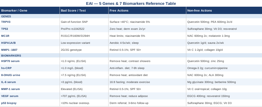
This article takes that deeper angle. The biomarker section identifies seven measurable indicators that reveal what is actually happening at the cellular and systemic level — from heat shock proteins and oxidative DNA damage markers to vascular remodeling signals and cancer-risk flags. The genetics section covers five genes whose variants shape heat sensitivity, pigmentation response, collagen integrity, and DNA repair. Neither section offers a cure. What both sections offer is the kind of specific, actionable information that generic advice never reaches — information that can point you toward a more targeted, and genuinely more useful, path forward.
Summary
This article goes well beyond the usual "stop using your laptop in bed" advice. In the biomarker section, you will find seven measurable indicators that reveal what is actually happening inside your skin — including a heat-stress protein that doubles as an early warning signal, an oxidative DNA damage marker you can test in urine, a tumor-risk flag detectable by biopsy, and a vascular marker that explains why some lesions deepen over time. Each biomarker comes with a practical improvement plan, including supplement options with dosing, cycling, and side-effect notes. In the genetics section, five genes are profiled — including one that controls how aggressively your skin reacts to infrared heat and another linked to collagen degradation speed. A condensed Huberman podcast summary covers the biology of heat stress proteins and how they can be leveraged therapeutically. The complementary section rounds things out with the three modalities that have the clearest evidence for supporting skin repair and reducing systemic inflammation in this context. If you have ever wondered why your skin reacted the way it did, or why it is not recovering as expected, the answers are likely in one of these sections.
The biomarkers section below is the core of this article — seven measurable signals, each telling a different part of the story.
7 Biomarkers That Reveal What Erythema Ab Igne Is Actually Doing to Your Skin
Biomarkers do not replace the clinical step of removing the heat source. But they reveal the biological cost of what happened and, critically, whether the damage is progressing silently. The seven markers below span different domains — stress response, inflammation, oxidative damage, structural degradation, vascular remodeling, and malignant potential — and together they give a far more complete picture than a visual skin exam alone.
Biomarker 1: Serum HSP70 (Heat Shock Protein 70)
Why it matters
HSP70 is the cell's primary damage-control protein. When tissue is exposed to heat, cells upregulate HSP70 to stabilize damaged proteins and prevent cell death. In acute, controlled heat exposure — such as sauna therapy — this response is transient and beneficial. In chronic low-grade infrared exposure, as in erythema ab igne, HSP70 remains persistently elevated in and around affected tissue, signaling that the cellular stress response has not resolved. Persistently elevated HSP70 also drives pro-inflammatory signaling through toll-like receptor pathways, which helps explain why inflammation persists long after heat exposure stops.
How to measure it
Serum HSP70 is measured via enzyme-linked immunosorbent assay (ELISA) through specialty labs. It is not yet part of standard panels, so you will need to request it specifically. Cost ranges from $80 to $150 through functional or integrative medicine labs. Reference ranges vary by lab; most set an upper normal around 0.5–1.0 ng/mL, though values in chronic skin injury can exceed this.
Research on HSP70 as a skin inflammation biomarker (PubMed)
If the score is elevated — plan without supplements
The most effective first intervention is complete and sustained removal of the triggering heat source — not reduction, complete elimination. Cold water immersion or contrast showers (alternating 2 minutes cold / 1 minute warm for 10–15 minutes, 3–4x/week) can help normalize HSP expression by providing a brief, controlled counterweight to chronic heat-induced activation. Moderate aerobic exercise (30 minutes, 4–5x/week) also modulates HSP expression toward a more adaptive, episodic pattern rather than chronic baseline elevation. Sleep optimization matters here: HSP70 expression follows circadian rhythms, and poor sleep prolongs stress protein elevation.
If the score is elevated — plan with supplements and equipment
Quercetin is one of the best-studied natural modulators of heat shock factor 1 (HSF1), the transcription factor controlling HSP70 expression. Dose: 500–1000 mg/day with a fat-containing meal (quercetin is fat-soluble). Cycle: 6 weeks on, 2 weeks off. Side effects are rare at this dose; mild digestive discomfort occasionally reported. Zinc supports cellular stress protein regulation and wound healing pathways: 15–25 mg/day with food, taken away from copper-containing foods. L-Glutamine (5 g/day in water, first thing in the morning) supports gut barrier integrity and modulates peripheral HSP expression. Use a sauna as a deliberate tool — brief, high-temperature sessions (15–20 minutes at 80–100°C) 2x/week create acute HSP pulses that paradoxically normalize the chronic low-level elevation pattern, a form of adaptive hormesis.
Biomarker 2: High-Sensitivity C-Reactive Protein (hs-CRP)
Why it matters
hs-CRP is the most accessible and affordable indicator of systemic low-grade inflammation. In erythema ab igne, chronic infrared radiation activates NF-κB signaling in keratinocytes, driving interleukin release that eventually elevates systemic CRP. While CRP is not specific to skin inflammation, it serves as a reliable proxy for the overall inflammatory burden the body is carrying. Persistently elevated hs-CRP (above 1.0 mg/L, and especially above 3.0 mg/L) has been associated by researchers including Peter Attia with accelerated tissue aging, impaired skin barrier function, and slower wound repair — all directly relevant to EAI recovery.
How to measure it
Standard blood draw. Widely available through any primary care physician or direct-to-consumer labs. Cost: $15–$40. Optimal target: below 0.5 mg/L. Values above 3.0 mg/L indicate systemic inflammation requiring intervention.
If the score is elevated — plan without supplements
Anti-inflammatory dietary shift is the foundation: eliminate ultra-processed foods, refined seed oils (soybean, canola, corn), and refined carbohydrates. Replace with extra-virgin olive oil, fatty fish 3x/week, abundant leafy greens and berries. Sleep 7–9 hours consistently (CRP rises measurably with both sleep restriction and fragmentation). Reduce chronic psychological stress through structured recovery time. Avoid repetitive heat source exposure.
If the score is elevated — plan with supplements and equipment
Omega-3 fatty acids (EPA + DHA, 2–3 g/day combined) reduce IL-6 and NF-κB signaling. Use a triglyceride-form fish oil for best absorption. Cycle: continuous use is supported for up to 12 months, then reassess. Side effects: mild fishy breath, loose stools at high doses. Curcumin with piperine (500 mg curcumin + 5 mg piperine, 2x/day): blocks NF-κB activation and directly reduces IL-6 production. Cycle: 8 weeks on, 3 weeks off. Side effects: mild GI discomfort; caution with blood thinners. Vitamin D3 with K2 (3000–5000 IU D3 with 100–200 mcg MK-7 K2, daily): low vitamin D is an independent driver of elevated CRP. Monitor serum 25(OH)D; target 50–80 ng/mL. Side effects at this dose are minimal if calcium intake is not excessive.
Biomarker 3: 8-Hydroxy-2'-deoxyguanosine (8-OHdG)
Why it matters
8-OHdG is a direct marker of oxidative DNA damage — it forms when reactive oxygen species (ROS) attack guanine bases in DNA. Infrared radiation generates ROS through mitochondrial disruption in skin cells, creating the same oxidative fingerprint seen in UV exposure. Elevated urinary 8-OHdG indicates that DNA repair systems are being overwhelmed, which in the context of EAI is particularly significant: this is the same mechanism that precedes p53 mutations and, in chronically heat-damaged skin, has been linked to squamous cell carcinoma development in case series and pathology reviews.
Research on 8-OHdG and skin oxidative damage (PubMed)
How to measure it
Urinary 8-OHdG via ELISA or LC-MS/MS; available through functional medicine labs (e.g., Genova Diagnostics). First-morning urine collection preferred. Cost: $80–$150. Results are typically normalized to creatinine. Optimal values: below 7.5 ng/mg creatinine for most reference ranges.
If the score is elevated — plan without supplements
Remove the heat source completely and immediately. Maximize dietary antioxidant intake: aim for 8–10 servings daily of colorful vegetables and fruits (blueberries, pomegranate, spinach, kale, red peppers). Reduce glycemic load in the diet, as hyperglycemia independently drives 8-OHdG production. Protect all affected skin from additional UV exposure — the already-compromised DNA repair capacity makes sun exposure significantly more damaging in EAI-affected areas.
If the score is elevated — plan with supplements and equipment
N-Acetylcysteine (NAC) is the most direct intervention for replenishing intracellular glutathione, the cell's primary antioxidant defense against ROS. Dose: 600 mg, 2x/day with food. Cycle: 8 weeks on, 2 weeks off. Side effects: occasional nausea, rare headache. Alpha-lipoic acid (ALA) regenerates vitamins C and E and directly quenches mitochondrial ROS. Dose: 300–600 mg R-ALA (the natural form)/day. Side effects: hypoglycemia risk in diabetics; take with food. Vitamin C (liposomal, 1–2 g/day) supports DNA repair and reduces urinary 8-OHdG directly in clinical studies. Astaxanthin (4–12 mg/day, from algae-based source): a carotenoid with particularly high antioxidant potency in skin tissue; shown to reduce UV and IR-induced oxidative markers. Cycle: continuous use supported up to 12 months.
Biomarker 4: Serum Interleukin-6 (IL-6)
Why it matters
IL-6 is a pleiotropic cytokine centrally involved in erythema ab igne's inflammatory mechanism. Heat stress and infrared-induced keratinocyte damage trigger IL-6 release, which amplifies melanogenesis signals, promotes angiogenesis through VEGF upregulation, and activates systemic acute-phase response. Chronically elevated IL-6 in EAI contributes to the persistence of hyperpigmentation, the degradation of dermal collagen, and the risk of keratinocyte dysregulation. Unlike CRP, IL-6 reflects a more upstream inflammatory signal and responds faster to interventions.
How to measure it
Serum IL-6 via electrochemiluminescence assay. Available through specialty labs; some primary care physicians will order it. Cost: $50–$100. Normal range: 0–7 pg/mL in most lab references. Values consistently above 3 pg/mL in the absence of active infection warrant investigation.
If the score is elevated — plan without supplements
Intermittent fasting (16:8 model, daily) consistently reduces circulating IL-6 in clinical studies. Time-restricted eating from 8 AM to 4 PM has shown stronger effects than later eating windows. Daily moderate-intensity exercise (not high-intensity, which can transiently spike IL-6) at 30–45 minutes reduces baseline IL-6 over 8–12 weeks. Reduce dietary saturated fat from ultra-processed sources while maintaining saturated fat from whole-food sources (eggs, dairy if tolerated).
If the score is elevated — plan with supplements and equipment
Magnesium glycinate (300–400 mg at night): magnesium deficiency is highly prevalent and independently drives IL-6 elevation. Side effects: loose stools at high doses. Berberine (500 mg, 2x/day with meals): activates AMPK, which directly suppresses IL-6 and NF-κB. Cycle: 8 weeks on, 4 weeks off. Side effects: GI discomfort, particularly in first two weeks; mild hypoglycemic effect in diabetics. Curcumin as noted above. Red and near-infrared light therapy devices (630–850 nm, 10–20 mW/cm², 10–20 minutes 3–4x/week) at sub-heating intensities have demonstrated IL-6 modulation in controlled skin trials — note that power density must be kept well below thermal threshold to avoid worsening EAI.
Biomarker 5: Matrix Metalloproteinase-1 (MMP-1)
Why it matters
MMP-1 is the primary collagenase enzyme responsible for degrading type I and III collagen. Both ultraviolet and infrared radiation upregulate MMP-1 expression in dermal fibroblasts. In erythema ab igne, chronically elevated MMP-1 explains the progressive collagen loss that contributes to skin thinning, loss of elasticity, and the persistence of the reticulate pattern. The MMP-1 promoter polymorphism (-1607 1G/2G) is a key genetic variable in this pathway and is covered in the genetics section below. Serum MMP-1 is a useful tracking tool for monitoring dermal structural response to treatment.
How to measure it
Serum MMP-1 via ELISA through specialty or research-oriented labs. Less commonly ordered than inflammatory markers; functional medicine practitioners are familiar with it. Cost: $80–$160. Reference ranges vary by sex and age; post-treatment comparison (baseline vs. 3 months) is more useful than a single absolute value.
If the score is elevated — plan without supplements
Topical retinol (0.5–1.0%) applied 3–5 nights per week is among the most evidence-backed interventions for reducing MMP-1 activity in photoaged and heat-damaged skin. Start at low concentration and build up over 6 weeks. Strict daily SPF 50+ sunscreen over affected areas prevents further MMP-1 induction from UV. An anti-glycation diet (reducing high-AGE processed foods) also slows collagen cross-linking and MMP-1 activation.
If the score is elevated — plan with supplements and equipment
Vitamin C (1–2 g/day oral + topical L-ascorbic acid 10–15%): directly stimulates collagen synthesis to counterbalance MMP-1 degradation and inhibits MMP-1 expression at the gene level. Marine collagen peptides (10 g/day, hydrolyzed type I/III): clinical trials have shown improved skin elasticity and collagen density with 8–12 weeks of use. Side effects: minimal; some report mild GI bloating initially. Copper (2 mg/day from copper glycinate): cofactor for lysyl oxidase, the enzyme that cross-links new collagen into stable fibers. Pair with zinc at a 15:1 zinc-to-copper ratio to avoid competition. Silica (from orthosilicic acid, 10 mg/day): supports collagen precursor pathways.
Biomarker 6: Vascular Endothelial Growth Factor (VEGF)
Why it matters
VEGF drives new blood vessel formation in damaged tissue. In erythema ab igne, chronic heat exposure upregulates VEGF expression in keratinocytes and fibroblasts, contributing to the vascular prominence that deepens the reticulate pattern and the reddish-brown discoloration. Elevated serum VEGF is also a concern because it promotes a tissue environment that may support keratinocyte dysplasia — one of the early steps in the progression toward squamous cell carcinoma in chronic EAI cases. Tracking VEGF gives indirect insight into the angiogenic state of affected tissue without requiring a biopsy.
How to measure it
Serum VEGF via ELISA. Available through specialty labs; cost $80–$130. Reference range typically 62–707 pg/mL in adults, though interpretation depends on clinical context. Trends over time are more meaningful than single readings.
If the score is elevated — plan without supplements
Complete and sustained heat source elimination is the most impactful intervention. Anti-inflammatory diet as described above reduces VEGF expression through NF-κB suppression. Reduce adipose tissue if overweight, as adipocytes are significant producers of VEGF. Avoid external factors that independently upregulate VEGF: smoking, excessive alcohol, poorly managed blood glucose.
If the score is elevated — plan with supplements and equipment
EGCG (Epigallocatechin gallate) from green tea extract is among the best-studied natural VEGF inhibitors. Dose: 400–800 mg standardized EGCG/day with food. Cycle: 8 weeks on, 2 weeks off to prevent tolerance. Side effects: nausea if taken on an empty stomach; rare liver-enzyme elevation at very high doses (above 800 mg/day). Resveratrol (150–300 mg/day, trans-resveratrol form): reduces VEGF expression and NF-κB activity. Cycle: 8 weeks on, 4 weeks off. Side effects: estrogen-mimetic activity at high doses is a consideration for hormone-sensitive conditions. Quercetin (as noted for HSP70): has additional VEGF-inhibiting properties through PI3K/mTOR pathway modulation.
Biomarker 7: p53 Expression (Biopsy-Based Assessment)
Why it matters
p53 is the most important tumor suppressor gene in the body. Its protein product regulates DNA repair, apoptosis of damaged cells, and prevention of malignant transformation. Both ultraviolet and infrared radiation generate the kind of oxidative DNA damage that produces p53 mutations. Published case series and pathology reviews have documented p53 mutations in erythema ab igne lesions — the same type of p53 alterations seen in UV-induced actinic keratosis and squamous cell carcinoma. This biomarker requires a skin biopsy of the affected area and is therefore reserved for cases with long-standing EAI, changing lesion appearance (ulceration, rapid darkening, induration, or new nodularity), or high cumulative heat exposure history.
Research on EAI, p53, and squamous cell carcinoma (PubMed)
How to measure it
Punch or shave biopsy of the affected area, processed with immunohistochemical staining for p53 protein overexpression (a surrogate for TP53 mutation). Performed by a dermatologist or dermatopathologist. Cost: $150–$400 depending on facility. Nuclear p53 overexpression in more than 10% of basal keratinocytes is considered abnormal and warrants closer follow-up.
If the score is elevated — plan without supplements
This is the one biomarker that demands immediate specialist referral rather than self-directed management. Dermatological monitoring every 3–6 months is the standard of care for confirmed p53 overexpression in EAI. All heat exposure to the affected area must stop permanently. Full sun protection over affected skin (SPF 50+, UPF 50+ clothing) is non-negotiable, since UV and infrared damage are additive on already-stressed p53 pathways.
If the score is elevated — plan with supplements and equipment
Sulforaphane from broccoli sprouts is the most robustly studied natural inducer of Nrf2, the transcription factor that activates a wide array of carcinogen-detoxification and DNA-repair enzymes. Dose: 30–60 mg/day of sulforaphane (or 50–100 g of fresh broccoli sprouts). Cycle: continuous use is well-tolerated in most people. Side effects: strong taste; mild GI discomfort in sensitive individuals. EGCG (as above): modulates p53-related apoptotic signaling. Vitamin D3 (with K2): supports DNA repair capacity and keratinocyte differentiation. Target serum 25(OH)D of 60–80 ng/mL. All supplements here are supportive measures — they do not replace dermatological monitoring and must never be used as an excuse to delay biopsy or specialist follow-up.
With these seven biomarkers mapped, the logical next layer is genetic — understanding which inherited variants made you more susceptible in the first place and how to compensate for them.
The Genetic Layer: 5 Genes That Shape Your Erythema Ab Igne Risk and Recovery
Genetic testing for the variants below is available through platforms like 23andMe, AncestryDNA, or clinical-grade panels through functional medicine practitioners. Raw genetic data can be uploaded to tools like Genetic Genie, SelfDecode, or Stratagene for interpretation. None of these findings are diagnostic — they are probabilistic. A single variant does not determine outcome; it shifts probability. The value is in knowing where your individual biology is most stressed, so compensatory strategies can be targeted rather than generic.
Gene 1: TRPV3 — The Heat-Sensing Channel in Your Keratinocytes
What it does
Transient receptor potential vanilloid channel 3 (TRPV3) is a temperature-activated ion channel expressed predominantly in skin keratinocytes. It is activated at warm temperatures (31–39°C), placing it squarely in the range of chronic heat exposure relevant to EAI. Upon activation, TRPV3 triggers downstream melanogenesis signaling through prostaglandin E2 and α-MSH pathways, which is one of the primary mechanisms behind heat-induced hyperpigmentation.
Research on TRPV3 and heat-induced melanogenesis (PubMed)
Variants that matter
Gain-of-function variants in TRPV3 (including several rs SNPs identified in genome-wide studies) lower the activation threshold — meaning the channel fires at lower temperatures than normal. Individuals with these variants may develop EAI-type pigmentation changes at heat exposures that leave others unaffected.
If the gene variant is unfavorable — plan without supplements
Lower your personal heat threshold significantly below what you think is necessary. The activation window of a sensitized TRPV3 channel starts well below skin-reddening temperature. Use laptop cooling pads, never place heat sources directly against skin, and check surface temperatures with an infrared thermometer — keeping skin-contact surfaces below 40°C. Topical niacinamide (5%) applied to historically exposed areas reduces prostaglandin-mediated pigmentation downstream of TRPV3 activation.
If the gene variant is unfavorable — plan with supplements and equipment
Quercetin has demonstrated TRPV channel-modulating properties in in vitro research. Dose: 500 mg/day with fat. Palmitoylethanolamide (PEA) at 600 mg twice daily has shown activity in downregulating TRPV channel sensitivity in inflammatory contexts. Cycle: 12 weeks on, 4 weeks off. Side effects: generally very well tolerated. An infrared thermometer for monitoring skin-contact surfaces ($20–$40) is a low-cost, high-value tool for TRPV3-sensitive individuals.
Gene 2: TP53 — Your Tumor Suppressor Efficiency
What it does
The TP53 gene encodes p53, the protein discussed in the biomarker section above. Several common polymorphisms affect p53's efficiency as a tumor suppressor. The Pro72Arg variant (rs1042522) is the most studied: the Arg/Arg genotype is associated with faster apoptosis of damaged cells (protective), while the Pro/Pro genotype is associated with slower response and potentially higher risk of DNA damage accumulation in chronically stressed tissue. This is directly relevant to EAI because infrared-driven DNA damage in skin is the same type of damage p53 is tasked with resolving.
If the gene variant is unfavorable — plan without supplements
Zero tolerance for ongoing heat exposure to affected areas. Bi-annual dermatological exams with dermatoscopy of affected areas. Aggressive sun protection — a Pro/Pro TP53 individual has both compromised UV-response and compromised IR-response capacity, making combined exposures cumulative risk. Avoid other known p53 stressors: smoking, alcohol, and extensive UV tanning.
If the gene variant is unfavorable — plan with supplements and equipment
Sulforaphane (30–60 mg/day from sprouts or supplement, continuous) activates Nrf2-dependent DNA repair independently of p53, providing a compensatory protective layer. Vitamin D3 (target serum 60–80 ng/mL) supports keratinocyte differentiation and helps maintain DNA repair gene expression. Resveratrol (150–300 mg/day) activates SIRT1, which modulates p53 deacetylation and DNA damage response. Cycle: 8 weeks on, 4 weeks off to avoid tolerance. Side effects: mild estrogen activity at high doses.
Gene 3: MC1R — The Pigmentation Wildcard
What it does
The Melanocortin 1 Receptor (MC1R) gene controls the type of melanin produced in response to UV and infrared signals. The eumelanin pathway (active MC1R) produces photoprotective dark pigment. Loss-of-function MC1R variants — notably R151C, R160W, and D294H — shift production toward pheomelanin, a less photoprotective pigment that also generates more ROS upon radiation exposure. Individuals with these variants tend to have red or auburn hair, fair skin, and high freckling density. In the context of EAI, impaired MC1R function means both more oxidative stress per unit of heat exposure and less effective barrier through pigmentation.
If the gene variant is unfavorable — plan without supplements
MC1R variant carriers have lower baseline antioxidant capacity in skin and need stricter behavioral precautions: lower heat thresholds (similar to TRPV3 guidance), year-round sun protection, and immediate dermatological evaluation of any changing EAI lesion. Topical niacinamide (5%) daily reduces melanin transfer between melanocytes and keratinocytes, improving cosmetic outcome even when MC1R is dysfunctional.
If the gene variant is unfavorable — plan with supplements and equipment
N-Acetylcysteine (NAC) at 600 mg 2x/day replenishes glutathione depleted by pheomelanin-derived ROS. Melatonin (1–3 mg at night): beyond its sleep role, melatonin is a potent antioxidant within skin cells and is particularly relevant for MC1R variant carriers who have reduced melanocyte-level antioxidant defense. Side effects: morning grogginess if dose is too high; start at 0.5 mg and titrate. Vitamin E (mixed tocopherols) at 200 IU/day provides lipid-phase antioxidant coverage in the melanocyte membrane layer.
Gene 4: HSPA1A / HSPA1B — Your HSP70 Production Capacity
What it does
HSPA1A and HSPA1B are the two genes encoding the inducible HSP70 isoforms (HSP70.1 and HSP70.2). Polymorphisms in the promoter and coding regions of these genes affect how quickly and robustly HSP70 is induced in response to heat stress. Reduced induction capacity means cells are slower to protect themselves against heat damage — they accumulate more protein misfolding and oxidative injury before the stress response catches up. This directly modulates how much damage accumulates in EAI-affected tissue and how quickly it resolves.
If the gene variant is unfavorable — plan without supplements
Regular moderate-intensity exercise is the most powerful lifestyle intervention for upregulating basal HSP70 expression — even in individuals with low-expression polymorphisms, aerobic exercise (4–5x/week, 30–40 minutes at 65–75% max heart rate) significantly increases baseline HSP70 mRNA levels over 8–12 weeks. Prioritize consistent sleep (the HSP70 circadian peak occurs during deep sleep).
If the gene variant is unfavorable — plan with supplements and equipment
Quercetin (500–1000 mg/day, cyclically) directly activates HSF1 to compensate for reduced constitutive HSP70 expression. L-Glutamine (5–10 g/day) is a cellular energy substrate that supports HSP70 induction under stress. Deliberate sauna use (80–100°C, 15 minutes, 2x/week) creates brief, high-intensity HSP70 induction pulses that compensate for chronically blunted baseline expression — a hormetic approach that only applies after all pathological heat sources have been eliminated.
Gene 5: MMP1 Promoter Polymorphism — Collagen Degradation Speed
What it does
The MMP1 gene contains a well-characterized promoter polymorphism at position -1607 (1G/2G). The 2G/2G genotype creates a binding site for the transcription factor Ets that significantly increases MMP-1 expression in response to inflammatory and radiation-based triggers. In practical terms, individuals with 2G/2G produce more MMP-1 per unit of infrared exposure — faster collagen degradation, deeper reticulate pattern, slower structural recovery.
If the gene variant is unfavorable — plan without supplements
Topical retinol (0.5%–1.0%, 4 nights per week) is the most evidence-supported intervention for reducing MMP-1 activity and stimulating compensatory collagen synthesis in photoaged skin — directly applicable to heat-damaged skin. Begin cautiously: use a pea-sized amount with a buffer moisturizer and increase concentration over 8 weeks. Strict daily SPF 50+ application — UV exposure is the most potent trigger of MMP-1 in 2G/2G carriers.
If the gene variant is unfavorable — plan with supplements and equipment
Vitamin C (1–2 g oral/day plus topical L-ascorbic acid 10–20%): both forms suppress MMP-1 gene expression and stimulate collagen synthesis. Marine collagen peptides (10 g/day) provide the amino acid substrate for accelerated collagen remodeling — particularly important when MMP-1 is degrading faster than baseline. Astaxanthin (4–12 mg/day): reduces MMP-1 upregulation from oxidative triggers and provides skin structural protection. A professional radiofrequency or microneedling treatment (performed by a dermatologist after EAI is no longer active) can mechanically stimulate collagen remodeling in MMP-1-depleted tissue — effective for reticulate scarring that has not resolved after 12+ months.
These five genes form the biological context behind individual susceptibility. Understanding the heat biology more deeply — including how to use heat therapeutically rather than incidentally — is the subject of the next section.
What the Huberman Lab Episode on Deliberate Heat Exposure Reveals About Erythema Ab Igne Biology
The Huberman Lab episode titled Using Deliberate Heat Exposure to Improve Health and Performance is not about erythema ab igne specifically, but it is one of the most rigorous public-facing breakdowns of heat stress biology available — and it provides the mechanistic framework that explains both why EAI does what it does and how heat can be redirected therapeutically. These are the ten most impactful things covered, reframed through the lens of EAI management.
1. The Dose Makes the Poison With Heat
Huberman distinguishes clearly between therapeutic acute heat stress (short, high-intensity, controlled) and pathological chronic heat exposure (prolonged, low-grade, repetitive). EAI results from the second type. The damage in EAI is not from high temperature per se but from the cumulative infrared load below the burn threshold — making it easy to underestimate. People continue harming themselves because it never hurts in the moment.
2. HSP70 Is the Central Protagonist
The episode details how heat shock proteins — particularly HSP70 — are the cell's primary defense against heat-induced protein misfolding. Brief sauna sessions spike HSP70 acutely and allow it to return to baseline. Chronic low-grade heat from laptops and heaters maintains HSP70 in a state of chronic low-level elevation that eventually overwhelms the system, reduces its responsiveness, and shifts it from protective to inflammatory.
3. Sauna Can Be Part of Recovery — Done Correctly
One of the counterintuitive points in the episode: after pathological heat exposure stops, brief, high-temperature deliberate heat exposure (sauna, properly managed) can restore adaptive HSP expression that chronic, moderate heat exposure had dysregulated. This only applies when the original heat source is gone and skin is no longer actively inflamed. Timing matters.
4. Cardiovascular Physiology Is Affected by Heat Load
Regular heat exposure at therapeutic levels improves vascular compliance and reduces resting heart rate through repeated VEGF upregulation and plasma volume expansion. This context matters for EAI: the VEGF elevation seen in EAI-affected tissue is a dysregulated version of a normally beneficial vascular signal. Understanding this distinction helps explain why VEGF-modulating supplements work for EAI without suppressing beneficial vascular adaptation in healthy tissue.
5. Growth Hormone Is Acutely Released by Heat
The episode covers how acute sauna sessions at 80–100°C trigger 2–16x spikes in growth hormone (GH), which supports tissue repair and skin collagen synthesis. This is another argument for redirecting heat exposure — from chronic low-grade (damaging) to deliberate acute (therapeutic) — as part of EAI recovery once the lesions have stabilized.
6. Timing of Heat Exposure Matters for Circadian Biology
Evening heat exposure (from heating pads, electric blankets, laptops) disrupts body temperature circadian descent, interfering with deep sleep architecture. Since HSP70 expression peaks during deep sleep, this creates a double burden for EAI patients: the same evening heat habits that cause skin damage also impair the nighttime repair cycle that could be healing it.
7. Infrared vs. Steam — Different Physiological Penetration
Huberman discusses the difference between dry heat, steam, and infrared sauna. Infrared specifically penetrates several centimeters into subcutaneous tissue — much deeper than surface warming. This directly explains EAI's deeper dermal involvement compared to surface heat burns. Infrared exposure from near-IR-emitting devices (including older laptops and incandescent heating elements) penetrates to the dermal level where fibroblasts, VEGF signals, and collagen reside.
8. The Role of Deliberate Cold Exposure in Calibrating Heat Response
Cold exposure (cold showers, cold-water immersion) is discussed as a powerful counterpart to heat that normalizes thermal signaling pathways. For EAI patients with dysregulated HSP70, deliberate cold exposure 3–4x/week can help recalibrate thermal stress response, reduce baseline inflammation, and improve norepinephrine-mediated skin barrier tone. Contrast protocol: 2 minutes cold / 1 minute warm, 3–4 rounds.
9. Oxidative Stress Is a Transient Feature of Therapeutic Heat, Not a Chronic One
Brief heat stress produces a controlled ROS spike that activates antioxidant defense enzymes — a hormetic benefit. Chronic low-grade heat keeps oxidative pathways persistently activated without triggering the full antioxidant compensatory response. This explains why 8-OHdG remains elevated in EAI patients long after a single exposure: the oxidative damage accumulated gradually without triggering an adequate adaptive response.
10. Mindset and Tolerance Are as Important as Intensity
One of Huberman's practical points is about building tolerance to heat discomfort by staying in deliberate heat environments when the urge to leave is strongest. Translated to EAI: patients who habitually tolerate uncomfortable heat exposures — without registering the cumulative toll because it never burns — are the highest-risk group. Developing deliberate awareness of skin temperature and implementing environmental changes before discomfort appears is the most underused preventive tool.
Complementary Approaches With Evidence Relevant to Erythema Ab Igne
The following three modalities have meaningful human clinical evidence relevant to EAI's core pathological features — not necessarily to EAI as a named diagnosis (the condition is too rare to be the subject of dedicated randomized trials), but to the underlying mechanisms it shares with other better-studied inflammatory and skin conditions.
Low-Level Laser Therapy / Photobiomodulation
Photobiomodulation (PBM) uses non-thermal light energy — typically at 630–850 nm — to modulate cellular metabolism in skin tissue without generating heat. It is relevant to EAI primarily through two mechanisms: first, it stimulates mitochondrial cytochrome c oxidase, increasing ATP production and cellular repair capacity in heat-damaged keratinocytes and fibroblasts; second, clinical studies have documented significant improvement in post-inflammatory hyperpigmentation (PIH) with PBM, which shares the same melanogenesis pathway that EAI activates.
A systematic review published in PubMed-indexed literature on PBM and skin pigmentation disorders has documented significant improvement in post-inflammatory hyperpigmentation using 830 nm infrared-wavelength PBM, 2–3 sessions per week, over 8–12 weeks. Review of PBM and hyperpigmentation evidence (PubMed)
For practical EAI application: at-home PBM devices (660 nm + 850 nm panels) used at non-heating power densities (10–30 mW/cm²) for 10–20 minutes, 3–4x/week over affected areas. Begin only after all thermal exposure has been eliminated and any active inflammation has resolved. Power density must remain well below the threshold that generates skin surface heat — use devices with confirmed non-thermal output ratings. Full results typically require 12–16 weeks of consistent use.
Mindfulness Meditation / MBSR
Mindfulness-based stress reduction (MBSR) is an 8-week structured program developed by Jon Kabat-Zinn that combines body scanning, sitting meditation, and gentle movement. Its relevance to EAI is systemic rather than local: multiple controlled trials have demonstrated that MBSR reduces serum IL-6 and CRP in healthy adults and in populations with inflammatory skin conditions. Given that chronic inflammation is central to EAI persistence and hyperpigmentation depth, any intervention that durably reduces systemic inflammatory markers supports faster skin resolution.
A landmark randomized controlled trial by Creswell et al. (2016) published in Brain, Behavior, and Immunity found that MBSR training in stressed adults produced significant reductions in IL-6 and loneliness-driven inflammatory signaling compared to an active control. Research on MBSR and inflammatory cytokines (PubMed)
For practical application: commit to the full 8-week MBSR program (free versions are available through the UMass Medical School online resources). The body scan practice is particularly relevant for EAI: it builds the habit of noticing skin temperature, heat discomfort, and early signs of heat stress before they become cumulative. For maintenance after the 8-week program, 20 minutes of daily meditation is sufficient to sustain inflammatory reductions observed in trials. Evidence for EAI specifically is indirect; the anti-inflammatory benefit is the mechanism.
Microbiome-Directed Therapies
The gut-skin axis is a well-documented bidirectional communication system — dysbiosis in the gut microbiome can amplify systemic inflammatory signaling, including IL-6, TNF-α, and CRP, through increased intestinal permeability and lipopolysaccharide (LPS) translocation into the bloodstream. This systemic inflammatory amplification worsens skin conditions with inflammatory components, including EAI. Probiotic and prebiotic interventions that restore microbiome balance and reduce intestinal permeability can therefore reduce the systemic inflammatory background that keeps EAI lesions from resolving.
Clinical trials in atopic dermatitis, psoriasis, and acne have consistently shown that Lactobacillus rhamnosus and Bifidobacterium longum supplementation reduces serum IL-6, improves skin barrier integrity markers, and reduces inflammatory cytokine production in affected skin. Research on probiotics and skin inflammation via gut-skin axis (PubMed)
For practical EAI application: begin with a dietary foundation — daily fermented foods (100–150 g kefir, sauerkraut, or kimchi) plus 25–30 g of prebiotic fiber (leeks, garlic, onions, green banana flour, cooked-and-cooled starchy foods). Add a targeted probiotic containing Lactobacillus rhamnosus GG and Bifidobacterium longum BB536 (at least 10 billion CFU/day, taken with food). Run a 12-week trial and monitor hs-CRP and IL-6 at weeks 0 and 12. Side effects: initial bloating in the first 2–3 weeks is common; start with half dose and increase. Evidence is indirect for EAI specifically; the anti-inflammatory mechanism is the rationale.
Conclusion
Erythema ab igne is not simply a cosmetic inconvenience to be managed by moving your laptop. It is a skin injury driven by measurable biological processes — heat stress protein dysregulation, oxidative DNA damage, inflammatory cascades, collagen degradation, and in some cases, early mutations in the same tumor suppressor pathways implicated in UV-induced skin cancers. The biomarkers in this article give you concrete, measurable targets to understand what is happening in your specific case. The genetic layer explains why your individual biology responded the way it did. And the complementary strategies offer the most evidence-supported routes toward reducing systemic inflammation, supporting skin repair, and preventing progression.
The smartest next step is not necessarily to order every test at once. Start with what is most accessible and most relevant to your situation: hs-CRP and 8-OHdG are the most affordable entry points. If you have long-standing EAI, changing lesions, or a history of significant cumulative heat exposure, a dermatologist visit for evaluation — and potentially a biopsy for p53 status — is worth prioritizing before any other intervention. Work with a physician who is comfortable ordering functional biomarkers if your primary care provider is not. The information is available; it just requires knowing what to ask for.
Skin: Inflammatory Skin Conditions Pigmentation Conditions
Autoimmune: Inflammatory Conditions
Cancer & Oncology: Skin Cancer