This article contains AI generated content.
· UpdatedErythema Nodosum — 4 Genes and 6 Biomarkers to Track
Introduction
Erythema nodosum doesn't arrive quietly. One morning you notice warm, tender nodules on your shins — sometimes on the forearms or elsewhere — that are deep red, painful to press, and alarming enough to prompt a doctor visit. The answer you usually receive is medically accurate: it's an inflammatory panniculitis, it should self-resolve in four to eight weeks, rest and ibuprofen are the main tools. That answer is correct as far as it goes.
Where it falls short is for the roughly 30–40% of people who experience recurrences, and for those whose erythema nodosum is a visible expression of something running deeper — Crohn's disease, sarcoidosis, a persistent streptococcal focus, or an immune system that over-reacts to triggers that wouldn't trouble someone else. For these people, the "wait and rest" approach doesn't address the driver, and the nodules return.
The emerging science around erythema nodosum is quietly useful here. Specific inflammatory biomarkers can tell you how active the process is, which cytokine pathways are most involved, and whether an organ system beyond the skin needs attention. Genetic variants — particularly in immune recognition and cytokine regulation genes — can explain individual susceptibility and predict flare severity. Neither angle is a guarantee, but together they give you something more useful than a waiting game: a framework for tracking what's actually happening and acting on it precisely.
This article focuses on two complementary approaches. The primary one covers six key biomarkers — including inflammatory markers, a cytokine panel, and an organ-specific enzyme — along with what to do when each is out of range, with or without supplementation. The second covers four genetic variants consistently found in EN research and what each means in practical terms. Further in, you'll find a deep look at the gut-immune connection that most EN discussions ignore entirely, three complementary approaches with real clinical evidence, and an anti-inflammatory protocol specifically designed for autoimmune-adjacent conditions. Better data leads to sharper questions, and sharper questions lead to care that is actually useful.
What This Article Covers — At a Glance
Here's a quick map before the deep content. The main focus is on six biomarkers: high-sensitivity CRP, ESR, TNF-alpha, IL-6, ferritin, and ACE. Measured as a set, these give a multi-angle view of the inflammatory activity behind EN — including which cytokine pathways are dominant, whether sarcoidosis is likely, and whether subclinical inflammation is persisting between flares. For each marker, you'll find a target range, how to measure it and what it costs, a lifestyle-based protocol for improving it, and a supplementation protocol with specific doses, cycling schedules, and side effect considerations.
The second major section covers four genetic variants: HLA-DRB1*03 and HLA-B8 (relevant especially in sarcoidosis-associated EN), the TNF-alpha promoter polymorphism -308G>A, NOD2/CARD15 mutations (critical when Crohn's disease is a possibility), and IL1B gene variants. Beyond genetics and biomarkers, the article also covers what one of the most evidence-backed gut-immune frameworks says about EN-class inflammatory conditions, three complementary modalities with meaningful human clinical data, and a structured nutritional protocol for autoimmune-pattern inflammation. The goal throughout is precision over reassurance.
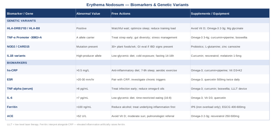
Six Biomarkers to Track When You Have Erythema Nodosum
Biomarkers don't diagnose erythema nodosum — that's clinical. But they do something arguably more useful: they help explain why you have it, how active the underlying process is, and whether any intervention (lifestyle, supplement, medication) is working. The six markers below were selected because they are measurable at standard labs, clinically meaningful for the conditions most commonly underlying EN, and genuinely actionable — meaning an abnormal result points to something you can do about it.
1. High-Sensitivity C-Reactive Protein (hs-CRP)
Why it matters
C-reactive protein is produced by the liver in response to inflammatory cytokine signals — primarily IL-6 — and rises dramatically during active EN flares, often reaching 50–150 mg/L or higher when a bacterial infection (such as streptococcal pharyngitis) is involved. The high-sensitivity version of the test (hs-CRP) is more valuable for monitoring purposes because it detects low-level systemic inflammation between flares — the kind that may not produce symptoms but signals ongoing immune activation.
Peter Attia, whose longevity medicine practice treats hs-CRP as a front-line inflammatory biomarker, advocates for a target below 0.5 mg/L as optimal — significantly stricter than the conventional clinical cut-off of 3 mg/L. For someone with recurrent EN, tracking hs-CRP quarterly can reveal whether the underlying process is truly resolved or simply below the threshold of clinical presentation. A persistently elevated hs-CRP between episodes is a meaningful signal that the trigger has not been addressed.
How to measure it
hs-CRP is available at virtually any clinical or private lab (Quest, LabCorp, and most national services). Cost ranges from $15–$40 out-of-pocket. It is often included in cardiovascular risk panels or can be ordered individually. Fasting is not required, but consistent testing conditions (same time of day, similar fasting state) improve comparability over time.
If the score is elevated, the plan without supplements
The priority is identifying and treating the underlying EN trigger — no lifestyle modification substitutes for this. That said, several non-supplemental strategies demonstrably reduce hs-CRP independently:
- Eliminate refined sugars and ultra-processed foods: adipose tissue produces IL-6 in response to hyperinsulinemia, which directly drives CRP. Even moderate reductions in dietary sugar intake reduce hs-CRP within 4–6 weeks. - Mediterranean-style diet with emphasis on oily fish: 2–3 servings per week of sardines, mackerel, or wild salmon provides dietary EPA and DHA, which compete with arachidonic acid pathways. - Sleep optimization: even one night of poor sleep elevates CRP. A consistent 7–9 hours, with regular sleep and wake timing, is anti-inflammatory in a direct, measurable sense. - Moderate aerobic exercise: 150 minutes per week of moderate activity reduces systemic inflammation. During active EN flares, avoid intense training over the affected limbs — it can worsen local inflammation — but walking and light activity are generally tolerated. - Treat upstream causes actively: throat culture when pharyngitis is present; gastroenterology referral if IBD symptoms coexist; chest imaging if sarcoidosis is in the differential.
If the score is elevated, the plan with supplements or equipment
Once lifestyle foundations are addressed, the following have human clinical evidence for reducing hs-CRP:
- Omega-3 (EPA + DHA combined): 2–4g daily. Take with a fat-containing meal for absorption. Safe long-term; may mildly prolong bleeding time — note before any surgery or invasive procedure. No cycling required; continuous use is standard. - Curcumin standardized extract with piperine: 500–1000 mg twice daily. Piperine (black pepper extract) increases curcumin bioavailability by approximately 20-fold. Cycle 6–8 weeks on, 2 weeks off. Potential interaction with anticoagulants (warfarin, aspirin therapy) — consult a physician if relevant. - Vitamin D3 with K2: if serum 25-OH-D is below 40 ng/mL, supplementing to a 40–60 ng/mL range reduces systemic inflammatory markers. Standard dose: 2000–5000 IU D3 combined with 100–200 mcg MK-7 form K2 daily. Important exception: if sarcoidosis is suspected or confirmed, avoid supplemental vitamin D without physician guidance (explained in the ACE section below). - Magnesium glycinate: 200–400 mg before bed. Magnesium deficiency independently elevates inflammatory markers; the glycinate form has the best GI tolerability. Continuous use is appropriate; monitor for loose stools at higher doses.
2. Erythrocyte Sedimentation Rate (ESR)
Why it matters
The ESR — the rate at which red blood cells settle in a standardized tube — is a classic, nonspecific marker of systemic inflammation. Unlike CRP, which rises and falls quickly (within 24–48 hours), ESR changes more slowly over days. This makes it a useful complement to CRP for understanding the trajectory of an EN episode rather than its peak intensity. ESR above 40–50 mm/hr during an active flare is common, and tracking it monthly during and after an episode helps establish whether the process is genuinely resolving.
ESR is also traditionally used to monitor conditions commonly underlying EN, including sarcoidosis and IBD. An ESR that remains elevated well after the skin nodules have resolved is a flag worth investigating.
How to measure it
ESR is among the least expensive inflammatory tests available: $10–$25 at most labs. Normal values are approximately <15 mm/hr for men under 50, <20 mm/hr for men over 50, <20 mm/hr for women under 50, and <30 mm/hr for women over 50. ESR rises with age, in pregnancy, and with anemia or polycythemia — interpret results in context rather than in isolation.
If the score is elevated, the plan without supplements
Because ESR is nonspecific, an elevated result primarily calls for investigation. Key steps:
- Pair ESR with hs-CRP to understand the pattern. A high CRP alongside high ESR suggests acute, active inflammation. A persistently elevated ESR with normal CRP points toward different processes — early connective tissue disease, mild anemia, or early sarcoidosis — worth further evaluation. - Investigate for chronic infections relevant to the clinical picture: tuberculosis and brucellosis in appropriate exposure contexts, persistent streptococcal carriage if EN has recurred. - All anti-inflammatory lifestyle strategies that reduce CRP — dietary refinement, sleep quality, exercise — also reduce ESR over a longer time horizon of 6–12 weeks.
If the score is elevated, the plan with supplements or equipment
The complete anti-inflammatory supplement stack described in the CRP section (omega-3, curcumin, vitamin D3/K2, magnesium) also reduces ESR over time. One addition specific to this context:
- Quercetin: 500 mg twice daily has anti-inflammatory properties through inhibition of histamine release, NF-κB signaling, and mast cell activation. Cycle 4–6 weeks on, 2 weeks off. Generally well-tolerated; potential interaction with some antibiotics (fluoroquinolones) and immunosuppressants.
3. TNF-Alpha (Tumor Necrosis Factor-Alpha)
Why it matters
TNF-alpha sits at the center of EN pathogenesis. Biopsy studies of EN lesions consistently show elevated TNF-alpha production by macrophages within the inflamed fat lobules. TNF-alpha drives the neutrophil recruitment cascade that produces the tissue destruction characteristic of panniculitis. This is not merely academic — it is the mechanistic reason why anti-TNF biologics (infliximab, adalimumab), prescribed for IBD, reliably resolve EN in those patients. Treating the TNF-alpha signal treats the skin.
Serum TNF-alpha is not routinely ordered in standard EN workups, but it is available at specialty labs and provides a more specific picture of the dominant cytokine environment. Measuring it during a flare and again during remission helps characterize your personal inflammatory pattern and may inform treatment decisions, particularly if recurrences are frequent.
How to measure it
Serum TNF-alpha is available at Quest Diagnostics, LabCorp, and similar national services. Cost: $80–$200 out-of-pocket. Reference range is typically undetectable to <8 pg/mL at baseline, though this varies by lab and assay. Request it proactively — many physicians are not accustomed to ordering it in the EN context, but it is a legitimate clinical test.
If the score is elevated, the plan without supplements
Since TNF-alpha is produced by activated macrophages responding to an upstream trigger, the most effective non-supplement approach is eliminating that trigger:
- Treat infectious triggers early and completely: streptococcal pharyngitis is among the most common EN precipitants. Aggressive early antibiotic treatment — and a post-treatment throat culture to confirm clearance — can prevent downstream TNF-alpha escalation. - Reduce dietary TNF-alpha drivers: trans fats, excess omega-6 polyunsaturated oils (corn, soybean, sunflower), and alcohol all stimulate macrophage TNF-alpha production. Replacing vegetable oils with olive oil, avocado oil, and butter is a practical step. - Cold exposure: short-duration cold immersion has been shown to acutely reduce TNF-alpha in human studies. Begin with 30–60 seconds of cold at the end of a shower and gradually work up to 2–3 minutes. Not a substitute for treating the underlying cause, but a useful complementary tool with a good safety profile in otherwise healthy individuals. - Sleep quality: monocyte TNF-alpha production is specifically upregulated by sleep deprivation. This is a significant, non-obvious connection worth taking seriously.
If the score is elevated, the plan with supplements or equipment
Several natural compounds have documented TNF-alpha-reducing properties in human studies:
- Omega-3 EPA+DHA (3–4g daily): EPA competes with arachidonic acid for the same enzymatic pathways, reducing the substrate available for TNF-alpha-driving eicosanoid synthesis. Continuous use; same notes on bleeding time as above. - Curcumin with piperine: inhibits NF-κB, the primary transcription factor for TNF-alpha gene expression. 500–1000 mg twice daily. Cycle 6–8 weeks on, 2 off. (See CRP section for interaction notes.) - Boswellia serrata (AKBA-standardized extract): 300–500 mg daily. Specifically inhibits 5-lipoxygenase, reducing leukotriene production and TNF-alpha secretion. Cycle 8–12 weeks on, 2–4 weeks off. Well-tolerated; mild GI effects (nausea, loose stools) in some individuals. - Low-level laser therapy (photobiomodulation): devices operating at 630–850 nm wavelengths have anti-inflammatory effects via mitochondrial cytochrome c oxidase activation and documented TNF-alpha reduction in several human trials. Consumer-grade devices suitable for home use range from $200–$1000. Apply over surrounding tissue (not directly over active, broken lesions). Typically used in sessions of 5–20 minutes, 3–5 times per week during a flare.
4. Interleukin-6 (IL-6)
Why it matters
IL-6 is upstream of CRP — the liver synthesizes CRP in direct response to IL-6 signals from activated immune cells. Measuring IL-6 directly gives an earlier and more sensitive view of immune activation than CRP alone, and it adds mechanistic specificity. In EN, IL-6 is elevated both locally within the skin lesions and systemically in circulation. Chronically elevated IL-6 also drives fatigue, anemia of chronic disease, and muscle catabolism — phenomena that EN patients often experience as a persistent tiredness and weakness that outlasts the visible skin findings.
How to measure it
Serum IL-6 is available at most major labs. Cost: $50–$150. Reference range is typically <7 pg/mL, though thresholds vary by assay. As with TNF-alpha, serial measurement — at flare onset, peak, and during established remission — is more informative than a single snapshot.
If the score is elevated, the plan without supplements
- Restrict high-glycemic and high-sugar foods: adipose tissue is a major IL-6 source, and hyperinsulinemia amplifies adipose IL-6 secretion. Even moderate carbohydrate restriction demonstrably reduces IL-6 within 3–6 weeks. - Reduce visceral fat: visceral adiposity is the strongest dietary-modifiable driver of resting IL-6. Caloric restriction combined with aerobic exercise reduces visceral fat and IL-6 in parallel over 12–16 weeks. - Increase aerobic fitness: chronically trained individuals mount lower resting IL-6 responses to immune challenges. This is a medium-term strategy requiring consistent weekly investment. - Time-restricted eating: a 14–16 hour daily fasting window suppresses NLRP3 inflammasome activity, which is one upstream activator of IL-6. Start with a 12-hour overnight fast and gradually extend.
If the score is elevated, the plan with supplements or equipment
- Omega-3 EPA+DHA: reduces IL-6 secretion from adipose and immune cells. Same protocol as above. - Vitamin D3: vitamin D receptors on immune cells, when adequately activated, suppress IL-6 transcription. Target serum 25-OH-D of 40–60 ng/mL. (Sarcoidosis exception applies — see ACE section.) - Quercetin 500 mg twice daily: documented IL-6-lowering effects in randomized trials. Cycle 4–6 weeks. (See ESR section for interaction notes.) - Intermittent fasting (16:8 protocol): not technically a supplement, but it functions as one of the most mechanistically direct levers on IL-6. A 2022 randomized controlled trial in overweight adults showed significant IL-6 reduction after 8 weeks of 16:8 time-restricted eating independent of caloric restriction.
5. Serum Ferritin (as an Inflammatory Signal)
Why it matters
Most people encounter ferritin as a proxy for iron stores — which it is. But ferritin is also an acute phase reactant: the liver upregulates ferritin production during inflammation, regardless of actual iron status. This creates a clinically important trap: ferritin can appear falsely normal or even elevated during an EN flare because inflammation is driving it up, masking what might actually be iron-deficient erythropoiesis underneath. Conversely, an EN patient who is also iron-depleted may have ferritin held artificially higher than true iron stores would suggest.
The clinically useful practice is always measuring ferritin alongside CRP. When CRP is elevated, ferritin is not a reliable iron-status marker until inflammation resolves. Very high ferritin (above 500 ng/mL) alongside high CRP can indicate severe systemic inflammation warranting additional investigation for conditions that overlap with EN clinically, including adult-onset Still's disease.
How to measure it
Ferritin is one of the most cost-accessible lab markers: $20–$40 out-of-pocket, often included in iron panels or metabolic screens. The laboratory reference range is intentionally broad (typically 12–300 ng/mL for women, 12–400 ng/mL for men). A functional target more commonly used in root-cause clinical practice is 50–100 ng/mL — high enough to support immune function and energy, low enough that it isn't tracking active inflammation.
If the score is elevated in an inflammatory context, the plan without supplements
When ferritin is elevated alongside high CRP, the target is the inflammation, not the ferritin. The ferritin will normalize as inflammation resolves. Additional strategies:
- Reduce alcohol intake: alcohol directly stimulates hepatic ferritin production and is independently a recognized EN trigger. Eliminating alcohol during and after an EN episode has dual benefit. - Blood donation (only for confirmed iron overload, not inflammation-driven elevation): when ferritin is persistently high in the context of normal inflammatory markers — suggesting true iron excess — regular blood donation is the safest non-pharmacological approach to reducing iron stores. This should not be applied during acute inflammatory states.
If the score is elevated, the plan with supplements or equipment
- IP6 (inositol hexaphosphate) 1–2g daily on an empty stomach: has iron-chelating properties studied in hereditary hemochromatosis. Appropriate only when ferritin elevation is confirmed to reflect iron overload rather than inflammation. Cycle 8–12 weeks; monitor ferritin and full iron panel. - EGCG (green tea extract) 400–600 mg daily: mild iron-chelating activity; take separately from iron-containing meals. Long-term high-dose use carries a theoretical liver enzyme elevation risk — limit to 12-week cycles with a 4-week break and check ALT if using beyond 3 months.
6. Angiotensin-Converting Enzyme (ACE)
Why it matters
ACE deserves its own marker slot because it directly addresses one of the most important and commonly missed EN scenarios: sarcoidosis. Sarcoid granulomas produce ACE in excess, and serum ACE is elevated in approximately 60–70% of patients with active pulmonary sarcoidosis. EN combined with bilateral hilar lymphadenopathy (seen on chest X-ray) and a raised ACE defines Löfgren syndrome — a clinically distinct, often favorable variant of sarcoidosis. Knowing this matters enormously because Löfgren syndrome tends to resolve spontaneously, especially in HLA-DRB1*03 carriers, and overly aggressive treatment is not only unnecessary but counterproductive.
If your EN has recurred more than once with no obvious infectious trigger identified, an ACE level and a chest X-ray together represent a low-cost, high-yield investigation.
How to measure it
Serum ACE is available at most standard labs. Cost: $30–$60. Normal range is typically 8–52 U/L, though this varies by assay and age. Levels above 70–80 U/L, particularly in the right clinical context, are highly suspicious. ACE can also be elevated in other granulomatous diseases — tuberculosis, histoplasmosis, berylliosis — so a raised result is a prompt for further evaluation, not a standalone diagnosis.
If the score is elevated, the plan without supplements
An elevated ACE in the context of EN warrants physician-led evaluation for sarcoidosis. The workup typically includes chest CT, spirometry, and sometimes bronchoscopy. If sarcoidosis is confirmed:
- Strongly avoid vitamin D supplementation: this is the most important contraindication to get right. Sarcoid granulomas express 1-alpha-hydroxylase and convert vitamin D to its active 1,25-dihydroxy form independently of renal regulation, creating a risk of hypercalcemia and nephrocalcinosis if supplemental D3 is added. This is one of the few situations where a commonly recommended supplement can cause direct harm. - Moderate sun exposure: for the same reason. Particularly important in summer months or in people who spend significant time outdoors. - Work with a pulmonologist for any pulmonary involvement. Löfgren syndrome in HLA-DRB1*03 carriers often requires only monitoring, but other sarcoid patterns may need corticosteroid or immunosuppressant management.
If the score is elevated, the plan with supplements or equipment
In confirmed sarcoidosis-associated EN, supplementation should be conservative:
- Omega-3 2–3g EPA+DHA daily: anti-inflammatory; no interaction with sarcoid-specific pathophysiology. Safe for continuous use. - Resveratrol 250–500 mg daily: small pilot studies in sarcoidosis suggest anti-inflammatory and immunomodulatory effects, particularly in granuloma regulation. Cycle 8–12 weeks. Well-tolerated; theoretical mild estrogen-agonist activity is worth noting in hormone-sensitive individuals. Some anticoagulant interaction potential. - Avoid aggressive fat-soluble vitamin supplementation in general (vitamins A, D, K in high doses) pending full diagnostic clarification — several fat-soluble nutrients interact with sarcoid metabolism in ways that require physician guidance.
With the inflammatory biomarker picture established, the natural next question is why some people's immune systems produce this pattern in the first place. That's where genetics enters the conversation.
What Your DNA May Reveal About Erythema Nodosum Risk
Not everyone exposed to a streptococcal infection develops erythema nodosum. Not everyone with Crohn's disease develops skin nodules. Genetic variation doesn't explain all of this difference, but in EN it explains a meaningful portion of who gets it, how severely, and how often. The four variants below appear consistently in EN-relevant research — each has practical implications beyond being a data point on a genetic report.
Gene 1: HLA-DRB1*03 and HLA-B8 — Predicting EN Course in Sarcoidosis
What this gene does
The HLA region encodes proteins that present foreign antigens to T cells — the critical interface between innate pattern recognition and adaptive immune responses. The 8.1 ancestral haplotype, which includes HLA-B8 and HLA-DRB1*03, is associated with a range of autoimmune and inflammatory conditions, but in the EN context its most important role is as a predictor of Löfgren syndrome course. Research by Grunewald, Eklund and colleagues demonstrated that patients with Löfgren syndrome who carry HLA-DRB1*03 have significantly higher rates of spontaneous complete remission compared to non-carriers — in some analyses, over 90% of carriers achieved remission within 2 years without immunosuppressive treatment.
This makes HLA-DRB1*03 one of the genuinely useful genetic markers in EN: it can shift a physician's approach from aggressive treatment to watchful monitoring in the sarcoidosis context.
You can assess HLA status through direct-to-consumer genetic testing (23andMe includes some HLA data) or through clinical HLA typing ordered by a physician (more accurate, typical cost $100–$400 through immunology or transplant labs).
If the gene is relevant, the plan without supplements
If you carry HLA-DRB1*03 and have sarcoidosis-associated EN:
- Favor watchful waiting over aggressive immunosuppression in discussion with your physician. The prognosis is favorable, and the risks of unnecessary corticosteroid treatment (metabolic effects, bone density loss, immune suppression) often outweigh the benefit in this subgroup. - Protect sleep and reduce physiological stressors: intense exercise training and chronic psychological stress can extend Löfgren syndrome activity. During active phases, reduce training load and prioritize recovery. - Monitor rather than treat with serial ACE levels, chest imaging, and inflammatory markers. Knowing your trajectory over 3–6 months often makes the management decision clear.
If the gene is relevant, the plan with supplements or equipment
- Omega-3 2–3g daily: supports anti-inflammatory resolution without vitamin D concerns. - Magnesium glycinate 300–400 mg nightly: safe in sarcoidosis, supports sleep quality and immune regulation. - Avoid vitamin D supplementation without physician-confirmed 25-OH-D levels and guidance — the sarcoidosis-vitamin D interaction detailed in the ACE section is most relevant here.
Gene 2: TNF-Alpha Promoter Polymorphism (-308G>A)
What this gene does
The TNFA gene promoter contains a well-studied single nucleotide polymorphism at position -308. Carriers of the A allele (as opposed to the more common G allele) show higher transcriptional activity from the TNF-alpha promoter — meaning their macrophages produce more TNF-alpha in response to the same inflammatory stimulus. This isn't a disease-causing mutation; it's a variation in immunological reactivity. The A allele is present in roughly 20–30% of individuals of European ancestry.
In the EN context, carrying the -308A allele may translate to more severe and longer-lasting flares in response to the same triggers that produce milder episodes in GG homozygotes. It may also explain recurrence — a persistently sensitized immune system maintaining low-level TNF-alpha output in the absence of obvious clinical relapse.
If the gene is unfavorable, the plan without supplements
Carriers of the -308A variant should focus especially on avoiding or rapidly treating known TNF-alpha-activating triggers:
- Early, complete antibiotic treatment of streptococcal infections: given that strep is among the top EN triggers, and that -308A carriers mount amplified TNF-alpha responses, early treatment and confirmed clearance are particularly important in this genotype. Don't wait to see if pharyngitis resolves spontaneously. - Gut integrity: the gut-immune axis drives systemic TNF-alpha production. Dietary diversity, regular fermented food intake, and minimizing gut-disruptive exposures (chronic NSAID use, alcohol, ultra-processed food) are especially relevant for this genotype. - Stress management: psychological stress activates the HPA axis and specifically increases monocyte TNF-alpha production. Mindfulness-based stress reduction (MBSR) has documented anti-inflammatory effects in human trials. - Consider oral contraceptive alternatives if relevant: estrogen-containing oral contraceptives are a recognized EN trigger. In -308A carriers with a history of recurrent EN, discussing non-estrogen-containing contraceptive options with a physician is reasonable.
If the gene is unfavorable, the plan with supplements or equipment
- Omega-3 3–4g EPA+DHA daily: EPA competes with arachidonic acid for the substrate of TNF-alpha-driving eicosanoid production. The higher end of the dosing range is appropriate for this genotype. - Curcumin with piperine: directly inhibits NF-κB transcription factor activity, suppressing TNF-alpha gene expression at the level of the promoter. 500–1000 mg twice daily, cycled 6–8 weeks on, 2 off. - Boswellia serrata (AKBA-standardized) 300–500 mg daily: 5-lipoxygenase inhibition, reduces downstream TNF-alpha secretion. 8–12 weeks on, 2–4 weeks off. - Quercetin 500 mg twice daily: additional NF-κB modulation. Stacks well with curcumin; generally well-tolerated.
Gene 3: NOD2/CARD15 Mutations
What this gene does
NOD2 (also known as CARD15) is an intracellular pattern recognition receptor that detects bacterial cell wall-derived components — specifically muramyl dipeptide. Loss-of-function mutations in NOD2 are among the strongest known genetic risk factors for Crohn's disease; the three most common variants (R702W, G908R, and Leu1007fsinsC) are found at significantly elevated frequencies in Crohn's patients compared to controls. This is directly relevant to EN because EN affects 2–10% of Crohn's patients and strongly correlates with gut disease activity. EN in Crohn's is not an independent skin disease — it is a manifestation of the bowel inflammation, and it typically improves when the Crohn's is treated.
If you have had more than one EN episode without a clear infection trigger, and if any gastrointestinal symptoms are present (irregular bowel habits, cramps, unexplained weight loss, blood in stool), NOD2 mutation status provides context that — combined with clinical features — should prompt a formal GI evaluation. NOD2 variants can be identified through clinical genetic IBD panels or through consumer SNP arrays (23andMe includes some of these variants).
If the gene is unfavorable, the plan without supplements
NOD2 mutations primarily compromise bacterial sensing and gut barrier integrity. The most effective non-supplement strategies:
- Maximize dietary microbiome diversity: people with NOD2 variants tend to have less diverse gut microbiomes. Targeting 30+ different plant foods per week (the threshold associated with microbiome diversity in the American Gut Project data) is the most practical population-level intervention. - Add regular fermented foods: kefir, kimchi, sauerkraut, miso, and natural yogurt all increase microbiome diversity and reduce inflammatory markers in human trials. - Remove gut permeability triggers: chronic NSAID use is a particularly important one — it directly damages the intestinal barrier and is ironically the most common medication prescribed for EN pain. Discuss alternatives (acetaminophen, rest, compression) with your physician. - Pursue GI evaluation if indicated: in the presence of NOD2 variants and GI symptoms, colonoscopy with biopsies is the appropriate next step. EN in Crohn's responds to treating the Crohn's.
If the gene is unfavorable, the plan with supplements or equipment
- Multi-strain probiotics: evidence in Crohn's specifically is mixed, but certain strains (VSL#3, Lactobacillus rhamnosus GG) show benefits for gut barrier function and may reduce systemic inflammatory burden. Take daily with food; safe long-term in the absence of severe immunosuppression. - L-glutamine 5–10g daily: the primary fuel for enterocytes; supports tight junction integrity and gut barrier repair. Well-tolerated; can be taken continuously. - Zinc carnosine 75 mg twice daily: specific gut barrier-supporting properties demonstrated in several human trials. Cycle 8–12 weeks. Take separate from high-phytate foods. - Prebiotic fiber (inulin, FOS, or a diverse plant-based diet): feeds short-chain fatty acid-producing bacteria (Bifidobacterium, Akkermansia-supporting species) that are often depleted in NOD2 mutation carriers. 10–20g daily from food or supplemental sources; introduce gradually to avoid gas and bloating.
Gene 4: IL1B (Interleukin-1 Beta) Gene Variants
What this gene does
IL-1β is one of the primary alarm signals of the innate immune system — it amplifies local inflammation, drives fever, recruits neutrophils, and initiates further cytokine cascades. Several common variants in the IL1B gene (particularly rs16944 and rs1143634) are associated with higher or lower baseline IL-1β production in macrophages. High-producer variants translate to more intense inflammatory responses to both infectious stimuli and sterile triggers.
In EN, IL-1β is produced within the inflamed fat lobules and contributes to the transition from early macrophage activation to full panniculitis. The specific contribution of IL1B variants to EN has not been characterized in large dedicated cohorts, but mechanistic data from related inflammatory skin conditions and from IBD genetics consistently implicate high IL-1β output as a disease severity modifier.
If the gene is unfavorable, the plan without supplements
- Low-glycemic, whole-food diet: glucose spikes specifically activate the NLRP3 inflammasome, which is the primary processing complex for IL-1β maturation. A low-glycemic, low-processed-food diet directly reduces NLRP3-driven IL-1β output. - Cold exposure: cold thermogenesis suppresses NLRP3 inflammasome activity in both animal and human models. Start with 30–60 seconds of cold at the end of a shower, gradually working to 2–3 minutes of cold immersion several times per week. - Time-restricted eating (14–16 hours daily): fasting suppresses NLRP3 activity through ketone body production (specifically beta-hydroxybutyrate, a direct NLRP3 inhibitor). Start with a 12-hour overnight fast and extend progressively.
If the gene is unfavorable, the plan with supplements or equipment
- Curcumin (500–1000 mg twice daily): directly inhibits NLRP3 inflammasome assembly and IL-1β processing. Same protocol as above. - Omega-3 EPA+DHA: EPA reduces NLRP3 activity through competing fatty acid pathways. Same protocol. - Resveratrol 250–500 mg daily: inhibits NLRP3 inflammasome in multiple human cell and some in vivo studies. Cycle 8–12 weeks. Mild estrogen-agonist activity warrants noting in hormone-sensitive individuals. - Melatonin 1–5 mg before bed: beyond sleep regulation, melatonin has documented NLRP3 inflammasome-suppressing effects. Start at 0.5–1 mg and titrate slowly. Side effects include vivid dreams and morning grogginess at doses above 3 mg. Take 30–60 minutes before intended sleep time.
Understanding your genetic profile adds depth to the biomarker picture. But there is a third angle — one that most EN management plans don't address — that connects both layers: the role of the gut and its microbial ecosystem in driving the immune reactivity that makes both biomarker elevations and genetic vulnerabilities express themselves as skin disease.
The Gut-Immune Connection: What This Framework Changes About EN
Emeran Mayer, MD, a gastroenterologist and neuroscientist at UCLA, has spent decades documenting the bidirectional relationship between gut health and systemic immune function. His 2021 book The Gut-Immune Connection synthesizes decades of research on how the gut microbiome regulates immune responses far beyond the intestine — including skin, joints, and lungs. The framework is particularly relevant for erythema nodosum because EN is so frequently triggered by gut-origin conditions and because the gut-skin immune axis is increasingly well-documented in the scientific literature.
The following ten insights from this body of work are among the most practically important for anyone trying to understand why their immune system is producing EN and what can be changed:
1. Most of the Immune System Lives in the Gut
Approximately 70–80% of immune cells reside in gut-associated lymphoid tissue (GALT). The state of the gut microbiome directly determines the activation threshold and reactivity of these cells — which then circulate throughout the body. A dysbiotic gut creates a primed immune system more likely to mount disproportionate inflammatory responses to triggers like streptococcal infections or minor gut barrier breaches.
2. The Gut-Skin Axis Is Bidirectional and Real
Skin inflammatory conditions are increasingly documented to correlate with gut microbiome composition. This isn't metaphorical — shared cytokine pathways (TNF-alpha, IL-6, IL-1β) connect gut and skin immune activation. Treating the gut is not an alternative to treating the skin; it's upstream intervention.
3. Dietary Diversity Is the Most Powerful Microbiome Lever
The American Gut Project found that individuals eating 30 or more different plant foods per week had significantly greater microbiome diversity than those eating fewer than 10. Microbiome diversity correlates inversely with systemic inflammatory marker levels. This single number — plant food variety — is one of the most evidence-backed dietary targets for immune health.
4. Fermented Foods Reduce Systemic Inflammation in Human Trials
A 2021 Stanford randomized controlled trial (published in Cell) showed that a high-fermented-food diet increased microbiome diversity and reduced 19 inflammatory proteins, including IL-6, over 10 weeks. The effect was independent of caloric intake and was larger than a matched high-fiber intervention alone. Daily fermented food intake (yogurt, kefir, kimchi, sauerkraut) is not a folk remedy — it is one of the more rigorously supported nutritional interventions for systemic inflammation.
5. Ultra-Processed Food Specifically Damages Gut Barrier Integrity
Emulsifiers, thickeners, and ultra-processed food ingredients used in industrial food production disrupt the intestinal mucus layer and increase gut permeability in ways that natural food ingredients do not. Lipopolysaccharide (LPS) leakage from a permeable gut directly activates TLR4 receptors on macrophages, which upregulates TNF-alpha production — the same cytokine pathway central to EN.
6. Chronic NSAID Use Is a Double-Edged Problem in EN
NSAIDs are the most common prescription for EN symptom management, yet chronic NSAID use damages the small intestinal barrier, increases gut permeability, and can worsen gut dysbiosis. In people with Crohn's disease or NOD2 mutations, NSAIDs may trigger IBD flares. Using the lowest effective dose for the shortest necessary duration — and exploring acetaminophen or compression for pain management — is a more gut-aware strategy.
7. Stress Directly Compromises Gut Integrity
The brain-gut axis is not metaphorical. Psychological stress activates corticotropin-releasing factor (CRF) in the gut wall, increasing intestinal permeability within hours. Chronic stress therefore maintains a state of mild gut permeability and LPS leakage, sustaining a low-grade inflammatory signal. This may explain why stress is a known EN trigger even in the absence of a detectable infection — the mechanism runs through the gut, not just the HPA axis alone.
8. Early Life Gut Microbiome Programming Matters
Antibiotic exposure in infancy, birth mode, and early dietary patterns durably influence microbiome composition and immune regulatory capacity. While this is not directly modifiable in adulthood, it contextualizes why some people's immune systems seem fundamentally more reactive — and why restorative gut strategies require sustained effort rather than quick fixes.
9. Specific Bacterial Species Regulate TNF-Alpha and IL-6 Output
Faecalibacterium prausnitzii and Akkermansia muciniphila are among the most intensively studied gut bacteria for their anti-inflammatory properties. Both produce short-chain fatty acids that inhibit NF-κB activity in gut-associated immune cells, directly reducing TNF-alpha and IL-6 output. F. prausnitzii is consistently reduced in active IBD. Feeding these bacteria — through prebiotic fiber, polyphenol-rich foods, and fermented food intake — is a practical intervention with emerging evidence.
10. Psychological Interventions Reduce Gut and Systemic Inflammation
Mind-body interventions — including mindfulness-based stress reduction, cognitive behavioral therapy for GI conditions, and gut-directed hypnotherapy — have measurable effects on gut permeability, inflammatory cytokine levels, and symptom burden in multiple randomized trials. This is not about positive thinking; it's about interrupting the neurological signaling pathway that keeps the gut barrier compromised under psychological stress.
Complementary Approaches with Meaningful Evidence
Three complementary modalities stand out as particularly relevant to erythema nodosum based on their mechanisms and their clinical evidence base. None of these replaces identifying and treating the underlying EN driver, but each addresses a documented pathway that the standard NSAID-and-rest approach leaves untouched.
Mindfulness-Based Stress Reduction (MBSR)
Psychological stress is a well-documented EN trigger, and the mechanism — HPA axis activation, gut permeability increase, and upregulation of TNF-alpha production in monocytes — gives this observation clinical teeth. MBSR is a structured 8-week program developed by Jon Kabat-Zinn at the University of Massachusetts, combining mindfulness meditation, body scan practice, and yoga, typically in 2.5-hour weekly sessions plus daily home practice.
A 2014 randomized controlled trial by Creswell et al. demonstrated that MBSR reduced circulating IL-6 and NF-κB activity in lonely older adults — a population with chronically elevated inflammatory markers. Multiple subsequent meta-analyses confirmed reductions in CRP, IL-6, and cortisol in MBSR-trained participants compared to controls. The effects are moderate but reproducible.
For practical application in EN: formally structured MBSR programs are available online and through hospitals (search for MBSR programs through the UMass Center for Mindfulness). The evidence supports daily practice of 20–45 minutes as the minimum effective dose. For EN patients who identify stress as a clear flare trigger, beginning an MBSR course during or immediately after an episode is well-timed — the flare provides motivational context, and 8 weeks of practice creates a neurological and inflammatory foundation before the next potential trigger appears.
Microbiome-Directed Therapies
Given the strong association between EN and gut-origin conditions — Crohn's disease, ulcerative colitis, celiac disease, and gut infections — microbiome-directed interventions have direct mechanistic relevance. The gut microbiome modulates systemic cytokine production (TNF-alpha, IL-6, IL-1β) through multiple pathways, including short-chain fatty acid production, LPS barrier containment, and direct immune cell training in gut-associated lymphoid tissue.
A 2021 Stanford randomized trial published in Cell by Sonnenburg and colleagues showed that a 10-week high-fermented food diet significantly increased microbiome diversity and reduced 19 immune proteins including IL-6 in healthy adults. This provides direct evidence that dietary microbiome intervention can alter the same cytokine environment dysregulated in EN. In IBD patients specifically, several controlled trials have shown that microbiome-modulating interventions (dietary fiber, specific probiotic strains including VSL#3, and fecal microbiota transplant in certain UC contexts) reduce disease activity and associated extraintestinal manifestations.
For practical application: begin with the most evidence-supported dietary changes — 30+ plant varieties per week, daily fermented food intake (2–3 servings), and elimination of gut-disruptive foods (emulsifiers, industrial seed oils, excess alcohol). Add a multi-strain probiotic (look for at least 4–5 species, 10–50 billion CFU) taken consistently with a meal. FMT remains an investigational therapy outside of C. difficile infection and is not recommended for EN management without specialist oversight, but the underlying science supporting it reinforces the gut-EN connection.
The Autoimmune Protocol (AIP) by Sarah Ballantyne
Erythema nodosum, particularly in the context of recurrence or association with autoimmune or inflammatory conditions, fits within the category of immune-mediated inflammatory disease for which the Autoimmune Protocol (AIP) was specifically designed. Developed by Dr. Sarah Ballantyne (also known as The Paleo Mom), a PhD research scientist and autoimmunity researcher, AIP is a structured nutritional and lifestyle intervention built on an elimination phase followed by careful reintroduction. It is more restrictive than a standard Mediterranean or anti-inflammatory diet — removing grains, legumes, dairy, eggs, nightshades, nuts, seeds, and alcohol — with the stated purpose of removing potential gut irritants and antigenic food triggers while emphasizing nutrient density.
Human clinical evidence for AIP exists in the IBD and autoimmune thyroid literature. A 2017 open-label trial published in Inflammatory Bowel Diseases (Konijeti et al.) showed that AIP resulted in clinical remission in 73% of IBD patients who completed the 11-week protocol, with significant reductions in inflammatory markers including CRP and calprotectin. For EN, the relevance is direct in the IBD-associated subgroup; in idiopathic or autoimmune-adjacent EN, the mechanisms (gut permeability reduction, inflammatory trigger elimination, microbiome improvement) are plausible even if direct EN-specific trial data does not exist.
For practical application: the AIP elimination phase is typically 60–90 days, followed by a systematic reintroduction phase that identifies individual triggers. Ballantyne's book The Paleo Approach provides the full protocol with scientific rationale. The most clinically supported implementation includes not just dietary changes but also the lifestyle components Ballantyne emphasizes: sleep of 8–9 hours, circadian rhythm alignment, stress management, and moderate movement. Beginning AIP during a resolved EN episode (not during an active flare with significant pain and mobility limitation) is more practical. Working with a registered dietitian familiar with elimination diets significantly improves adherence and reintroduction accuracy.
Conclusion
Erythema nodosum is rarely just about the skin. Behind the nodules — whether they arrive once or repeatedly — there is an immune system responding to something: an infection, a gut-origin inflammatory process, a genetic predisposition to cytokine over-reaction, or a combination of these. Waiting for each episode to resolve while leaving the underlying driver unexamined is not a strategy; it's a hope.
The six biomarkers covered here — hs-CRP, ESR, TNF-alpha, IL-6, ferritin, and ACE — give you and your physician a view of what's actually active and which systems to investigate further. The four genetic variants — HLA-DRB1*03, TNF-alpha -308G>A, NOD2/CARD15, and IL1B — explain a meaningful part of why your immune system reacts the way it does. Neither layer is a diagnosis by itself, but together they replace guesswork with direction.
The clearest next step: request an hs-CRP and ESR at your next visit, and ask whether an ACE level is appropriate given your history. If gut symptoms are present at all, raise the possibility of IBD evaluation and NOD2 testing with your physician. And regardless of what the labs show, the gut-immune connection and the lifestyle foundations covered here — dietary quality, microbiome support, sleep, stress management — reduce the inflammatory substrate that makes EN flares possible. That's the kind of progress that doesn't depend on a single answer.
Digestive: Intestinal Conditions
Skin: Inflammatory Skin Conditions
Autoimmune: Inflammatory Conditions
Infectious: Bacterial Infections