This article contains AI generated content.
· UpdatedErythropoietic Protoporphyria - 6 Biomarkers And 3 Genes To Track
Introduction
Living with erythropoietic protoporphyria means navigating a world designed around sunlight. A few minutes near a window, a walk to the car at noon, a afternoon drive with the sun angled through the glass — any of these can trigger burning pain that lasts for hours and leaves you exhausted long after the exposure ends. People around you rarely understand. The condition is real, measurable, and mechanistically understood, yet so poorly recognized that many patients spend years before getting the right diagnosis, and even longer before getting management that genuinely helps.
What makes EPP particularly difficult is that it varies significantly from one person to the next. The same diagnosis can mean one person who tolerates brief outdoor exposure with careful timing, and another who cannot sit near a window without pain. That difference is not arbitrary — it reflects which genetic variant is present, how efficiently the liver handles protoporphyrin excretion, whether iron stores are optimal, and several other measurable variables that standard "avoid the sun" advice does not address.
Generic guidance from a generalist clinician often ends at photoprotection and beta-carotene. But the research of the past two decades has opened more precise doors. Genetic profiling can explain why disease severity varies between patients with the same diagnosis. Biomarker tracking can monitor variables that predict complications before they become emergencies — particularly the liver complications that represent the most serious long-term risk in EPP.
This article takes a more targeted approach. The main section covers the six most useful biomarkers to track in EPP: what each one reveals, how to measure it affordably, and what you can do when a number trends in the wrong direction. A genetics section follows, covering three key genes and how their variants shape disease expression and management. Additional sections explore what emerging light biology research suggests for EPP management, and which complementary modalities have genuine clinical backing for this condition. Better information enables better conversations with your specialist — and, over time, better outcomes.
Summary
This article maps the six most actionable biomarkers for EPP monitoring — including free erythrocyte protoporphyrin, liver enzymes, ferritin, vitamin D, complete blood count, and fecal protoporphyrin — with specific measurement protocols, cost ranges, and evidence-based response plans for both supplement and non-supplement approaches. It then profiles the three genetic drivers of EPP severity (FECH, ALAS2, and the FECH hypomorphic allele), explaining what each variant does to heme biosynthesis and how to build a practical compensation plan around your specific genotype.
Beyond the core diagnostics, the article covers ten insights from recent light biology research that challenge standard EPP management thinking — including why glass windows are not the safe haven most patients assume, how afamelanotide works at a mechanistic level, and the overlooked role of circadian rhythm in heme synthesis regulation. Three complementary modalities with meaningful clinical evidence close the article: mindfulness-based stress reduction for pain and sun-exposure anxiety, microbiome-directed strategies for reducing hepatic protoporphyrin load, and breathing-based techniques for acute episode management.
If you are living with EPP or supporting someone who does, the goal here is to give you sharper tools — not promises, but actionable knowledge.
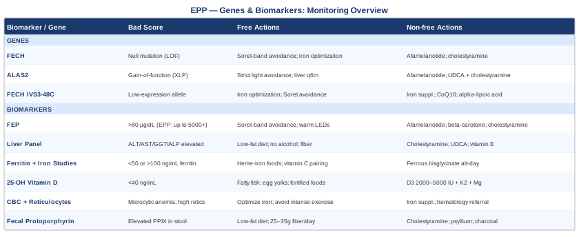
6 Biomarkers to Track in Erythropoietic Protoporphyria
Biomarker tracking in EPP serves two distinct purposes. The first is monitoring disease activity — particularly free erythrocyte protoporphyrin, which reflects how much PPIX is accumulating in red blood cells and determines both photosensitivity severity and hepatic risk. The second is monitoring the downstream consequences of the disease and its management — iron status, vitamin D, liver function — because EPP creates secondary nutritional and metabolic vulnerabilities that are avoidable with early detection. The six markers below represent the most informative, most accessible, and most actionable panel for this condition.
1. Free Erythrocyte Protoporphyrin (FEP)
Why it matters and what it reveals
Free erythrocyte protoporphyrin is the signature biomarker of EPP and the number that most directly connects to the photosensitivity you experience. In healthy red blood cell precursors, ferrochelatase (the FECH enzyme) inserts iron into protoporphyrin IX to form heme. When FECH activity is reduced — which is the defining defect in EPP — protoporphyrin IX accumulates in red blood cells in its metal-free form. This distinction is critical: free PPIX, unlike zinc protoporphyrin (which predominates in iron deficiency anemia), absorbs light maximally at approximately 400–410nm, in the violet-blue visible range known as the Soret band. When skin absorbs that wavelength, photoactivated PPIX generates singlet oxygen and reactive oxygen species that destroy cell membranes and nerve endings — producing the burning, stinging, and edema patients experience.
Elevated FEP also predicts hepatic risk. Because protoporphyrin is not water-soluble, it cannot be renally excreted and must transit through the liver into bile. When circulating FEP is very high, the liver receives a proportionally high PPIX load — which can crystallize in bile canaliculi and hepatocytes, forming the basis of EPP-associated liver disease. FEP is therefore not only a measure of symptom burden but an early warning indicator for the most serious long-term complication in EPP.
Normal FEP is typically below 70–80 µg/dL. In EPP, values can range from 300 µg/dL to well above 5,000 µg/dL in severe cases. Periodically published case series suggest that patients with consistently very high FEP — particularly above 2,000–3,000 µg/dL — warrant more aggressive hepatic monitoring.
How to measure it
FEP is measured from whole blood. The critical detail is to request fluorescence-based fractionation that distinguishes free (metal-free) protoporphyrin from zinc protoporphyrin — not all labs default to this fractionation, and without it you cannot confirm the EPP diagnosis or monitor correctly. Specialty labs in the US such as ARUP Laboratories and Mayo Clinic Laboratories run porphyrin panels with fractionation. Cost ranges from approximately $80–$250 depending on fractionation type and insurance. With a confirmed EPP diagnosis, insurance typically covers this test.
Testing frequency: every 6–12 months for stable patients. If FEP trends upward across two consecutive tests, or if it has been elevated above 2,000 µg/dL on any test, hepatology consultation is warranted.
If the score is bad, the plan without supplements
The core non-pharmacological lever is reducing erythropoietic drive and PPIX exposure simultaneously. Behavioral strategies that reduce circulating PPIX load include: strict avoidance of the Soret band wavelengths, not just UV. This means regular glass windows are not safe — glass blocks most UV (UVA and UVB) but transmits violet and blue visible light, which is the range that activates PPIX. Window films rated for Soret-band blocking (some automotive films qualify) meaningfully reduce indoor exposure near windows. Tight-woven, physically opaque fabrics are more protective than UV-blocking-rated textiles alone, for the same reason. Time outdoor activity before 9am or after 6pm during summer months in northern climates, when the Soret-band solar irradiance is lowest. Avoid fluorescent and halogen lighting in workspaces — LED lighting with a warm (2700–3000K) color temperature is safer because it emits far less in the 400–410nm range.
Maintaining iron stores in the mid-normal range (see biomarker 3) is also important because iron deficiency independently raises free PPIX production.
If the score is bad, the plan with supplements or equipment
Afamelanotide (Scenesse) — 16mg subcutaneous implant every 60 days: This is the most effective pharmacological intervention for managing high FEP-related photosensitivity. It is a synthetic alpha-melanocyte-stimulating hormone analogue that stimulates melanocyte production of eumelanin — the broadband photoprotective pigment — across a spectrum that includes the Soret band. Clinical trials (including the pivotal EU and US trials that led to approval) showed patients gained a median of 69 additional minutes of direct sun exposure per day without symptoms during peak treatment periods. It does not lower FEP itself but reduces the clinical consequences of PPIX photoactivation. FDA approved in the US in 2019 for EPP. Side effects: nausea in the first days after implant, generalized skin darkening, implant-site reactions, occasional fatigue. Administered by a physician; cycling is every 60 days during high-sun-exposure months or year-round depending on geography and severity.
Beta-carotene (120–180mg/day for adults): Historically the first-line option before afamelanotide became available, beta-carotene was theorized to quench singlet oxygen generated by photoactivated PPIX. Controlled trial evidence is mixed — some patients report meaningful benefit while others see none. It is still used by patients who responded to it before afamelanotide was available, and by those without access to afamelanotide. Effect builds over 4–6 weeks of consistent use. Side effect: skin yellowing (carotenemia), which is cosmetically significant but not dangerous. Continuous use; no standard cycling protocol.
Cholestyramine (4g twice daily before meals): A bile acid sequestrant that interrupts the entero-hepatic recirculation of protoporphyrin by binding PPIX in the gut before reabsorption. This gradually reduces systemic PPIX burden and is one of the most direct non-transplant interventions for managing hepatic protoporphyrin load. Side effects: constipation and malabsorption of fat-soluble vitamins (A, D, E, K) — supplement these if using long-term. Activated charcoal (25–50g per day) is an over-the-counter alternative with a similar mechanism and more accessible cost, though with weaker evidence; it must be taken well separated from meals and medications to avoid interfering with nutrient and drug absorption.
2. Liver Function Panel (ALT, AST, GGT, ALP, Bilirubin)
Why it matters and what it reveals
EPP-associated liver disease is the most serious complication of EPP, affecting approximately 2–5% of patients over their lifetime. However, subclinical liver enzyme elevation is more common, appearing in a broader subset of patients with high sustained PPIX loads. Protoporphyrin IX is excreted almost exclusively via the liver into bile — it cannot be cleared renally. When hepatic PPIX concentrations are high, crystalline deposits form in bile canaliculi and hepatocytes, triggering cholestasis, inflammation, and over time, fibrosis and cirrhosis. The danger is that progression can be clinically silent until advanced stages.
Tracking all five liver markers rather than relying on a single enzyme provides meaningfully more information. ALT is most hepatocyte-specific and sensitive to hepatocellular damage. AST rises alongside ALT in hepatocellular injury but is less specific. GGT is particularly sensitive to bile duct stress and is often the first marker to rise in cholestatic disease — which is the pattern most characteristic of EPP-associated liver injury. ALP also reflects cholestasis. Bilirubin (both total and direct) rises when bile excretion is significantly impaired. Peter Attia's framework for comprehensive liver assessment advocates tracking all five rather than a simplified panel, for exactly this reason — different markers reflect different mechanistic pathways.
How to measure it
A comprehensive metabolic panel or dedicated liver function test is available at any standard laboratory and is covered by most insurance with appropriate diagnostic codes. Without insurance, the full panel costs approximately $30–$80. Testing frequency: annually as baseline for all EPP patients; every 3–6 months if any value is elevated above the reference range on two consecutive measurements.
If the score is bad, the plan without supplements
Abnormal liver enzymes in EPP require immediate attention. Non-pharmacological priorities include: reducing PPIX delivery to the liver (stricter sun avoidance reduces erythrocyte PPIX production), lowering dietary fat intake (fat stimulates bile secretion and increases PPIX flux through bile ducts), eliminating alcohol entirely (it independently stresses the liver and accelerates damage), and increasing dietary soluble fiber to bind bile acids and PPIX in the gut. Referral to a hepatologist experienced in metabolic liver disease is warranted when ALT or AST exceeds twice the upper limit of normal on two consecutive tests, or when GGT is persistently elevated alongside ALP.
If the score is bad, the plan with supplements or equipment
Cholestyramine (4g twice daily): The most directly evidence-based intervention for reducing hepatic PPIX load short of bone marrow transplant or liver transplantation. Use requires attention to fat-soluble vitamin supplementation with a multivitamin or individual A/D/E/K supplementation at low to moderate doses, taken at least 4 hours away from cholestyramine doses.
Ursodeoxycholic acid / UDCA (13–15mg/kg/day): A naturally occurring bile acid that improves bile flow and reduces hepatocellular toxicity from hydrophobic bile components. It is used empirically by hepatologists managing EPP-associated cholestasis; evidence in EPP specifically is case-series level, but it is well-established in other cholestatic liver diseases and is generally well tolerated. Side effects: loose stools, occasional mild nausea. Use is continuous under hepatologist supervision.
Vitamin E (400–800 IU/day, alpha-tocopherol): As a fat-soluble antioxidant, vitamin E may reduce oxidative liver damage from reactive oxygen species generated by PPIX photoactivation. Direct EPP-specific evidence is limited, but antioxidant liver support is rational in any cholestatic process. More critically relevant if cholestyramine is being used (which depletes fat-soluble vitamins including vitamin E). Side effects at these doses are minimal; do not exceed 1,000 IU/day long-term without monitoring.
3. Serum Ferritin and Iron Studies (TIBC, Transferrin Saturation, Serum Iron)
Why it matters and what it reveals
The relationship between iron and EPP is counterintuitive and frequently mismanaged. FECH — the deficient enzyme — inserts iron into protoporphyrin IX to form heme. When iron is insufficient, the enzyme has less substrate to work with, the PPIX-to-heme conversion becomes even less efficient, and free protoporphyrin accumulates further. Iron deficiency worsens EPP, sometimes dramatically. Yet because the anemia in EPP is mild and microcytic, it is often attributed to dietary iron deficiency or menstrual losses without recognizing the underlying enzymatic context. Correcting iron deficiency in EPP is one of the most impactful and underutilized interventions.
At the same time, iron overload shifts heme synthesis dynamics in complex ways and introduces its own hepatic risk. The goal is not maximum iron, but optimal iron — a mid-normal range that supports FECH function without excess.
How to measure it
Order a full iron panel: serum iron, total iron binding capacity (TIBC), transferrin saturation, and serum ferritin. Ferritin alone is insufficient because it is an acute-phase reactant — it can be falsely elevated during infection, inflammation, or liver stress, masking true iron depletion. Transferrin saturation (serum iron divided by TIBC, expressed as a percentage) is a more direct real-time measure of iron availability. Normal range: 20–45%. Cost: $40–$100 for the full panel. A reasonable working target for ferritin in EPP patients is 50–100 ng/mL — mid-normal rather than the lower end of the reference range, given the enzyme's dependence on adequate iron substrate.
If the score is bad, the plan without supplements
For low ferritin or low transferrin saturation: increase heme iron dietary sources — red meat, organ meats, and dark poultry provide iron in a form absorbed 2–3 times more efficiently than non-heme plant sources. Pair non-heme sources (legumes, dark leafy greens) with vitamin C at the same meal to enhance absorption. Avoid tea, coffee, dairy, and calcium supplements within 1 hour of iron-rich meals, as polyphenols and calcium competitively inhibit iron absorption. Cooking in cast iron modestly increases dietary iron.
For high ferritin without an inflammatory explanation: investigate for hereditary hemochromatosis (HFE gene testing), which occasionally co-occurs with EPP and would require different management (phlebotomy).
If the score is bad, the plan with supplements or equipment
Ferrous bisglycinate (25–50mg elemental iron every other day): Better tolerated than ferrous sulfate (less GI side effects) with comparable or superior absorption. Every-other-day dosing exploits the hepcidin cycle — by allowing hepcidin levels to normalize between doses, iron absorption per dose is meaningfully enhanced compared to daily dosing, as demonstrated in a randomized crossover study in premenopausal women. Start low (25mg every other day) and titrate based on ferritin response. Monitor ferritin every 8–12 weeks during supplementation. Side effects: occasional constipation, nausea, darkened stool. Cycle: supplement until ferritin reaches 50–100 ng/mL, then reassess need.
Important caution: Iron supplementation in EPP should be undertaken with medical oversight because, in rare cases, aggressive iron repletion in EPP can transiently alter PPIX dynamics. The goal is optimization, not aggressive loading.
4. 25-OH Vitamin D
Why it matters and what it reveals
Vitamin D deficiency is nearly universal in EPP patients who practice strict sun avoidance, and represents one of the most entirely avoidable secondary burdens of the disease. The primary behavioral intervention — avoiding sunlight — eliminates the main route of vitamin D synthesis for most humans. Chronic vitamin D insufficiency affects bone density, immune regulation, muscle function, mood, and has been associated in population studies with increased risk of several inflammatory conditions. In a patient already managing significant pain burden and quality-of-life limitations from EPP, adding a preventable vitamin D deficiency compounds the picture unnecessarily.
How to measure it
Measure 25-hydroxyvitamin D (25-OH-D) — not 1,25-dihydroxyvitamin D, which reflects kidney activation rather than total body stores. Cost: $30–$80, and covered by most insurance when a deficiency diagnosis or risk factor is documented. Functional optimal range: most evidence, including the framework advocated by Peter Attia, supports 40–60 ng/mL (100–150 nmol/L) as the range associated with maximal benefit across outcomes without reaching supraphysiological levels. EPP patients should test at minimum once per year, and twice per year if previously deficient or if supplementation is being adjusted.
If the score is bad, the plan without supplements
Dietary sources of vitamin D can provide approximately 400–800 IU/day with consistent effort: fatty fish (salmon, mackerel, sardines) consumed three times weekly, egg yolks regularly, and fortified foods. This is helpful but rarely sufficient to maintain optimal levels in the complete absence of UV exposure. Still, building a foundation with dietary sources reduces the supplementation dose required to reach target levels.
If the score is bad, the plan with supplements or equipment
Vitamin D3 (cholecalciferol, 2,000–5,000 IU/day): D3 is the preferred form (vs D2/ergocalciferol) — it is more effective at raising and sustaining 25-OH-D levels. Always co-supplement with vitamin K2 (MK-7 form, 100–200 mcg/day) — K2 directs calcium toward bones and away from soft tissue, which is essential when raising D3 significantly. Retest 25-OH-D after 12 weeks to assess response. Side effects at 2,000–5,000 IU are minimal; toxicity is rare below 10,000 IU/day but requires monitoring with annual testing. Cycling: generally continuous; taper to maintenance (1,000–2,000 IU/day) once 40–60 ng/mL is achieved and confirmed on a retest.
Magnesium (glycinate or malate form, 200–400mg/day): Magnesium is a required cofactor for vitamin D activation in the kidneys (conversion of 25-OH-D to the active 1,25-dihydroxy form). Magnesium is commonly insufficient in modern diets, and patients with high stress loads and limited sun exposure are particularly prone to depletion. Magnesium glycinate is well tolerated and does not cause diarrhea at these doses. Taking it in the evening also supports sleep quality. Continuous use.
5. Complete Blood Count with Reticulocytes
Why it matters and what it reveals
Mild microcytic hypochromic anemia is a classic but frequently underappreciated finding in EPP, present in approximately 30–60% of patients. It results from impaired heme synthesis in red blood cell precursors — the bone marrow produces cells that are smaller and contain less hemoglobin than normal. The reticulocyte count reflects the compensatory response: an elevated reticulocyte count with persistent anemia signals that the bone marrow is working harder to maintain red blood cell numbers, which in EPP means increased erythropoietic drive and therefore increased PPIX production. Tracking both the CBC and the reticulocyte count together gives more information than either alone.
How to measure it
A complete blood count with differential and reticulocyte count is a routine blood test available at any laboratory. Cost: $15–$50. Key values in EPP: hemoglobin and hematocrit (anemia severity), MCV (mean corpuscular volume — watch for values below 80 fL indicating microcytosis), MCH (mean corpuscular hemoglobin — reduced in EPP anemia), and reticulocyte percentage or absolute count. Annual monitoring is sufficient for stable patients; more frequent if hemoglobin is trending downward over serial tests.
If the score is bad, the plan without supplements
Mild anemia in EPP rarely requires specific treatment beyond addressing iron deficiency if present. Avoiding unnecessary erythropoietic stimulation is the key behavioral principle: very high-intensity aerobic exercise regimens that significantly stress red blood cell turnover may not be ideal. If hemoglobin is below 10 g/dL, formal hematology evaluation is warranted. Altitude travel or relocation can increase erythropoietic drive and worsen PPIX accumulation — worth considering in any relocation decision.
If the score is bad, the plan with supplements or equipment
If anemia is confirmed and iron studies show deficiency, iron supplementation as described in biomarker 3 is the first intervention. If anemia persists despite adequate iron, formal hematology evaluation is required. Notably, X-linked protoporphyria (caused by ALAS2 gain-of-function mutations rather than FECH mutations) frequently presents with a more pronounced sideroblastic component and may require different management strategies. Erythropoiesis-stimulating agents are contraindicated in EPP — they increase red blood cell production and therefore worsen PPIX accumulation.
6. Fecal Protoporphyrin
Why it matters and what it reveals
Unlike most porphyrins, protoporphyrin IX is not water-soluble and cannot be excreted in urine — it is cleared almost entirely through the liver into bile and then eliminated in feces. Measuring fecal protoporphyrin provides a direct window into how much PPIX the liver is actually handling and how efficiently it is excreting it. Elevated fecal PPIX indicates high hepatic protoporphyrin load. When fecal PPIX is markedly elevated alongside rising liver enzymes, it signals that PPIX excretion is becoming impaired — a meaningful early warning of the cholestatic process that precedes EPP liver disease. This marker adds prognostic information that blood FEP alone cannot provide.
How to measure it
Fecal porphyrin testing requires a 24-hour stool collection or a random stool sample (protocol varies by lab). It is available through specialty reference labs (ARUP Laboratories, Mayo Clinic Laboratories). Cost: approximately $100–$200. This test is most useful in patients with known elevated FEP who are being monitored for hepatic risk, or those with abnormal liver enzymes of uncertain cause. It does not need to be part of annual monitoring for all EPP patients — it is most valuable as a triggered test when liver markers rise.
If the score is bad, the plan without supplements
Elevated fecal PPIX should trigger a low-fat diet (fat stimulates bile secretion and increases PPIX flux through bile ducts), complete alcohol avoidance, and hepatology referral. Increasing dietary soluble fiber — oats, psyllium husk, legumes, cooked vegetables — to 25–35g of total daily fiber can meaningfully bind bile acids and PPIX in the gut, reducing reabsorption before excretion. This is a no-risk, high-value strategy.
If the score is bad, the plan with supplements or equipment
Cholestyramine (4g twice daily before meals): Gold standard for interrupting entero-hepatic PPIX recirculation. Same protocol as above with fat-soluble vitamin supplementation. Continuous under medical supervision.
Activated charcoal (25g taken 2–3 times per week, well separated from meals and medications): A more accessible adjunct with a similar binding mechanism. Must be separated by at least 2 hours from any food, medication, or supplement as it is non-selective. Not appropriate for daily long-term use without monitoring due to risk of nutritional interference. Best used episodically or under physician guidance.
Psyllium husk (5–10g/day with adequate water): As a soluble fiber supplement, psyllium binds bile acids and PPIX in the gut lumen, reduces reabsorption, and supports healthy bowel transit. Gentle, well-tolerated, inexpensive, and appropriate for continuous daily use. Takes effect over 2–4 weeks of consistent use.
Genetics and the Root Cause: 3 Key Genes
Biomarkers track what is happening. Genetics explains why it is happening the way it is for you specifically. Most EPP patients carry a mutation in the FECH gene, but the severity of disease expression, the likelihood of complications, and even the management response depend on which type of mutation is present and what appears on the second copy of the gene. A smaller but important subset of patients carries a different genetic cause entirely. Understanding your specific genetic situation changes how you prioritize monitoring and what interventions are most likely to help.
Gene 1: FECH (Ferrochelatase)
What FECH does
The FECH gene on chromosome 18q21.31 encodes ferrochelatase, a mitochondrial enzyme that catalyzes the final step of heme biosynthesis: the insertion of iron (Fe²⁺) into protoporphyrin IX to form heme. This step is the enzymatic bottleneck in EPP. When FECH activity falls below approximately 25–35% of normal, protoporphyrin IX can no longer be efficiently converted and accumulates in erythrocytes, plasma, and eventually reaches the liver at dangerous concentrations.
FECH mutations associated with EPP are varied: they include nonsense mutations, frameshift mutations, splice site mutations, and missense mutations scattered across the gene. Most disease-causing mutations are null (loss-of-function) alleles that produce no functional enzyme from that copy. EPP follows an autosomal recessive inheritance pattern, but with a twist described in gene 3 below.
What a bad FECH variant means clinically
The severity of EPP from FECH mutations depends on how much residual ferrochelatase activity remains across both alleles combined. A patient with one severe null mutation and one moderately hypomorphic allele may have 15–20% residual activity — enough for mild-to-moderate disease. A patient with two severe mutations would theoretically have near-zero activity, but this combination is extremely rare and is associated with the most severe form of the disease including hydrops fetalis. Most EPP patients have one null allele plus the hypomorphic IVS3-48C allele (see gene 3), which allows enough residual enzyme activity for patients to survive but produces a consistent clinical picture.
If the gene is bad, the plan without supplements
The behavioral plan for FECH mutations focuses on minimizing PPIX photoactivation and accumulation:
Light management: Install Soret-band-blocking window film in home and workplace. Replace fluorescent and halogen lighting with warm-spectrum LEDs (2700–3000K). Plan outdoor activities outside peak solar hours. Carry a portable UV-blocking umbrella for unexpected outdoor needs. Wear physically opaque, tightly woven fabrics on sun-exposed areas — look for UPF 50+ with physical weave density rather than relying solely on chemical UV treatment, since Soret-band wavelengths are in the visible range.
Iron optimization: Monitor ferritin and maintain mid-normal iron stores as described above — this is arguably the highest-yield non-pharmacological intervention for patients with FECH mutations.
Dietary fiber loading: 30–35g/day of mixed dietary fiber (soluble and insoluble) to support healthy entero-hepatic PPIX transit. This is sustainable, low-risk, and directly relevant to hepatic PPIX management.
If the gene is bad, the plan with supplements or equipment
Afamelanotide (Scenesse, 16mg implant every 60 days): The primary pharmacological intervention for FECH-mutation EPP patients with significant photosensitivity. Best used during high-sun seasons or year-round in high-latitude, high-UV environments. See biomarker 1 section for full protocol.
Cholestyramine (4g twice daily) or activated charcoal (25g, 2–3x/week): For FECH-mutation patients with elevated FEP and any concern about liver enzyme trajectory, PPIX enterohepatic recirculation interruption is a meaningful long-term protective intervention.
Beta-carotene (120–180mg/day): May be tried as an adjunct if afamelanotide is unavailable or not tolerated. Evidence is inconsistent but some patients respond. Worth a 12-week trial with objective assessment. Continuous use; no standard cycling.
Gene 2: ALAS2 (Delta-Aminolevulinic Acid Synthase 2)
What ALAS2 does
ALAS2 on chromosome Xp11.21 encodes the erythroid-specific form of ALA synthase, which catalyzes the first step in heme biosynthesis in red blood cell precursors: the condensation of glycine and succinyl-CoA into delta-aminolevulinic acid (ALA). Normally, ALA synthase activity is the rate-limiting step and is tightly regulated — it produces just enough ALA to meet heme demand.
In X-linked protoporphyria (XLP), the situation is the opposite of FECH deficiency. Instead of losing enzyme function, patients have gain-of-function mutations in ALAS2 — the enzyme is constitutively overactive, producing far more ALA than the downstream pathway can handle. This floods the protoporphyrin synthesis pathway with substrate, overwhelming ferrochelatase even when FECH activity is normal, and resulting in massive PPIX accumulation. XLP accounts for approximately 2–5% of protoporphyria cases but is associated with more severe anemia and a higher rate of liver complications than classic FECH-mutation EPP.
Why distinguishing ALAS2 from FECH matters
XLP patients often have a more pronounced sideroblastic picture on CBC, higher free PPIX levels, and a greater risk of liver disease. The clinical phenotype overlaps significantly with FECH-mutation EPP, which is why genetic testing is necessary to make the distinction — it cannot be done from biomarkers alone. Knowing you have an ALAS2 mutation means more aggressive hepatic surveillance from diagnosis, and different considerations around iron.
If the gene is bad, the plan without supplements
All of the light management strategies for FECH mutations apply equally to ALAS2/XLP, with the additional note that the higher PPIX production rates in XLP mean the photoprotection window before symptoms is even narrower — the behavioral thresholds should be set more conservatively.
Hepatic monitoring should be more frequent from the outset: every 6 months for liver enzymes rather than annually, and fecal protoporphyrin testing annually rather than only as a triggered test.
Iron status management in XLP carries a subtlety: unlike FECH-mutation EPP where iron deficiency directly worsens PPIX accumulation, in XLP the iron-PPIX relationship is more complex because the pathway floods are substrate-driven rather than enzyme-limitation driven. Clinical guidance from a specialist familiar with XLP is essential before iron supplementation.
If the gene is bad, the plan with supplements or equipment
Afamelanotide: Approved for EPP broadly and used in XLP patients clinically, though formal trial evidence in pure XLP cohorts is more limited. Same dosing protocol: 16mg implant every 60 days. High priority given the more severe photosensitivity profile.
Liver-protective protocol (UDCA + cholestyramine): Given the higher baseline hepatic risk in XLP, proactive use of UDCA (13–15mg/kg/day) and cholestyramine (4g twice daily) is reasonable to discuss with a specialist even before liver enzymes become abnormal, particularly in patients with very high FEP levels.
Gene 3: The FECH Hypomorphic IVS3-48C Allele
What this allele is and why it matters
The third genetic element in EPP is not a disease-causing mutation in the traditional sense but rather a low-expression allele — a common variant in the general population that reduces FECH expression to approximately 65% of normal on its own. Known as the IVS3-48C allele (intronic variant at position -48 of intron 3), it affects splicing efficiency and reduces ferrochelatase mRNA and protein production by roughly 35%.
This allele is present in approximately 10% of European-ancestry individuals and is the key to understanding why EPP usually requires two defective FECH alleles to express but affects only 1 in 75,000–200,000 people rather than the proportion expected for a fully recessive disease. The most common EPP genotype is one severe null mutation on one FECH allele plus the hypomorphic IVS3-48C allele on the other. The hypomorphic allele reduces expression enough that combined FECH activity falls below the threshold for disease — typically 15–35% of normal. Carriers of the hypomorphic allele alone (without any severe mutation on the other chromosome) are completely unaffected.
What knowing your hypomorphic status tells you
If you have been genetically tested and your results show one severe FECH mutation plus the IVS3-48C allele, this is the classic EPP genotype and typically produces moderate disease. Patients with two severe null mutations (extremely rare) tend to have more severe disease. Patients carrying the hypomorphic allele as part of their EPP genotype are generally expected to have some residual FECH activity, which means they are candidates for interventions that further support residual enzyme function.
If the gene is bad, the plan without supplements
For carriers of the classic hypomorphic genotype, residual FECH activity is present, which means the enzyme can still function — it is simply operating at reduced capacity. Strategies that reduce the substrate burden on residual ferrochelatase are most directly relevant: iron optimization (the enzyme works more efficiently when iron substrate is adequate), Soret-band light avoidance (reducing photoactivation reduces erythrocyte PPIX turnover), and dietary strategies to reduce entero-hepatic recirculation.
Family screening is also relevant — siblings of EPP patients who carry the IVS3-48C allele have a 10% chance of also carrying it on their other FECH allele, and first-degree relatives with one severe FECH mutation have a meaningful probability of EPP. Early identification in family members can prevent diagnostic delays.
If the gene is bad, the plan with supplements or equipment
Iron optimization (as above): Directly supports the residual ferrochelatase activity that the hypomorphic genotype preserves. Maintaining ferritin at 50–100 ng/mL is particularly meaningful for this genotype.
Mitochondrial support: FECH is a mitochondrial enzyme — it operates on the inner mitochondrial membrane. There is emerging interest (primarily at the bench and early clinical level) in whether mitochondrial-targeted antioxidants can reduce the oxidative microenvironment that impairs FECH function. CoQ10 (ubiquinol form, 100–200mg/day) and alpha-lipoic acid (300mg/day) support mitochondrial electron transport chain efficiency and reduce mitochondrial oxidative stress. Direct evidence in EPP is lacking, but the mechanistic rationale is present and the risk profile is low. Cycling: CoQ10 continuous; alpha-lipoic acid can be cycled (8 weeks on, 4 weeks off) to avoid potential thiamine depletion with long-term use.
What Light Biology Research Is Teaching Us About EPP
The Huberman Lab podcast has produced a series of deeply research-grounded episodes on light — its biology, its effects on the skin, melanin, circadian biology, and UV physics. While no episode addresses EPP directly, the mechanistic science discussed across episodes on UV exposure, vitamin D, sleep, and mitochondrial function has direct and underappreciated implications for EPP management. Combined with recent research on heme synthesis rhythm, porphyrin pharmacology, and the gut-liver axis, ten insights emerge that challenge or refine standard EPP management thinking.
1. Glass is not the photoprotective barrier most EPP patients assume
This is one of the most practically important and overlooked points in EPP management. Standard window glass (soda-lime glass) blocks the UVB (280–315nm) and most UVA (315–400nm) spectrum effectively. But the Soret band — at 400–410nm — is in the violet end of the visible light spectrum, and most glass transmits visible light by design. This means that patients sitting near a closed car window, or next to a home or office window on a sunny day, are receiving meaningful Soret-band irradiance. The Huberman Lab episodes on UV and skin physiology make clear that these wavelength distinctions are rarely communicated to patients by clinicians. EPP-specific window film that blocks into the 400–420nm range (some automotive tints achieve this) is a meaningfully different intervention from standard UV-blocking.
2. Warm-spectrum LED lighting is meaningfully safer than fluorescent or halogen
Andrew Huberman discusses spectral composition of artificial lighting extensively in the context of circadian biology. For EPP, the practical implication is important: fluorescent tubes and halogen bulbs both emit non-trivial amounts of light in the 400–420nm range. Warm-spectrum LED bulbs (2700–3000 Kelvin color temperature) have their emission peak firmly in the red and orange range, with minimal output in the Soret band. Switching all workspace and home lighting to warm LEDs is a low-cost, durable environmental modification that reduces Soret-band exposure throughout the day without lifestyle compromise.
3. Melanin is the broadband photoprotector — and it can be induced
Eumelanin absorbs light across a broad spectrum including the violet and blue visible range — not just UV. This is the biological basis for afamelanotide's effectiveness in EPP: by stimulating melanin production through the MC1R pathway, it creates a natural filter at the dermal level that reduces Soret-band penetration to erythrocytes in superficial capillaries. Huberman has discussed the MC1R pathway and melanin biology in the context of skin cancer risk and photoprotection. The implication for EPP patients who cannot access afamelanotide is that interventions that support MC1R pathway activity — including very brief, strictly controlled morning UV-B exposure to stimulate basal melanogenesis — have theoretical relevance, though this must be approached with extreme caution in EPP patients whose threshold for phototoxicity is very low.
4. The heme synthesis pathway is circadian-regulated
Recent research has confirmed that ALAS1 (the hepatic isoform of ALA synthase, which drives heme synthesis in the liver) follows a circadian rhythm regulated by the core clock genes BMAL1, CLOCK, and CRY. This circadian regulation means that hepatic porphyrin production is not constant throughout the day. The implication for EPP — particularly for liver management — is that maintaining a consistent sleep-wake cycle and avoiding circadian disruption (irregular sleep, late-night light exposure) may support more predictable hepatic porphyrin handling. Huberman's extensive work on circadian entrainment applies here: consistent morning light exposure (non-phototoxic wavelengths for EPP patients — infrared lamps rather than direct sunlight), consistent sleep timing, and minimizing bright blue-spectrum light after 8pm.
5. Infrared and red light do not photoactivate protoporphyrin
PPIX absorbs maximally at 400–410nm and has secondary absorption peaks at approximately 500–630nm (the Soret and Q bands respectively), but absorbs essentially no light above 670nm. Red light (620–700nm) is near the upper edge of PPIX activation, and NIR (near-infrared, 700–1200nm) is entirely outside the activation spectrum. This has two practical implications: (1) red/NIR light-based photobiomodulation (LLLT) is theoretically safe from a PPIX-activation standpoint, unlike blue or UV light — relevant for the complementary therapies section below; (2) activities conducted in environments with low-intensity, warm or red-spectrum illumination (candlelight, infrared sauna at safe temperatures) may be tolerated by many EPP patients, providing quality-of-life options that typical advice does not mention.
6. Vitamin D photosynthesis and EPP are mechanistically incompatible at the source
Vitamin D3 synthesis in the skin requires UVB irradiance — specifically 280–315nm — which is a completely separate wavelength range from the PPIX Soret band. This means, theoretically, that brief UVB-only exposure (morning sun, which has a lower Soret-band intensity than midday sun but still carries UVB) might be considered as a source of vitamin D for EPP patients. In practice, however, solar spectra are not cleanly separable in real-world conditions — the same sun that provides UVB at 9am also provides some violet light that activates PPIX. Supplemental vitamin D3, combined with vitamin K2 and magnesium, is the practical solution. This distinction matters because some general practitioners mistakenly suggest brief "safe sun exposure" for vitamin D without appreciating that EPP patients cannot practically exploit this window.
7. Iron-heme crosstalk is bidirectional — iron status affects PPIX toxicity, not just synthesis
Beyond FECH's dependence on iron as a substrate, emerging research has clarified that iron regulatory pathways influence the stability and photoactivation potential of accumulated PPIX. Specifically, iron-saturated transferrin and ferroportin expression in erythrocyte precursors affect how PPIX is partitioned. Low iron states shift PPIX toward a form more susceptible to light-induced oxidation. This adds another mechanistic layer to the well-established clinical observation that iron deficiency worsens EPP — the effect operates both at the synthesis level (less substrate for FECH) and at the photodynamic level (more photoactivatable PPIX per unit).
8. The gut microbiome influences porphyrin metabolism in the entero-hepatic cycle
Several gut bacterial species — including certain Firmicutes and Bacteroidetes — encode enzymes that can cleave and modify porphyrins in the gut lumen. This means the composition of the gut microbiome influences how much biliary PPIX is reabsorbed versus excreted fecally. Dysbiosis (an imbalanced microbiome) may theoretically increase entero-hepatic recirculation of PPIX, while a diverse, fiber-rich microbiome may support greater fecal excretion. This is emerging science — primarily from animal and in vitro data — but it provides mechanistic support for the dietary fiber and prebiotic recommendations discussed elsewhere in this article.
9. Oxidative stress from PPIX photoactivation is localized — antioxidant delivery matters
The reactive oxygen species generated by photoactivated PPIX are produced at the dermal capillary level, not systemically. Antioxidant interventions are most relevant if they reach dermal and vascular compartments. Topical antioxidants (vitamin C serum, vitamin E oil applied to exposed skin) may provide some protection by neutralizing ROS before they can cause membrane damage. This is not a substitute for photoprotection but is a rational adjunct for patients who must tolerate brief unavoidable exposure. Oral antioxidants (vitamin E, vitamin C, CoQ10) also contribute at the vascular level but the delivery efficiency to superficial capillaries is lower.
10. Stress amplifies photosensitivity responses — the nervous system is a variable
Huberman's extensive research synthesis on the stress response is relevant here: acute psychological stress activates the HPA axis and increases systemic inflammation, which sensitizes nociceptors and lowers pain thresholds. EPP pain is driven by a photochemical and inflammatory mechanism, but the perception and severity of that pain is modulated by baseline nervous system state. Patients report anecdotally that stress, sleep deprivation, and anxiety make their EPP episodes feel more severe. This is not psychosomatic — it is a real neurobiological amplification. Practices that support parasympathetic tone (consistent sleep, breathwork, physical movement within tolerable limits, and stress management) may not reduce PPIX levels but can reduce the subjective severity of episodes, which has real quality-of-life implications.
Complementary Approaches Worth Considering
Three modalities from the clinical evidence base stand out as particularly relevant for EPP: mindfulness-based stress reduction for managing the psychological burden and pain amplification that accompany chronic photosensitivity, microbiome-directed strategies for supporting hepatic PPIX handling through the gut-liver axis, and breathing-based techniques for managing the acute distress of phototoxic episodes. None of these replace medical management, but each addresses a real and underserved aspect of the EPP experience with meaningful clinical support.
Mindfulness-Based Stress Reduction (MBSR)
Living with EPP involves not only physical pain but a pervasive anxiety about sunlight that structures every outdoor activity, every social commitment, and every travel plan. This anticipatory anxiety is rational — the consequences of an exposure miscalculation are genuinely painful — but over time it contributes to hypervigilance, social withdrawal, and depression. Mindfulness-based stress reduction (MBSR), the structured 8-week program developed by Jon Kabat-Zinn, is one of the most rigorously studied psychological interventions for chronic pain, and it works not by eliminating pain but by changing the relationship to it — reducing catastrophizing, improving pain acceptance, and breaking the anxiety-pain-avoidance cycle.
A landmark JAMA Internal Medicine meta-analysis (Goyal et al., 2014) of randomized controlled trials found that mindfulness meditation programs produced moderate reductions in anxiety, depression, and pain across chronic conditions. In the chronic pain literature, MBSR consistently outperforms control conditions in reducing pain interference (how much pain disrupts daily function) even when it does not reduce pain intensity itself — a meaningful distinction for EPP patients whose goal is better functioning despite ongoing constraint.
MBSR programs are available in-person at medical centers and universities, and digitally through platforms such as Palouse Mindfulness (free) and the Mindfulness-Based Stress Reduction app. The standard format is 8 weekly 2.5-hour group sessions with 45 minutes of daily home practice — a significant commitment but one with robust outcome data. For EPP patients, the body scan and sitting meditation practices are particularly relevant for building equanimity around physical sensation. Start with 10-minute sessions if the full format feels overwhelming.
Microbiome-Directed Strategies
As discussed in the light biology section, emerging evidence suggests that gut microbiome composition influences the entero-hepatic handling of porphyrins. While most of this research is preclinical, it provides mechanistic support for microbiome-directed interventions as a component of managing hepatic PPIX load. The specific mechanism involves bile acid transformation: certain gut bacteria deconjugate and modify bile acids in ways that affect how bile (and biliary PPIX) is reabsorbed in the terminal ileum. A microbiome that supports efficient fecal PPIX excretion rather than reabsorption may be a meaningful long-term protective factor.
A 2021 systematic review in Cell Host & Microbe (Lavelle and Sokol) documented the significant influence of gut microbiome composition on bile acid metabolism in humans, confirming that diet-driven microbiome shifts translate to measurable changes in bile acid profiles and hepatic load. While EPP-specific microbiome studies do not yet exist in the published literature, the mechanistic pathway is biologically plausible and intervention is low-risk. Dietary strategies — high fiber, fermented foods, minimizing ultra-processed foods — consistently shift microbiome composition toward profiles associated with better bile acid management and lower inflammatory markers.
Practically: target 30+ different plant foods per week (associated with higher microbiome diversity in population studies), include 1–2 servings of fermented foods daily (unsweetened yogurt, kefir, kimchi, sauerkraut), and maintain the soluble fiber intake (25–35g/day) recommended throughout this article. For patients with significant dysbiosis markers (bloating, irregular bowel habits, low fecal protoporphyrin transit despite high fiber intake), a microbiome-literate gastroenterologist or dietitian can guide more targeted probiotic or prebiotic protocols. Strain-specific probiotics for porphyrin metabolism are not yet established, so broad-spectrum dietary diversity remains the most evidence-grounded approach.
Breathing-Based Techniques for Acute Episodes
During an acute EPP phototoxic episode, pain can be severe, the duration unpredictable (typically several hours, sometimes longer), and the psychological distress significant. Conventional pain management is limited — non-steroidal anti-inflammatories have modest efficacy for this type of photochemically-driven pain. Breathing-based techniques that activate the parasympathetic nervous system offer a practical, immediately accessible tool for reducing both the distress and the neurological amplification of pain during acute episodes.
Physiological research on respiratory regulation and pain has consistently shown that slow, controlled breathing (4–6 breath cycles per minute) activates vagal tone, reduces sympathetic arousal, and lowers cortisol — all of which reduce pain intensity via top-down modulation of nociception. A 2021 review in Pain Medicine found that respiratory regulation techniques, including diaphragmatic breathing and box breathing, significantly reduced acute pain scores across multiple chronic pain conditions. While no EPP-specific trial exists, the physiological mechanism is condition-independent.
The most accessible protocol for acute EPP episodes is box breathing: inhale for 4 counts, hold for 4, exhale for 4, hold for 4. Repeat for 5–10 minutes at the onset of a reaction. This specific pattern is used clinically to quickly shift autonomic balance toward parasympathetic predominance. A second option is the physiological sigh (double inhale through the nose followed by a long exhale through the mouth) — Huberman's research has identified this as the fastest single breath pattern for reducing acute anxiety and physiological arousal. Practice these techniques daily during non-crisis periods so they are available as a reliable tool during episodes rather than something being attempted for the first time in distress.
Conclusion
Erythropoietic protoporphyria is a condition where precise knowledge pays dividends in a way that general advice does not. Knowing your specific FECH or ALAS2 variant tells you how much residual enzyme activity to expect and how aggressively to monitor for liver involvement. Tracking free erythrocyte protoporphyrin, liver enzymes, ferritin, and vitamin D gives you the data to intervene before problems become crises. Understanding why glass windows are not photoprotective in EPP, why iron status directly affects PPIX accumulation, and why the gut-liver axis matters for PPIX excretion opens practical management avenues that "stay out of the sun" does not capture.
The next smart step is to review which of these biomarkers you are already tracking and identify any gaps — then bring a specific testing request to your specialist. If you have not had genetic testing to confirm your exact EPP genotype, that conversation is worth having, as it changes monitoring priorities. And if the psychological burden of living with EPP is not being addressed alongside the physical management, the evidence for MBSR and breathing practices is strong enough to act on without waiting for EPP-specific trials. Precision monitoring, combined with targeted interventions, is where the most meaningful progress for EPP patients is likely to come from.
Skin Mental Health Endocrine & Metabolic
Digestive: Liver & Gallbladder Conditions
Autoimmune: Inflammatory Conditions