This article contains AI generated content.
· UpdatedExtensor Mechanism Disruption — 6 Genes And 7 Biomarkers To Track
Introduction
There is a particular kind of frustration that comes with extensor mechanism disruption. The injury itself — whether a quadriceps tendon tear, patellar tendinopathy, patellar tendon rupture, or chronic patellofemoral instability — is already limiting enough. But what makes it especially wearing is the uncertainty: why did this happen, why is it not healing as expected, and why does the same generic advice keep circling back without actually moving things forward?
The standard prescription — rest, ice, physical therapy, perhaps a course of anti-inflammatories — treats the structure but ignores the biology surrounding it. Two people with nearly identical MRI findings and the same rehabilitation program can have dramatically different trajectories. One recovers well and returns to full activity within four months. The other stalls, re-injures, or deals with chronic pain that lingers well beyond what the imaging would predict. Much of that difference lives in the biochemical environment: circulating inflammation levels, collagen synthesis rates, hormonal balance, genetic variants in connective tissue proteins. These factors rarely come up in a ten-minute clinical appointment.
This article takes a different approach. It focuses on the measurable, modifiable signals that determine whether your body is in a state that supports tendon healing or one that is quietly working against it. Blood biomarkers give you actionable data points — things you can test, track, and improve over weeks and months. Genetic markers give you a deeper picture of your baseline vulnerabilities and explain why certain interventions need to be more deliberate for some individuals than others.
Both approaches offer real traction. Knowing your hs-CRP, IGF-1, vitamin D, and collagen synthesis markers can explain a plateau in recovery that physiotherapy alone cannot. Knowing whether you carry unfavorable variants in COL5A1, MTHFR, or ACTN3 helps you calibrate your load progression, supplementation strategy, and expected timeline with far more precision. Better information genuinely leads to better decisions — and for a condition as mechanically demanding as extensor mechanism disruption, that difference is meaningful.
7 Biomarkers That Reveal the True State of Your Extensor Mechanism
Most extensor mechanism injuries are treated as purely structural problems — a tendon that tore, a patella that tracked incorrectly, a tendon that became chronically inflamed. But the biochemical environment surrounding that structure is equally important. It determines how quickly the tissue recovers, how well it adapts to progressive loading, and whether the same injury keeps recurring. These seven biomarkers reflect that environment directly and offer the clearest window into why recovery is going well or stalling.
1. High-Sensitivity C-Reactive Protein (hs-CRP)
Why it matters
hs-CRP is the most accessible clinical marker of systemic inflammation. In tendon biology, inflammation plays a double role: acute, controlled inflammation after injury is necessary to initiate repair, but when hs-CRP remains persistently elevated — even mildly, and often without any obvious symptoms — it signals a chronic low-grade inflammatory state that degrades tendon collagen, impairs fibroblast function, and prolongs the pain phase of tendinopathy.
In people with recurrent patellar tendinopathy or slow-healing quadriceps tendon injuries, chronically elevated background inflammation is a frequently overlooked driver. It does not directly cause the injury, but it significantly slows every phase of the repair process and raises re-injury risk substantially.
How to measure it
hs-CRP is available through most GPs and commercial labs as a standard blood draw. Cost ranges from approximately $10 to $35 USD. It should be drawn fasted and without recent intense exercise (training transiently elevates CRP), ideally in the morning. Request high-sensitivity CRP specifically, not standard CRP, which lacks precision at the low ranges most relevant for chronic inflammation assessment.
Optimal range: below 1.0 mg/L for low systemic inflammation risk. Anything above 3.0 mg/L warrants investigation. Levels above 10 mg/L suggest acute illness or significant systemic inflammation and need medical attention before interpreting in a tendon recovery context.
If the score is bad, the plan without supplements
The most powerful non-supplemental interventions for sustained hs-CRP reduction are eliminating ultra-processed foods and refined seed oils, optimizing sleep to 7–9 hours of consistent, high-quality rest, reducing visceral adiposity through a whole-food dietary pattern, and establishing regular zone-2 cardiovascular training (150 or more minutes per week). Collectively, these interventions lower hs-CRP by 30–60% over 8–12 weeks in chronically inflamed individuals. Blood glucose stabilization — specifically avoiding post-meal glucose spikes — is significantly underrated as an inflammation driver and is directly addressable through meal composition and timing.
If the score is bad, the plan with supplements or equipment
Omega-3 fatty acids (EPA+DHA): 2–4g combined EPA and DHA daily, taken with meals. Strong evidence for reducing hs-CRP by 20–40%. Safe for continuous long-term use; GI upset is the main adverse effect at higher doses. Triglyceride-form products provide better bioavailability than ethyl ester forms.
Curcumin with piperine: 500–1000mg curcumin daily paired with 5–10mg piperine for absorption. Anti-inflammatory via NF-kB pathway inhibition. Cycle 8 weeks on, 2–4 weeks off. Not recommended alongside blood thinners without medical supervision.
Magnesium glycinate: 300–400mg nightly. Supports enzymatic reactions involved in inflammation resolution and improves sleep quality as a secondary benefit. Generally safe for continuous use.
Red light therapy panels (photobiomodulation): 10–20 minutes daily at 630–850nm over the knee. Emerging evidence for local reduction of inflammatory cytokines in tendon tissue. Devices range from $100 to $500 depending on power output.
2. 25-OH Vitamin D
Why it matters
Vitamin D is not primarily a bone mineral — it is a steroid hormone precursor with receptors in muscle fibers, tendon fibroblasts, and the immune cells that orchestrate tendon repair. Deficiency is strongly associated with reduced quadriceps strength, impaired neuromuscular coordination, and significantly slower tendon healing after injury or surgical repair.
Several studies have documented that athletes and orthopedic surgical patients with low vitamin D levels have worse functional outcomes after tendon procedures. The mechanism is multifactorial: impaired myosin expression in type II muscle fibers, reduced anti-inflammatory cytokine production, and diminished fibroblast proliferation in healing tendon tissue. The quadriceps — the primary motor of the extensor mechanism — is particularly sensitive to vitamin D status given its high proportion of type II fibers.
How to measure it
A standard 25-OH vitamin D blood test, cost approximately $30 to $80 USD commercially (often covered by insurance when musculoskeletal symptoms are present). Results typically within 1–5 days.
Optimal range: Clinicians focused on functional outcomes and longevity, including Peter Attia, recommend 40–60 ng/mL (100–150 nmol/L) as a working target — meaningfully above the conventional lab "sufficiency" cutoff of 20 ng/mL. In the context of tendon healing, the gap between "not deficient" and "optimized" is clinically significant.
If the score is bad, the plan without supplements
Regular midday sun exposure with face, arms, and legs uncovered for 15–30 minutes daily, adjusted for skin tone and latitude. This is genuinely difficult to achieve at latitudes above 40°N between October and March, and dietary sources (fatty fish, egg yolks, fortified foods) rarely raise levels meaningfully without sun or supplementation. Still, improving dietary sources and sun habits costs nothing and sets a better baseline.
If the score is bad, the plan with supplements or equipment
Vitamin D3 with K2: Most adults with deficiency below 30 ng/mL need 4,000–8,000 IU D3 daily to reach optimal levels. Always pair with 100–200mcg K2 in MK-7 form to direct calcium away from arteries and into bone. Retest after 8–12 weeks. Continuous supplementation at these doses is generally safe; toxicity risk is relevant above 10,000 IU daily sustained long-term.
Magnesium (300–400mg daily): Required as a cofactor for vitamin D conversion to its active form. Supplementing D3 without adequate magnesium can significantly stall the activation process.
3. IGF-1 (Insulin-Like Growth Factor 1)
Why it matters
IGF-1 is the primary anabolic signaling molecule for tendon and muscle tissue repair. It is produced mainly in the liver in response to growth hormone, and also locally within tendons themselves in response to mechanical loading. Low IGF-1 is associated with reduced tendon fibroblast proliferation, impaired extracellular matrix remodeling, and a diminished anabolic response to physiotherapy — meaning the body does not adequately capitalize on the exercise stimulus it is receiving.
IGF-1 declines naturally with age, but lifestyle factors — poor sleep, insufficient protein intake, sedentary behavior, chronic caloric restriction, and high stress — can suppress it well beyond what aging alone explains. In the context of extensor mechanism disruption, IGF-1 is a direct marker of whether the body has the anabolic capacity to rebuild.
How to measure it
Standard fasted blood draw. Cost ranges from $40 to $90 USD commercially. Because IGF-1 is age-referenced, interpretation requires comparing against age-matched norms. The target is the upper quartile of the age-matched reference range — not merely "within normal" — since healing and repair demands are high above baseline.
If the score is bad, the plan without supplements
Sleep quality: 80% of daily growth hormone pulsatility occurs during slow-wave sleep, which directly drives IGF-1 production. Consistently poor or short sleep suppresses IGF-1 substantially — this is the highest-leverage non-supplemental intervention available.
Progressive resistance training: Heavy compound movements involving the lower body — leg press, step-ups, terminal knee extensions, RDLs — are the most potent natural IGF-1 stimulants available. Three to four sessions per week, with progressive overload managed carefully within the constraints of the injury.
Protein intake: 1.6–2.2g per kg body weight daily, distributed across 3–4 meals. Leucine-rich protein sources (whey, eggs, red meat) provide the amino acid signaling that drives IGF-1-dependent protein synthesis.
Chronic stress reduction: Cortisol directly antagonizes IGF-1 signaling. Stress management is not a soft recommendation in this context — it has direct mechanistic relevance to healing capacity.
If the score is bad, the plan with supplements or equipment
Collagen peptides + vitamin C (30–60 minutes before loading exercise): 15g collagen hydrolysate with 50mg vitamin C prior to a loading session. Works synergistically with the post-exercise IGF-1-dependent synthesis window. See Shaw, Baar et al. (2017) in the American Journal of Clinical Nutrition (PMID 27852613).
Zinc bisglycinate (15–30mg daily): Zinc deficiency impairs IGF-1 synthesis. Take away from calcium-rich meals. Cycle 8 weeks on, 2 weeks off. Long-term zinc supplementation without copper can deplete copper stores — consider a zinc-copper combination product at 10:1 ratio.
Ashwagandha (KSM-66, 300–600mg daily): Reduces cortisol and has been shown in several trials to modestly raise IGF-1 as a secondary effect. Cycle 8–12 weeks on, 4 weeks off.
4. P1NP — Procollagen Type 1 N-Terminal Propeptide
Why it matters
P1NP is a collagen synthesis marker — a byproduct of type I collagen production that circulates in the blood. Because tendons are composed almost entirely of type I collagen, P1NP gives a direct snapshot of how actively your body is building the structural protein that makes tendons strong. Low P1NP indicates that collagen production is sluggish, which directly impairs tendon repair and matrix remodeling after injury or chronic overload.
Thomas Dayspring, one of the most clinically rigorous voices in biomarker medicine, has pointed to P1NP and its counterpart CTX-1 (a collagen breakdown marker) as underused tools in metabolic and musculoskeletal health assessment. Together, the ratio of P1NP to CTX-1 tells you whether synthesis or breakdown is winning — information that is not available from imaging or physical examination alone.
How to measure it
Available through specialized labs and some hospital systems. Cost ranges from $50 to $120 USD. A morning fasted draw is preferred for consistency. Interpret P1NP alongside CTX-1: high CTX-1 with low P1NP means breakdown is outpacing synthesis, which directly predicts poor healing trajectories.
Target: Upper half of the age-adjusted reference range. A P1NP:CTX-1 ratio favoring synthesis over breakdown is ideal.
If the score is bad, the plan without supplements
Increase dietary collagen precursors through slow-cooked bone broths, skin-on poultry, gelatin-rich dishes, and organ meats. A diet chronically high in refined sugar suppresses collagen synthesis via advanced glycation end-product (AGE) formation, which crosslinks and degrades existing collagen while impairing new synthesis. Reducing processed sugar is one of the fastest non-supplemental interventions for improving collagen biology systemically.
If the score is bad, the plan with supplements or equipment
Collagen hydrolysate (15–20g daily, 30–60 minutes pre-exercise): The evidence for timing this supplement before mechanical loading is among the strongest in tendon supplement research. The 2017 RCT by Shaw et al. mentioned above and subsequent work from Baar's lab support this protocol specifically. Daily use, long-term, no significant side effects.
Vitamin C (50–200mg co-administered with collagen): Essential cofactor for prolyl hydroxylase and lysyl hydroxylase, the enzymes responsible for collagen crosslinking. Megadosing above 200mg does not provide additional benefit for this purpose.
Glycine (3–5g daily): Glycine is the most abundant amino acid in collagen — every third residue. Direct supplementation at 3–5g daily supplies the limiting substrate for collagen synthesis. Well-tolerated long-term and secondarily improves sleep quality in some individuals.
5. Free Testosterone and Estradiol
Why it matters
Testosterone and estrogen have direct, opposing effects on tendon biology. Testosterone promotes tendon stiffness, fibroblast activity, and anabolic repair. Estrogen increases tendon laxity by altering collagen fibril structure — a physiologically important adaptation during reproductive years, but a liability when collagen is being rebuilt after injury. Rapidly fluctuating estrogen in perimenopausal women, low testosterone in aging men, or severely suppressed hormones in overtraining athletes directly alters extensor mechanism tissue quality and healing capacity.
This is not a niche concern. Low free testosterone in men significantly impairs the anabolic response to loading — the same stimulus that drives tendon fibroblast activity. In postmenopausal women, estrogen decline dramatically reduces tendon compliance and slows healing. Hormonal status belongs in any serious investigation of why extensor mechanism recovery is stalling.
How to measure it
Free testosterone, total testosterone, and estradiol (E2) via morning fasted blood draw. Include LH and SHBG for complete context — SHBG binds testosterone and determines how much is biologically available. Cost ranges from $60 to $150 USD for the full panel commercially.
Optimal ranges: Free testosterone in the upper-normal quartile for age; estradiol between 20–30 pg/mL in men; estradiol levels consistent with menstrual phase or menopausal status in women.
If the score is bad, the plan without supplements
Sleep duration and quality is the highest-leverage lever. Multiple studies have shown that restricting sleep to 5 hours for one week reduces free testosterone by 10–15% in healthy young men — a clinically relevant reduction without any other change. Alcohol, even at moderate intake, suppresses testosterone and upregulates aromatase, shifting the testosterone-to-estrogen ratio unfavorably. Visceral adiposity amplifies this effect — fat tissue is metabolically active and converts testosterone to estrogen via aromatase. Resistance training targeting large lower-body muscle groups produces reliable testosterone responses and should be the centerpiece of any loading rehabilitation protocol.
If the score is bad, the plan with supplements or equipment
Zinc (15–30mg daily as bisglycinate): Mild aromatase inhibitor and cofactor for LH-driven testosterone production. Pair with copper at 10:1 ratio for long-term use.
Boron (3–6mg daily): Small studies show reductions in SHBG and modest increases in free testosterone. Generally safe; continuous use is well-tolerated.
DHEA (25–50mg daily): A testosterone precursor most relevant in older adults with confirmed low DHEA-S levels. Test DHEA-S before supplementing. Can raise estrogen in women depending on conversion pathways. Cycle 8–12 weeks, retest.
For significant deficiency: Testosterone replacement therapy in men or hormone therapy in postmenopausal women, under endocrinologist supervision, is clinically meaningful for extensor mechanism healing — not merely performance optimization — and should not be dismissed as outside the scope of orthopedic recovery.
6. Ferritin and Iron Panel
Why it matters
Iron is the core component of hemoglobin, which delivers oxygen to healing tissue. Tendons are already relatively avascular structures; even modest reductions in oxygen-carrying capacity from suboptimal iron status can meaningfully impair the aerobic metabolism of tenocytes and the surrounding connective tissue. Ferritin is the storage form of iron and a more stable marker than serum iron alone. Low ferritin — even without clinical anemia — impairs physical recovery, reduces tissue oxygenation, and compromises the aerobic energy production required for sustained repair.
Iron overload is also directly relevant: elevated ferritin is associated with systemic inflammation and oxidative stress, which impairs tendon healing through a different mechanism. Both extremes have consequences and both are identifiable from a basic blood draw.
How to measure it
Complete iron panel: serum iron, ferritin, TIBC (total iron-binding capacity), and transferrin saturation. Standard blood test, cost $30–$70 USD commercially.
Optimal range: Ferritin between 60–100 ng/mL for healing and physical performance (the higher end of this range is supported by sports medicine and functional medicine practitioners for active individuals); transferrin saturation 20–35%.
If the score is bad, the plan without supplements
For low ferritin: increase dietary heme iron from red meat, organ meats, and dark poultry. Pair plant-based iron sources with vitamin C to enhance non-heme absorption. Reduce tea and coffee consumption at mealtimes — tannins substantially inhibit iron absorption. Investigate root causes (heavy menstrual bleeding, GI blood loss, malabsorption) before proceeding to supplementation, as supplementing without addressing the source is inefficient.
For high ferritin: rule out hereditary hemochromatosis with genetic testing. Reduce red meat intake. Regular blood donation is the most effective and medically supervised intervention for iron overload.
If the score is bad, the plan with supplements or equipment
Iron bisglycinate (25–50mg elemental iron daily): Taken on an empty stomach with vitamin C for deficiency. Monitor ferritin every 8 weeks. Bisglycinate form produces significantly fewer GI side effects than ferrous sulfate. Do not supplement without confirmed deficiency — excess iron generates oxidative stress and worsens systemic inflammation.
B12 (methylcobalamin) and folate: Co-deficiencies impair red blood cell production and amplify the functional effect of low iron, even when hemoglobin levels appear borderline normal. Test B12 and folate alongside ferritin if fatigue or poor recovery is prominent.
7. Homocysteine
Why it matters
Homocysteine is an amino acid byproduct of methionine metabolism. Elevated homocysteine — above 10 μmol/L — impairs collagen crosslinking, promotes oxidative stress in connective tissue, and has been associated in both observational and mechanistic research with higher rates of tendon and ligament fragility. The mechanism is precise: homocysteine inhibits lysyl oxidase, the enzyme responsible for forming the crosslinks that give type I collagen its tensile strength. A tendon built from poorly crosslinked collagen is biomechanically weaker and more susceptible to injury and re-injury under the same loading demands.
This is a frequently measured cardiovascular marker that most people do not realize is directly relevant to connective tissue structural quality. It costs almost nothing to test and is often abnormal in individuals with B vitamin insufficiency — a common and correctable finding.
How to measure it
Fasted plasma homocysteine. Cost $30–$80 USD commercially. A result above 10 μmol/L warrants investigation of B12, folate, and B6 status, as these are the primary metabolic cofactors for homocysteine clearance.
Optimal: Below 7–8 μmol/L for connective tissue benefit — not merely the conventional clinical cutoff of 15 μmol/L. The difference between "normal" and "optimized" is meaningful for collagen crosslinking biology.
If the score is bad, the plan without supplements
Increase leafy greens, legumes, and eggs (excellent sources of folate, B12, and choline). Reduce alcohol, which depletes B vitamins and raises homocysteine reliably. Investigate and improve gut health if B12 absorption is suspected (common in older adults, long-term PPI users, or those with GI pathology).
If the score is bad, the plan with supplements or equipment
Methylcobalamin B12 (500–1000mcg daily sublingual): Sublingual delivery bypasses GI absorption issues. Particularly important for older adults and PPI users.
5-MTHF methylfolate (400–800mcg daily): The pre-converted form of folate that bypasses the MTHFR enzyme step. For MTHFR variant carriers (see genetics section), standard folic acid is largely ineffective and methylfolate is essential.
P5P — Pyridoxal-5-Phosphate B6 (25–50mg daily): Active form of B6, cofactor for homocysteine transsulfuration.
TMG — Trimethylglycine/Betaine (1000–3000mg daily): Direct methyl donor that lowers homocysteine independently of B vitamins. Particularly useful as an add-on when B vitamins alone are insufficient to normalize levels. Generally well-tolerated; mild GI effects at higher doses. Continuous use is appropriate while monitoring homocysteine levels.
The Genetic Architecture Behind Extensor Mechanism Vulnerability
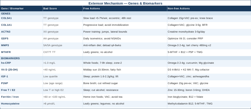
Understanding your genetic predisposition does not lock you into any outcome. But it changes the calibration of everything else. Someone with a favorable genetic profile for tendon integrity may recover well from a quadriceps tendon injury with standard physiotherapy and adequate protein intake. Someone with an unfavorable cluster of connective tissue variants may need a more systematic approach: more deliberate collagen support, more conservative load progression, and more attention to specific nutritional cofactors. The following six genes represent the strongest current evidence for tendon and extensor mechanism injury susceptibility.
COL5A1 — Collagen Type V Alpha-1
COL5A1 encodes the alpha-1 chain of type V collagen, which acts as a nucleating template that controls the diameter of type I collagen fibrils during fibril formation. Smaller, more uniform fibrils create stiffer, stronger tendons. The rs12722 C/T polymorphism has been consistently associated with Achilles tendinopathy, ACL rupture, and general tendon injury susceptibility across multiple case-control studies in diverse athletic populations. The TT genotype confers structurally less organized tendon collagen that fatigues faster under repetitive mechanical loading.
If the gene is unfavorable, the plan without supplements
Increase training volume more gradually than standard guidelines recommend — closer to 5–7% per week rather than the conventional 10% rule. Prioritize eccentric loading protocols, where the muscle lengthens under controlled load, to stimulate collagen remodeling without overwhelming structurally fragile fibrils. Allow 48–72 hours between tendon loading sessions. Avoid sudden changes in footwear, training surfaces, or impact volume, which TT carriers tolerate poorly.
If the gene is unfavorable, the plan with supplements or equipment
Collagen hydrolysate (15–20g, 30–60 minutes pre-exercise) with 50mg vitamin C: The timing protocol identified by Baar's research is particularly important for TT carriers because their fibrils are harder to organize — providing maximal substrate during the synthesis window is essential. Daily use, no relevant side effects.
Patellar tendon support braces and compression sleeves: Reduce peak stress on the extensor mechanism during loading. Structural protection during rehabilitation reduces fibril fatigue without preventing the mechanical stimulus needed for remodeling. Use during higher-load sessions; not needed at rest.
COL1A1 — Collagen Type I Alpha-1
COL1A1 encodes the predominant structural protein in tendons, ligaments, and bone. The Sp1 binding site polymorphism (rs1800012, G/T) reduces the efficiency of transcription factor binding, lowering type I collagen gene expression in TT carriers. Less COL1A1 expression means less collagen produced per anabolic stimulus — directly relevant for all tendon healing scenarios. The TT genotype has been associated in multiple studies with higher rates of tendon rupture, reduced bone mineral density, and impaired soft tissue healing after orthopedic procedures.
If the gene is unfavorable, the plan without supplements
Consistent, progressive mechanical loading is more important for COL1A1 TT carriers, not less — fibroblasts from low-producer genotypes need stronger mechanical signaling to produce adequate collagen. Avoid prolonged immobilization: bed rest or excessive offloading leads to faster collagen loss in low-producer genotypes, creating a recovery deficit that is hard to reverse.
If the gene is unfavorable, the plan with supplements or equipment
The same collagen plus vitamin C protocol applies, with added emphasis on glycine (3–5g daily) as a direct rate-limiting substrate for collagen synthesis. Additionally, blood flow restriction (BFR) training using pneumatic cuffs applied to the proximal thigh allows low-load knee extension work (20–30% of 1RM) to produce an anabolic and collagen-stimulating effect equivalent to heavy conventional loading — critical for COL1A1 TT carriers who need to maximize collagen stimulus at safer force levels during rehabilitation.
ACTN3 — Alpha-Actinin-3
ACTN3 encodes alpha-actinin-3, a structural protein found exclusively in fast-twitch (type IIX) muscle fibers. The R577X polymorphism creates a premature stop codon: RR carriers have full alpha-actinin-3 function, while XX carriers — approximately 18% of the general population — have no ACTN3 protein at all. XX carriers have lower intrinsic fast-twitch force generation capacity, trending toward a more endurance-oriented muscle profile.
For extensor mechanism function specifically, the quadriceps must produce rapid, high-force contractions during deceleration, landing, and rapid direction changes — the loading conditions most associated with extensor mechanism injury and re-injury. XX carriers have a structural disadvantage in these situations that is not corrected by standard rehabilitation protocols unless power training is explicitly prioritized.
If the gene is unfavorable, the plan without supplements
Power-focused training elements are not optional for ACTN3 XX carriers — they are compensatory. Ballistic exercises (jump squats at controlled intensity, step-up jumps, lateral bounds) should be introduced progressively once load tolerance allows. Eccentric quadriceps loading emphasizing speed of muscle lengthening further develops the fast-twitch neuromuscular capacity available despite the fiber-type disadvantage.
If the gene is unfavorable, the plan with supplements or equipment
Creatine monohydrate (3–5g daily, continuous): The strongest evidence base of any supplement for improving explosive lower-limb force output. Its benefits are disproportionately meaningful in individuals with less natural fast-twitch capacity. No cycling needed; long-term use at standard doses is well-studied and safe. Loading phase (20g daily for 5 days) is optional but accelerates tissue saturation.
GDF5 — Growth Differentiation Factor 5
GDF5 is a member of the TGF-beta superfamily with a key role in embryonic tendon and joint development and adult maintenance of tendon, ligament, and cartilage tissue. The rs143384 A/G polymorphism in the 5' UTR of GDF5 alters expression levels: the AA genotype is associated with significantly lower GDF5 expression, which has been linked in genome-wide association studies to higher rates of knee osteoarthritis and soft tissue injury. Reduced GDF5 expression impairs tenocyte differentiation and extracellular matrix organization — weakening the biological scaffolding of tendon and joint tissue throughout life.
If the gene is unfavorable, the plan without supplements
Daily isometric loading protocols — wall sits, Spanish squats, isometric leg press holds — stimulate GDF5 expression locally in tendon tissue via mechanotransduction, partially compensating for lower baseline expression. This is one of the few non-pharmacological mechanisms known to upregulate GDF5 in adult tendon. Avoiding chronic NSAID use is also important: evidence suggests long-term NSAID exposure suppresses tendon matrix synthesis pathways including those dependent on GDF5 signaling.
If the gene is unfavorable, the plan with supplements or equipment
Adequate vitamin D status (see biomarker section) is particularly important for GDF5 AA carriers: VDR signaling interacts with GDF5-dependent pathways in musculoskeletal tissue. Platelet-rich plasma (PRP) injections, while controversial as a standalone intervention, deliver concentrated TGF-beta family growth factors that interact with GDF5 signaling and may provide additive benefit when combined with structured loading rehabilitation — not as a replacement for it.
MMP3 — Matrix Metalloproteinase 3
MMP3 encodes stromelysin-1, an enzyme that degrades extracellular matrix components including collagen, fibronectin, and proteoglycans. Some matrix degradation is necessary for remodeling, but the 5A/6A promoter polymorphism (rs679620) affects MMP3 transcription rate: 5A/5A carriers have significantly higher baseline MMP3 expression, meaning more aggressive matrix degradation. Under conditions of high training volume, poor recovery, or elevated systemic inflammation, these individuals may tip the balance toward collagen breakdown faster than synthesis can replace it.
If the gene is unfavorable, the plan without supplements
Reducing systemic inflammatory load is the highest-priority intervention for 5A/5A carriers: inflammatory cytokines upregulate MMP3 expression further in these individuals, creating a compounding degradation cycle. Structured deload periods every 4–6 weeks are particularly important — allow matrix remodeling to catch up with degradation before resuming progressive loading.
If the gene is unfavorable, the plan with supplements or equipment
Omega-3 fatty acids (EPA+DHA, 2–4g daily): Shown to downregulate MMP-3 and MMP-9 activity in connective tissue. Continuous use, safe long-term.
Tart cherry extract (480mg twice daily): Contains anthocyanins that inhibit MMP activity and reduce post-exercise connective tissue degradation markers. Cycle 6–8 weeks on, 2 weeks off. Generally well-tolerated.
MTHFR — Methylenetetrahydrofolate Reductase
MTHFR is not typically listed among tendon-specific genes, but its direct connection to homocysteine metabolism makes it immediately relevant. The C677T and A1298C polymorphisms reduce MTHFR enzyme efficiency by 30–70% in heterozygous and homozygous carriers, impairing folate conversion and elevating circulating homocysteine. As described in the biomarker section, elevated homocysteine inhibits lysyl oxidase — the enzyme responsible for the crosslinks that give type I collagen its tensile strength. MTHFR C677T TT homozygotes are at significantly higher risk for elevated homocysteine and structurally weaker collagen throughout all connective tissue, including the extensor mechanism.
If the gene is unfavorable, the plan without supplements
Increase dietary methylated folate sources: leafy greens, legumes, liver. Eliminate alcohol. Investigate gut absorption if B12 levels are chronically low despite adequate dietary intake.
If the gene is unfavorable, the plan with supplements or equipment
The critical intervention is replacing standard folic acid with 5-MTHF (L-methylfolate, 400–1000mcg daily) — the pre-converted form that bypasses the impaired MTHFR enzymatic step. This is non-negotiable for TT carriers, as unconverted folic acid can actually accumulate and competitively inhibit methylfolate uptake. Pair with methylcobalamin B12 and P5P B6 as above. Add TMG (betaine, 1000–2000mg daily) if homocysteine remains elevated after 3–6 months on the B vitamin protocol. This targeted intervention can normalize homocysteine within 8–12 weeks and directly improves the structural integrity of newly synthesized collagen — a measurable, practical change with direct relevance to tendon healing.
What Keith Baar's Research Reveals About Tendons That Most Clinicians Don't Know
The Huberman Lab podcast featuring Keith Baar — tendon biologist at the University of California Davis and one of the most cited researchers in connective tissue physiology — synthesizes a body of mechanistic research that directly challenges standard clinical assumptions about tendon recovery. The following are the ten most impactful insights for anyone managing extensor mechanism pathology.
1. Tendons Are Metabolically Slow — and That Changes Everything About Recovery Timelines
Unlike skeletal muscle, which has rich vascularity and rapid protein turnover, tendons are largely avascular structures with collagen half-lives measured in years, not days. True structural remodeling is inherently slow. Expecting tendon tissue to be repaired in weeks when the biology operates on a months-to-years timescale leads to premature return to load and predictable re-injury. Patience calibrated to the actual biology — not to pain levels or activity tolerance — is the most underutilized clinical strategy.
2. The 6-Hour Collagen Synthesis Window
Baar's laboratory established that collagen synthesis in tendons peaks 5–6 hours after a mechanical loading stimulus. To capture two distinct synthesis peaks in the same day during intensive rehabilitation, loading sessions should ideally be separated by at least 6 hours. This is mechanistically important for those using twice-daily rehabilitation protocols.
3. Collagen and Vitamin C Before Exercise — the Timing Is the Mechanism
The Shaw et al. 2017 RCT showed that 15g of gelatin or collagen hydrolysate with 50mg vitamin C taken 30–60 minutes before loading exercise doubled collagen synthesis rates in tendon tissue compared to a placebo. Post-exercise timing is not equivalent. The pre-exercise window is when circulating amino acid precursors are available to be driven into tendon by the subsequent mechanical stimulus — the sequence matters.
4. Isometric Contractions Are the Best First-Line Pain Management Tool for Tendinopathy
Isometric contractions — sustained muscular contraction without joint movement — produce significant analgesic effects in chronic tendinopathy, with pain reduction in some protocols equivalent to NSAIDs in short-term trials, without the collagen synthesis suppression that chronic NSAID use can cause. For patellar tendinopathy specifically, isometric leg press holds at 45 degrees knee flexion and approximately 70% maximal voluntary contraction, five sets of 45 seconds, have a well-established clinical evidence base.
5. Not All Loading Is Equal — Compressive Loads Damage Tendons
Tendons are built to handle longitudinal tension along the fiber direction, which is generated during controlled eccentric and concentric movements. Shear and compressive loads, however, are poorly tolerated and are a primary driver of degenerative tendinopathy rather than repair. For patellar tendinopathy, this explains why deep squat positions with heavy load are often provocative — at high knee flexion angles, the patellar tendon wraps around the inferior pole of the patella and experiences compressive loading it is not built to sustain.
6. Estrogen Has a Larger Biological Effect on Tendons Than Most Clinicians Acknowledge
Estrogen alters collagen fibril structure and reduces tendon stiffness — a mechanism that explains the cyclical nature of tendon pain in premenopausal women, increased injury rates around ovulation, and the dramatically different injury profiles of female versus male athletes in identical sport activities. Post-menopausal estrogen decline significantly reduces tendon healing capacity. Baar is direct: hormonal context should be part of every tendon rehabilitation assessment, not an afterthought.
7. Complete Rest Is One of the Worst Strategies for Tendon Healing
Baar is unambiguous on this point: total offloading leads to fibril disorganization, cellular metabolic impairment, and ultimately weaker, less organized healed tissue. Tendons require mechanical load to direct collagen fibril alignment during repair. Even during acute pain phases, gentle sub-pain isometric loading is preferable to full rest. The goal is never zero loading — it is appropriately dosed loading calibrated below the provocation threshold.
8. Blood Flow Restriction Is a Legitimate Physiological Tool, Not a Shortcut
BFR training uses pneumatic cuffs to partially restrict venous return, allowing low-load exercises at 20–30% of 1RM to produce an anabolic and collagen-stimulating stimulus comparable to 60–80% 1RM conventional loading. For extensor mechanism rehabilitation where heavy loading is not yet tolerable, BFR is not a workaround — it is mechanistically well-supported and has RCT-level evidence for tendon applications. It allows training at stimulus levels that would normally require pain-provoking loads.
9. Growth Hormone and IGF-1 Are the Master Regulators — and Sleep Is Their Primary Driver
The GH-IGF-1 axis drives fibroblast proliferation and collagen matrix synthesis. Most of the daily GH pulsatility that sustains this axis occurs during deep slow-wave sleep. Sleep deprivation is not just a fatigue issue in the context of tendon healing — it is a direct suppressor of the primary anabolic system responsible for tissue repair. Baar identifies poor sleep as arguably the single most under-addressed variable in tendon recovery programs, with effects that no amount of supplementation or exercise can fully compensate for.
10. Early Symptom Resolution Does Not Equal Structural Healing
This is perhaps the most clinically consequential insight: early-stage tendon "adaptation" (within weeks of a loading program) is predominantly neural and cellular — stiffness improves and pain decreases via cell activation without significant structural collagen change. True structural remodeling — new collagen organized into mechanically competent fibrils — takes months and is not reflected in subjective pain scores. Athletes who feel recovered are often returning to load before the tissue is structurally ready, producing re-injury at the same site. Biomarkers like P1NP can help identify when structural healing is genuinely occurring, rather than relying solely on pain as a proxy for tissue status.
Complementary Approaches With Meaningful Clinical Evidence
Several complementary modalities have reasonable human evidence for tendon and extensor mechanism-adjacent pathology. The following three represent the best combination of clinical relevance and evidence quality for this specific condition.
Low-Level Laser Therapy / Photobiomodulation
Photobiomodulation (PBM) uses red and near-infrared light (630–1000nm) to stimulate mitochondrial activity, increase local ATP production, and reduce pro-inflammatory cytokine expression in target tissue. For extensor mechanism pathology — particularly chronic patellar tendinopathy, which is characterized by disorganized matrix and persistent local inflammation — PBM is mechanistically relevant because tendons' poor vascularity means any intervention that enhances cellular energy availability and reduces local cytokine burden may accelerate repair.
A systematic review and meta-analysis published in the British Journal of Sports Medicine (Tumilty et al.) found that PBM significantly reduced pain in Achilles tendinopathy compared to placebo, with optimal parameters at 904nm wavelength, 4–8 joules per point, applied 3 times per week. A subsequent RCT by Stergioulas et al. confirmed that PBM combined with eccentric loading produced superior outcomes to eccentric loading alone in Achilles tendinopathy. While the extensor mechanism literature is less extensive, the shared tendon biology makes these findings directly applicable.
For practical application: a clinical-grade device or a reputable consumer PBM panel (minimum 60–100mW output, 630–850nm range) applied directly over the patellar tendon and inferior quadriceps region for 10–20 minutes, five sessions per week. Warm the tissue first with gentle movement. Most improvements in pain and function are observed after 6–8 weeks of consistent application. Side effects are minimal at appropriate doses; do not apply over open wounds or compromised skin.
Massage Therapy
Deep transverse friction massage (DTFM) and myofascial release applied to the quadriceps and patellar tendon region have a specific rationale for extensor mechanism pathology: chronically tightened and fibrotic quadriceps increase resting tension through the extensor chain, amplifying load on the patellar tendon during movement and reducing blood flow to the already avascular tendon tissue. Manual therapy directed at the quadriceps, IT band, and patellar tendon adhesions reduces this mechanical compression and improves local circulation to the rehabilitation zone.
A randomized controlled trial published in the Journal of Orthopaedic and Sports Physical Therapy found that friction massage combined with stretching significantly improved pain and functional outcomes in patellar tendinopathy compared to stretching alone. The mechanism is partly mechanical (breaking down disorganized scar tissue and adhesions at the tendon surface) and partly neurological (reducing muscle spindle sensitivity in the quadriceps, decreasing resting tone through the extensor mechanism chain).
Realistically, 2–3 sessions per week of 30–45 minutes by a trained sports massage or physiotherapy practitioner are appropriate during the early-to-mid rehabilitation phase. DTFM specifically applied perpendicular to the patellar tendon fibers for 5–10 minutes per session is the most evidence-aligned technique. Self-massage with a foam roller (for quadriceps) and a Theragun or similar percussion device (for the tendon region) can maintain progress between professional sessions. Expect mild soreness for 24–48 hours after initial sessions — this resolves with consistent treatment.
Biofeedback
The vastus medialis oblique (VMO) — the teardrop-shaped muscle on the inner quadriceps that controls patellar tracking — is frequently underactivated in extensor mechanism dysfunction, both as a cause and a consequence of injury. When the VMO fires weakly or late relative to the vastus lateralis, the patella tracks laterally under load, increasing patellar tendon stress and patellofemoral contact pressure. Surface electromyographic (EMG) biofeedback allows individuals to see, in real time, whether the VMO is activating at the right time and magnitude during therapeutic exercises.
A systematic review in Physiotherapy Theory and Practice found that EMG biofeedback significantly improved VMO activation timing and pain outcomes in patellofemoral pain syndrome compared to conventional physiotherapy alone. The effect is not simply about the biofeedback signal itself — it accelerates the neuromuscular re-learning process by providing immediate, objective feedback that verbal cueing cannot match in precision.
Clinically, biofeedback is best applied during 3–4 weeks of supervised physiotherapy early in the rehabilitation process, using surface electrodes over the VMO during terminal knee extensions, step-ups, and leg press variations. The goal is to establish correct motor patterns under feedback guidance, which then transfer to unmonitored exercise. Home-use EMG biofeedback devices are available ($150–$400) for those without regular access to supervised sessions, though professional setup and baseline calibration remain important for accurate interpretation.
Conclusion
Extensor mechanism disruption is one of those conditions where the standard approach covers the structure but ignores the biology. The path forward is not more generic advice — it is more precise information. Knowing where your hs-CRP, vitamin D, IGF-1, P1NP, hormones, ferritin, and homocysteine actually stand tells you whether your body is in a state that supports repair or one that is quietly undermining it. Understanding your genetic variants in COL5A1, COL1A1, ACTN3, GDF5, MMP3, and MTHFR explains why your recovery may need to be more deliberate than average — and exactly how to adjust.
None of this replaces good rehabilitation programming or clinical oversight. But it adds a layer of precision that changes what you prioritize, what you supplement, and how you pace your loading progression. The next smart step is to get a basic biomarker panel done, review your results with a clinician who understands functional medicine or sports physiology, and build a protocol grounded in your specific numbers — not the average. That approach, applied consistently, gives you the clearest possible path back to full function.
Musculoskeletal: Joint Conditions Muscle Conditions Tendon & Ligament Conditions Sports Injuries
Autoimmune: Connective Tissue Conditions