This article contains AI generated content.
Extraskeletal Chondroma - 2 Genes and 7 Biomarkers to Track
Introduction
Finding out you have a slow-growing lump near a finger, toe, hand, or foot — and hearing the word "chondroma" attached to it — tends to trigger two competing reactions at once. Relief, because "benign" is in the sentence somewhere. And unease, because nobody handed you a clear explanation of why it formed, whether it will come back after removal, or what, if anything, you can actually do about it.
Most general advice about soft tissue lumps stops at "get it checked" and "surgery if it bothers you." That's not wrong, but it's thin. It treats every small mass the same way, when in fact extraskeletal chondroma is a specific, well-described entity with its own cell biology, its own pattern on imaging, and its own — admittedly narrow — genetic signature. Generic reassurance doesn't tell you what a pathology report is actually checking for, why a molecular test might get ordered, or which lab values are genuinely relevant versus which ones just sound relevant.
This article goes deeper on purpose. It walks through the biomarkers that show up in the real diagnostic workup for this condition, what each one is measuring, what it costs, and what — realistically — you can and can't influence about it. It also looks honestly at the genetic research behind extraskeletal chondroma, because that research exists, it's real, and it's more limited than headlines about "cancer genes" usually suggest. Where the evidence is thin, that will be stated plainly rather than dressed up.
None of this replaces a pathologist or a hand/foot surgeon. But better information changes the questions you ask, the follow-up you request, and how confidently you can tell the difference between what matters and what's just noise. The sections ahead cover the practical biomarkers to track, what the underlying genetics actually show, a cancer-biology podcast episode worth your attention, and a few complementary approaches with real, if adjacent, supporting evidence.
Summary
Extraskeletal chondroma is a rare, benign cartilage tumor that grows in soft tissue rather than bone — most often in a finger or toe — and its molecular story is smaller and more specific than most people expect. There are really only two genetic threads worth knowing: a chromosome rearrangement involving a gene called HMGA2, found in a subset of cases and shared with lipoma biology, and the near-total absence of the IDH1/IDH2 mutations that define its bony cousin, the enchondroma. Beyond genetics, the real diagnostic muscle comes from seven trackable biomarkers — spanning imaging, pathology, and a couple of blood tests used to rule out lookalike conditions — each with its own cost, its own reliability, and its own limits on what lifestyle changes can and can't touch. Read on to see exactly which markers a competent workup should include, why two of the most-cited "cancer markers" (S100, Ki-67) aren't actually validated for this specific tumor, what a leading cancer-immunology podcast episode adds to the bigger picture, and which complementary approaches have genuine — if imperfect — evidence behind them for the anxiety and recovery side of dealing with a soft tissue mass.
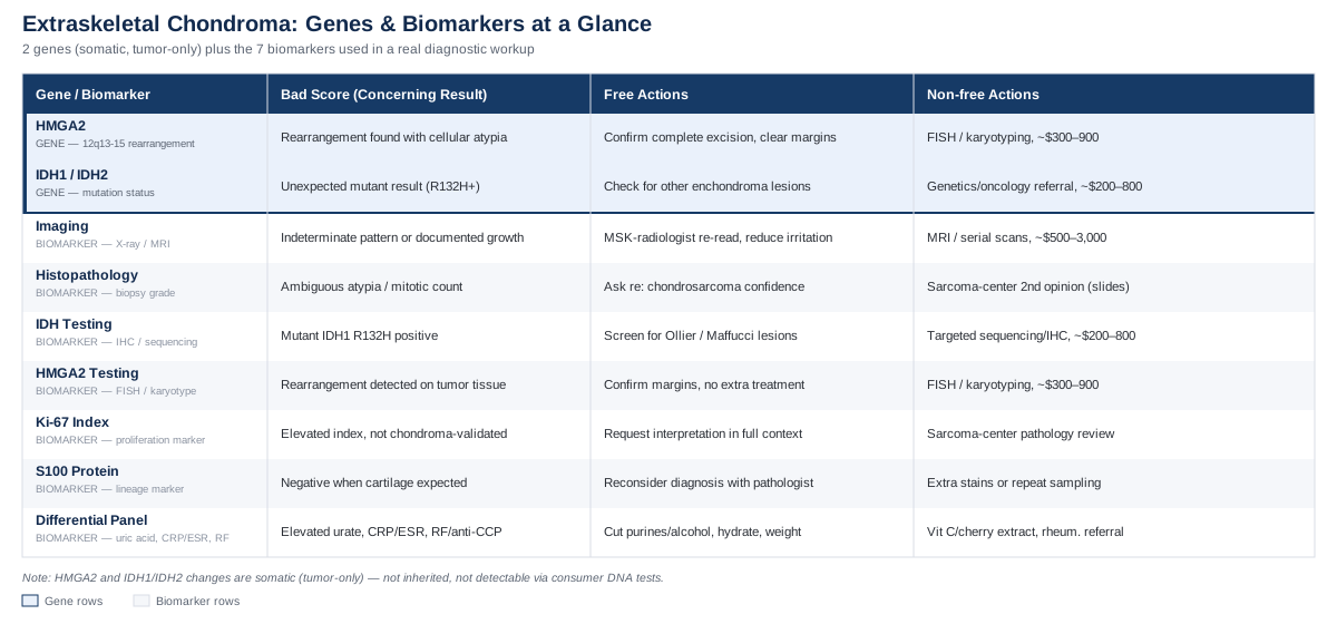
7 Biomarkers That Matter for Extraskeletal Chondroma
Because extraskeletal chondroma is rare and almost always localized, there's no single blood test that says "yes, this is it." The real diagnostic picture is built from a combination of imaging, tissue analysis, and — increasingly — targeted molecular testing, with a couple of blood markers used mainly to rule out other conditions that can mimic a small firm lump near a joint. What follows is the most useful and honest version of "biomarkers to track" for this specific tumor: what each one is, how it's actually measured, roughly what it costs, and what can (and can't) be done if a result raises concern.
Imaging appearance and growth pattern
Imaging is usually the first real "biomarker" in this story, because it's what turns a lump someone noticed into a lesion someone can characterize. On X-ray, extraskeletal chondroma often shows scattered or ring-like calcifications inside the soft tissue mass — present in roughly a third to two-thirds of cases — while MRI typically shows a well-circumscribed lesion with low-to-intermediate signal on T1 and high signal on T2, sometimes with septal enhancement after contrast. The catch, confirmed in published case reports, is that this appearance is nonspecific: benign and locally aggressive cartilaginous lesions can look similar enough on imaging alone that tissue sampling is usually still required before anyone can be confident about the diagnosis (Adaletli et al., soft tissue mass imaging case report).
How to measure it
A plain X-ray is the standard first step and is inexpensive, typically in the range of 50 to 200 dollars depending on location and insurance. If the lesion is deeper or the diagnosis is unclear, ultrasound (roughly 150 to 400 dollars) or MRI (commonly 500 to 3,000 dollars without contrast pricing adjustments) adds detail on size, borders, and internal architecture. Serial imaging — repeating the same study every 6 to 12 months — is how growth rate gets tracked over time, which matters more than any single snapshot.If the imaging looks concerning — the plan without supplements
There is no lifestyle intervention that changes what a cartilaginous lesion looks like on a scan. The realistic, non-supplement plan is: get the comparison imaging read by a musculoskeletal radiologist rather than a general reader, ask specifically whether the pattern is "typical for a benign cartilaginous lesion" or "indeterminate," and avoid repeated mechanical irritation at the site (tight footwear over a toe lesion, repetitive gripping over a finger lesion) since chronic local irritation is a plausible, if not rigorously proven, contributor to local tissue change in superficial lesions near tendons and joints. If growth is documented between two comparable scans, that's the trigger for biopsy or excision discussion, not a reason to "wait and monitor" indefinitely.If the imaging looks concerning — the plan with supplements or equipment
Supplements do not alter tumor imaging characteristics, and no equipment-based intervention (TENS units, compression devices, etc.) has evidence for shrinking a cartilaginous soft tissue mass. The only equipment genuinely relevant here is protective: a properly fitted shoe insert or a padded finger sleeve to reduce mechanical pressure on the lesion while imaging and specialist input are arranged. This is comfort and irritation-reduction, not treatment — worth stating clearly so no one mistakes it for one.Histopathologic grade at biopsy
Once tissue is obtained — usually via excisional biopsy, since these lesions are typically small enough to remove entirely — the pathologist's assessment of cellularity, mitotic count, and cytological atypia is the actual gold standard for confirming benignity. This is the core of the classic description of the entity from Chung and Enzinger's foundational 1978 series, which remains the largest published cohort of this rare tumor and established the criteria still used today to separate chondroma of soft parts from its malignant look-alikes (Chung & Enzinger, Chondroma of soft parts, 1978).
How to measure it
This requires a procedure — a needle or excisional biopsy performed by a surgeon, followed by histopathology review. Costs vary widely by country and setting, but a reasonable US range for the combined procedure and pathology work is 300 to 1,500 dollars before insurance, more if performed under general anesthesia in an operating room rather than local anesthesia in a clinic.If the biopsy result is ambiguous — the plan without supplements
Ask directly whether the pathologist felt confident distinguishing the lesion from chondrosarcoma, and if there's any hesitation, request a second opinion from a sarcoma-referral pathology center — this is standard practice for rare mesenchymal tumors and costs nothing extra beyond sending slides. There is no dietary or behavioral change that affects mitotic count or cellular atypia; this is purely a tissue-level finding that only additional expert review can clarify.If the biopsy result is ambiguous — the plan with supplements or equipment
None apply here, and it would be inaccurate to suggest otherwise. The only "equipment" relevant post-biopsy is standard wound care and, if excision was more extensive, a splint or supportive brace during healing — not a lever on the pathology result itself.IDH1/IDH2 mutation status
IDH1 and IDH2 mutations are one of the best-established findings in cartilage tumor biology — but they belong almost entirely to intraosseous (bone-based) cartilage tumors. A large screen of roughly 1,200 mesenchymal tumors found IDH1/IDH2 mutations were frequent in central and periosteal chondromas and central chondrosarcomas, and essentially absent across the hundreds of other bone and soft tissue tumor types tested alongside them (Amary et al., IDH1/IDH2 mutations in cartilaginous tumors, 2011). The same mutations, in mosaic form, drive Ollier disease and Maffucci syndrome, the multiple-enchondroma disorders that carry real malignant transformation risk (Pansuriya et al., IDH mutations in Ollier disease and Maffucci syndrome, 2011). For extraskeletal (soft tissue) chondroma specifically, a directly-published, chondroma-of-soft-parts-only dataset confirming IDH-wild-type status is thin — this is more an inference from the broader pattern than a single dedicated study, and it's worth knowing that distinction rather than treating it as settled fact.
How to measure it
IDH1/IDH2 testing is done on tumor tissue (not blood) via targeted sequencing or immunohistochemistry for the mutant IDH1 R132H protein, usually ordered by the pathologist when there's any diagnostic overlap with enchondroma or when multiple lesions raise suspicion for Ollier disease. Cost typically runs 200 to 800 dollars as an add-on to the pathology workup, though it isn't ordered routinely for a single, typical-appearing soft tissue lesion.If IDH status is unexpected — the plan without supplements
An unexpected IDH-mutant result in what looked like a simple soft tissue lesion should prompt a re-evaluation of location (is this actually a periosteal or intraosseous lesion presenting unusually?) and a check for other enchondroma-type lesions elsewhere in the skeleton, since that changes both surveillance frequency and specialist referral (genetics, orthopedic oncology). This is a workup-and-referral response, not a self-directed one.If IDH status is unexpected — the plan with supplements or equipment
There is no supplement, nutraceutical, or device that alters IDH1/IDH2 mutation status in existing tumor tissue. Claims to the contrary — and they do circulate in some longevity and biohacking spaces — are not supported by evidence for this tumor type and shouldn't guide decisions here.HMGA2 rearrangement
This is the most tumor-specific genetic finding for extraskeletal chondroma. Small cytogenetic series have found rearrangements involving chromosome band 12q13-15 in a subset of soft tissue chondromas, with several cases showing activation or fusion of the HMGA2 gene — the same gene implicated in lipoma and other benign mesenchymal tumors (Dahlén et al., HMGA2 in bone and soft tissue chondromas, 2003). Earlier case reports established that 12q13-15 rearrangement in chondroma was a novel but recurring finding (Mandahl et al., 1993), while other reported cases show entirely different chromosomal changes, including chromosome 6 and 11 involvement — meaning HMGA2 rearrangement is one recognized subgroup, not a universal feature (Dal Cin et al., 1997).
How to measure it
HMGA2 rearrangement is detected through cytogenetic karyotyping or FISH (fluorescence in situ hybridization) on tumor tissue, typically only performed at specialized sarcoma/soft-tissue-pathology reference labs rather than routine community pathology labs. Expect a cost range of roughly 300 to 900 dollars, and expect it to be ordered only when the diagnosis is genuinely unclear or research-relevant, not as a routine confirmatory test.If HMGA2 rearrangement is found — the plan without supplements
A positive finding is reassuring in one specific sense: it aligns the lesion with a benign mesenchymal tumor family (like lipoma) rather than a malignant one, and it doesn't change surgical management if the lesion has already been fully excised. The practical response is confirming complete excision with clear margins on the pathology report and standard follow-up imaging, not additional intervention aimed at "HMGA2" itself.If HMGA2 rearrangement is found — the plan with supplements or equipment
Because this is a somatic (tumor-only) chromosomal rearrangement rather than an inherited variant in your genome, no supplement or device changes it — it isn't a gene you're "born with" in the sense that a 23andMe-style report would flag. Where general connective tissue support has a role is unrelated to the mutation itself: adequate vitamin C intake (75–90 mg/day from diet, more only if genuinely deficient) supports normal collagen synthesis in healing tissue after excision, and this is reasonable supportive care, not tumor-genetics correction.Ki-67 proliferation index
Ki-67 is a nuclear protein marker used broadly across pathology to estimate what fraction of cells in a sample are actively dividing, and pathologists sometimes report it for soft tissue tumors as one piece of context. It's worth being direct here: a Ki-67 index specifically validated to distinguish extraskeletal chondroma from extraskeletal chondrosarcoma was not identified in the available literature. The Ki-67 data that does exist for cartilaginous soft tissue tumors relates mainly to extraskeletal myxoid chondrosarcoma, a molecularly distinct, fusion-driven malignant entity — not the benign chondroma this article is about (review of extraskeletal myxoid chondrosarcoma). If a Ki-67 value appears on your report, it should be interpreted by the reporting pathologist in context, not compared to a chondroma-specific cutoff — because no well-established one exists.
How to measure it
Ki-67 is assessed via immunohistochemical staining on the biopsy or excision sample, usually included as an add-on stain for 50 to 150 dollars on top of standard pathology processing.If Ki-67 comes back elevated — the plan without supplements
Request that the pathologist interpret the number in the context of the overall morphology (mitotic count, atypia, necrosis) rather than in isolation, and if there's any uncertainty, request sarcoma-center review. An isolated elevated Ki-67 without corresponding atypia or increased mitoses is not, on its own, a validated marker of malignancy in this tumor type.If Ki-67 comes back elevated — the plan with supplements or equipment
No supplement or device lowers a proliferation index in existing tumor tissue, and none should be pursued as a substitute for appropriate surgical and pathology follow-up.S100 protein expression
S100 is a lineage marker, not a malignancy marker — it stains positive in cells of neural crest and cartilaginous origin, which makes it useful for confirming that a lesion is genuinely cartilage-forming rather than something else entirely (a ganglion cyst, giant cell tumor of the tendon sheath, or an epidermal inclusion cyst can all present as a similar small firm lump). It does not reliably separate benign chondroma from malignant chondrosarcoma, and no chondroma-specific validation study establishing it as a discriminator between the two was found in the available literature — so its practical value here is confirming "this is cartilage," not "this is safe."
How to measure it
Like Ki-67, this is an immunohistochemical stain performed on the same tissue sample, typically 50 to 150 dollars as part of the pathology panel.If S100 is negative when cartilage was expected — the plan without supplements
A negative result in a lesion presumed to be cartilaginous should prompt reconsideration of the diagnosis altogether, since it suggests the mass may not be a chondroma at all. This is a "go back and re-evaluate the diagnosis" situation, handled through additional stains or repeat sampling, not a lifestyle response.If S100 is negative when cartilage was expected — the plan with supplements or equipment
Not applicable — this is a tissue-typing question, and no external intervention changes what's already in the sample.General differential-diagnosis panel
This is the one genuinely actionable blood-based category, and it exists to rule out — not confirm — extraskeletal chondroma. A small firm lump near a finger or toe joint has several common mimics with very different treatments: gout (elevated serum uric acid), rheumatoid nodules (positive rheumatoid factor or anti-CCP antibodies), and infected or inflamed soft tissue (elevated CRP or ESR). None of these tests diagnose chondroma directly, but a clinician will often check them when the clinical picture isn't perfectly clear-cut, precisely because these are more common than extraskeletal chondroma and some of them are far more modifiable through lifestyle and medication.
How to measure it
A standard blood draw covering uric acid, CRP or ESR, and, if joint inflammation is suspected, rheumatoid factor and anti-CCP antibodies, typically costs 50 to 250 dollars depending on which markers are included and whether it's bundled into a broader panel.If a mimicking condition is flagged — the plan without supplements
If uric acid is elevated, reducing alcohol (especially beer), limiting high-purine foods like organ meats and certain shellfish, staying well hydrated, and maintaining a healthy body weight are the standard first-line lifestyle measures, typically reassessed after 8 to 12 weeks. If inflammatory markers or rheumatoid antibodies are positive, referral to rheumatology for proper evaluation is the appropriate next step — not self-management.If a mimicking condition is flagged — the plan with supplements or equipment
For elevated uric acid specifically, some evidence supports vitamin C supplementation (500 mg daily) and tart cherry extract as modest adjuncts to lower urate levels, generally well tolerated, though vitamin C above 1,000 mg/day can cause gastrointestinal upset and cherry extract should be used cautiously alongside anticoagulant medications. These measures address a lookalike condition, not extraskeletal chondroma itself, and should be framed that way to whoever is managing your care.Taken together, these seven markers make the point clearly: the useful diagnostic work here is mostly done through imaging and tissue analysis, with blood tests playing a supporting role in ruling out other explanations rather than confirming this one. That distinction matters when the genetics research on this tumor gets discussed, because it's easy to assume "gene" means "something you can test in a cheek swab and optimize" — and for this condition, that assumption doesn't hold.
What the Genetics Research Actually Shows
The genetic story behind extraskeletal chondroma is real, but it's smaller and more specific than genetics coverage for common chronic conditions tends to be. Genomics researchers such as Ali Torkamani have spent their careers arguing for whole-genome-informed, individualized medicine, and biomarker educators like Gary Brecka have popularized the idea that a person's own biology holds actionable answers — both are reasonable starting frames for thinking about genetics in health. Neither, to be clear, has published specifically on this rare tumor, and no consumer genetic test (23andMe, Ancestry, or similar) will tell you anything about it, because the relevant mutations aren't the kind those tests look for.
Why this is different from a typical "gene report"
The two findings worth knowing about — HMGA2 rearrangement and IDH1/IDH2 status — are somatic mutations, meaning they occur only in the tumor's own cells, acquired during the tumor's formation. They are not inherited variants sitting in your germline DNA, and they won't show up on a saliva-based consumer test. This matters because most "gene optimization" content assumes you're dealing with an inherited variant (like MTHFR or APOE) that shows up everywhere in your body and can be nudged with diet, supplements, or lifestyle. Somatic tumor mutations don't work that way — they exist only in the tissue that was removed, and once it's gone, so is the mutated tissue.
HMGA2: the chromosome 12 connection
HMGA2 is a gene that, in its normal form, helps regulate cell growth during development and is typically switched off in most adult tissues. In a subset of soft tissue chondromas, a rearrangement at chromosome band 12q13-15 reactivates or fuses HMGA2, a pattern first documented in individual case reports in the early 1990s and later confirmed with molecular detail — including one case with an HMGA2-LPP gene fusion identical to a rearrangement seen in lipoma (Dahlén et al., 2003). A closely related lesion, extraskeletal osteochondroma, has shown a comparable HMGA2-SOX5 fusion in a small case series, reinforcing that this chromosome 12 pathway shows up repeatedly across benign cartilage-and-bone-forming soft tissue tumors, even though it's a different specific diagnosis (Panagopoulos et al., 2015). Not every case shows this pattern — other reported cases involve entirely different chromosomal changes — so HMGA2 rearrangement should be understood as one recognized subtype, not a universal feature of the tumor.
If this gene pattern shows up on a pathology report — the plan without supplements
Confirm complete surgical excision with clear margins, since HMGA2-rearranged lesions in this family (chondroma, lipoma) are benign and don't require additional treatment once removed. There's no monitoring regimen specific to HMGA2 status beyond standard post-excision follow-up for recurrence at the original site.If this gene pattern shows up on a pathology report — the plan with supplements or equipment
Nothing changes the rearrangement itself, since it exists only in tissue that has typically already been removed by the time this is discussed. General wound-healing support — adequate protein intake, vitamin C for collagen synthesis, avoiding smoking (which measurably slows soft tissue healing) — is reasonable supportive care for the surgical site, not a genetic intervention, and should be understood as such.IDH1/IDH2: the mutation this tumor mostly doesn't have
IDH1 and IDH2 mutations are one of the most consequential findings in cartilage tumor biology generally — they drive Ollier disease and Maffucci syndrome (multiple enchondromatosis conditions with real malignant transformation risk over a lifetime) and appear in the majority of central, bone-based enchondromas and central chondrosarcomas (Pansuriya et al., 2011). The same large screening study that established this also tested hundreds of other bone and soft tissue tumor types and found IDH mutations essentially confined to the intraosseous cartilage tumor group (Amary et al., 2011). For extraskeletal chondroma specifically, this supports — by strong inference rather than a single dedicated chondroma-only study — that it sits outside the IDH-driven, Ollier/Maffucci-related disease spectrum, which is one reason it doesn't carry the same lifetime malignant transformation risk that multiple enchondromatosis does.
If IDH testing is done and comes back wild-type (unmutated) — the plan without supplements
This is generally the expected and reassuring result for a soft tissue lesion, and the practical response is simply standard surgical follow-up rather than the enhanced surveillance used for Ollier disease or Maffucci syndrome patients, who do need lifelong monitoring for malignant transformation.If IDH testing is done and comes back wild-type (unmutated) — the plan with supplements or equipment
No supplement protocol is needed or supported here — a wild-type result is the baseline expectation, not a deficiency to correct.Understood together, these two genetic findings do something genuinely useful even without offering a "fix": they explain why extraskeletal chondroma behaves the way it does clinically — locally confined, low recurrence after complete excision, and without the systemic malignant-transformation risk seen in inherited enchondromatosis syndromes. That context is worth more than a false sense of genetic control would be, and it sets up a broader point about tumor biology that a recent podcast episode explores in more general terms.
10 Insights From a Leading Cancer Biology Podcast
Andrew Huberman's conversation with UCSF physician-scientist Dr. Alex Marson, "Avoiding, Treating & Curing Cancer With the Immune System", isn't about extraskeletal chondroma specifically — no mainstream podcast episode will be, given how rare this tumor is. What it offers instead is a clearer general framework for understanding how the body handles abnormal cell growth, which is directly useful background for anyone processing a tumor diagnosis, benign or not (Huberman Lab, Avoiding, Treating & Curing Cancer With the Immune System).
1. Most abnormal cells never become a problem
The immune system routinely identifies and clears cells with DNA damage or abnormal growth signals long before they form anything detectable. A visible tumor, even a benign one, represents cells that slipped past that surveillance — not evidence that the body's defenses failed broadly.2. Benign and malignant are genuinely different biological categories
The distinction isn't just severity — it's a difference in whether cells retain normal growth controls, invasive capacity, and metastatic potential. This is exactly why pathology (mitotic count, atypia, margins) carries more diagnostic weight than any single genetic finding for a lesion like extraskeletal chondroma.3. Genetic mutations are common; dangerous mutations are rare
Cells accumulate DNA changes constantly through normal replication and environmental exposure. The overwhelming majority are irrelevant or get repaired. A tumor forming from a specific chromosomal rearrangement, like the HMGA2 changes seen in some chondromas, reflects one of the rare combinations that actually alters cell behavior.4. Immunotherapy works by removing the brakes on the immune system, not by adding new tools
Modern immunotherapy approaches largely function by unblocking immune cells that were already capable of recognizing abnormal tissue but had been suppressed by signals the tumor itself produces. This reframes cancer treatment progress as restoring a natural process rather than inventing an entirely artificial one.5. Engineered immune cells (CAR-T) show that precision matters more than force
Re-engineering a patient's own T-cells to recognize a specific tumor marker has produced durable results in certain blood cancers by being highly targeted, rather than broadly toxic. It's a useful mental model for why molecular testing (like the HMGA2 or IDH testing discussed earlier) is increasingly valued: precision changes outcomes.6. Chronic inflammation can quietly support tumor growth
Sustained low-grade inflammation changes the local tissue environment in ways that can favor abnormal cell survival. This is one legitimate reason general inflammatory markers (like CRP, discussed in the biomarker section above) have relevance beyond just ruling out infection.7. Genetic testing is becoming more clinically actionable, but context still matters
As sequencing gets cheaper and more available, more tumors — even rare ones — are being molecularly characterized. But a result is only useful when interpreted against what's already known about that specific tumor type, which is why an IDH or HMGA2 result means something different for a soft tissue chondroma than for a bone-based enchondroma.8. Lifestyle factors influence immune surveillance, but modestly, not dramatically
Sleep, stress regulation, and metabolic health all influence how effectively the immune system polices abnormal cells over time. This supports general health maintenance as a reasonable background practice — not as a treatment for an existing, already-formed benign tumor.9. Most cancer breakthroughs come from mechanism, not from single "miracle" compounds
Meaningful progress has consistently come from understanding a specific biological pathway (immune checkpoints, T-cell engineering) rather than from a single supplement or intervention. This is a useful filter for evaluating any claim that a vitamin or compound "targets" tumor genetics.10. Rare tumors benefit from expert, not general, review
Because rare tumor types don't have the volume of data that common cancers do, getting pathology and imaging reviewed by clinicians who specifically handle sarcomas and rare mesenchymal tumors matters more than it does for common conditions — a point that applies directly to extraskeletal chondroma.The throughline across these ten points is that biology rewards precision over broad intervention — which is a fitting bridge into a set of approaches that, while they won't touch the tumor's genetics, have real evidence for making the surrounding experience — the anxiety, the recovery, the waiting — more manageable.
Complementary Approaches Worth Considering
No complementary or alternative modality has clinical evidence for shrinking, preventing, or resolving extraskeletal chondroma itself — that needs to be stated plainly before anything else. What does have reasonable, if general and adjacent, evidence is support for the anxiety and physical recovery that surround diagnosis and surgical removal, particularly for a lesion in a sensitive area like a finger or toe.
Guided imagery
Guided imagery involves focusing attention on detailed, calming mental scenes, often with recorded audio guidance, to reduce the physiological stress response before and after a medical procedure. For someone facing biopsy or excision of a soft tissue mass, this targets the anticipatory anxiety and post-procedure pain perception that are common, normal, and worth managing directly rather than pushing through.
A randomized study of children undergoing minor surgery found that relaxation-guided imagery significantly reduced both preoperative anxiety and postoperative pain compared to standard care (Vagnoli et al., 2019), and a separate trial found similar benefit for preoperative anxiety using guided imagery relaxation more broadly (guided imagery and preoperative anxiety RCT, 2018). It's worth noting the evidence isn't uniformly positive: a study in colorectal surgery patients found high satisfaction with guided imagery but no measurable difference in pain or physiological outcomes (Haase et al., 2005), so benefit appears more consistent for anxiety and subjective experience than for hard physiological endpoints.
Realistically, this means using a guided imagery recording (many are free through hospital patient-education programs or reputable apps) for 15 to 20 minutes in the days before a scheduled biopsy or excision, and again during early recovery if anxiety or pain perception feels disproportionate to the actual procedure. It costs nothing beyond time, carries no side effects, and is reasonable to try regardless of how the evidence eventually shakes out for you personally.
Progressive muscle relaxation
Progressive muscle relaxation (PMR) involves systematically tensing and then releasing muscle groups throughout the body, which reduces overall sympathetic nervous system activation and is one of the most-studied non-drug anxiety interventions in perioperative settings generally.
The same pediatric minor-surgery study cited above combined relaxation techniques with guided imagery and found meaningful reductions in both anxiety and pain scores, supporting the pairing of these two techniques rather than treating them as entirely separate practices (Vagnoli et al., 2019).
A practical approach is a 10 to 15 minute PMR session once or twice daily in the week leading up to a scheduled procedure, and as needed during recovery if muscle guarding or tension develops around the surgical site. There are no meaningful side effects for most people, though anyone with a recent injury near the treated muscle groups should ease pressure/tension on that specific area.
Massage therapy
Massage therapy applied to healed surgical scars and surrounding soft tissue is commonly recommended by hand and foot surgeons to reduce adhesions, improve tissue mobility, and desensitize a healing scar — relevant here because excision of a finger or toe lesion leaves a scar in a highly mobile, frequently-used area. A registered clinical trial has examined structured scar massage after surgery, reflecting genuine clinical interest in the question, though this is an evolving area rather than one with large, definitive trial results specific to hand or foot surgery scars.
The realistic application is gentle circular massage over a fully closed, surgeon-cleared scar for 5 minutes, two to three times daily, typically starting two to four weeks post-surgery once the incision has closed — always confirmed with the treating surgeon first, since starting too early risks disrupting healing tissue. There's no meaningful downside when timed correctly, beyond mild, temporary tenderness.
Mindfulness meditation
Mindfulness-based approaches train sustained, non-judgmental attention to present-moment experience, and have a growing evidence base specifically in postoperative orthopedic pain management, which is a reasonably close analogue to recovery from soft tissue tumor excision near a joint.
A systematic review and meta-analysis of mindfulness interventions in hip and knee arthroplasty patients found preliminary but encouraging evidence of benefit for postoperative pain and recovery experience (mindfulness and orthopedic postoperative outcomes meta-analysis), though the authors themselves describe the evidence as preliminary rather than conclusive.
A practical starting point is a structured 8-week mindfulness program (many are freely available through validated apps or hospital-affiliated programs) beginning around the time of diagnosis or biopsy scheduling, continued through recovery. Sessions of 10 to 20 minutes daily are typical, with no known side effects, though people with certain trauma histories should use trauma-informed programs or professional guidance rather than unsupervised apps.
Each of these approaches shares the same honest boundary: they support the human experience of dealing with a tumor diagnosis and its treatment, not the tumor's underlying biology. That distinction is worth carrying into any conversation with a treating clinician, so expectations stay grounded.
Conclusion
Extraskeletal chondroma is a rare, generally low-risk soft tissue tumor whose real diagnostic power comes from imaging and tissue-level pathology, not from a genetic test you can order yourself. The two genetic findings worth knowing — HMGA2 rearrangement and the general absence of IDH1/IDH2 mutations — are somatic, tumor-confined changes that help explain the tumor's benign, locally-contained behavior, but they aren't levers you can pull with supplements or lifestyle changes. The seven biomarkers covered here matter because they clarify what a competent workup should actually include, where blood tests genuinely help (ruling out gout, rheumatoid disease, or infection), and where commonly-cited markers like S100 or Ki-67 have real uses that fall short of what people sometimes assume they prove.
None of this replaces individualized care. If you're currently facing a diagnosis or waiting on a biopsy result, the most useful next step is concrete: ask your clinician directly which of these markers were actually checked, whether the imaging or pathology read came from someone with specific soft tissue tumor experience, and whether a second opinion is reasonable given how uncommon this tumor is. Track your own symptoms and any changes in size between visits, keep copies of your imaging and pathology reports, and treat the complementary approaches here as support for the process — not substitutes for the medical follow-up that actually determines the outcome.
Musculoskeletal: Bone Conditions Joint Conditions