This article contains AI generated content.
Extraskeletal Osteosarcoma Genes and Biomarkers: 6 Genes and 7 Biomarkers to Track
Extraskeletal osteosarcoma is uncommon enough that most people who go looking for information after a diagnosis, a suspicious biopsy, or a worrying family history end up with one of two extremes. Either the material is written for pathologists and oncologists, full of terms like "Huvos grading" and "MDM2 amplification" with no explanation of what any of it means for a real person, or it is written for a general cancer audience and reduces everything to "eat clean, reduce stress, boost your immune system." Neither one helps you make an actual decision about what to ask your oncology team, what to track between visits, or what a lab value on a printout is really telling you.
Generic advice struggles here because extraskeletal osteosarcoma behaves differently from the more familiar bone-based osteosarcoma, and because the handful of markers that matter are only useful when you understand what they measure, how reliable they are, and what a change actually means. A single elevated liver enzyme or an unfamiliar genetic term can trigger unnecessary alarm, while a genuinely important trend can be missed entirely if nobody explains why it deserves attention in the first place.
This article takes a more specific approach. It walks through the biomarkers that oncology teams actually use to monitor this disease, explains what each one reveals, how it is measured, what a cost range looks like, and what realistically can and cannot be influenced. It then adds context on the inherited genes and rare syndromes linked to osteosarcoma biology, summarizes a widely read book that reframes how early cancer detection should be approached, and reviews the complementary practices with genuine human evidence behind them.
None of this replaces oncologic care, and nothing here promises to reverse or cure a cancer diagnosis. What better information can do is help you ask sharper questions, understand what your care team is already tracking, and recognize which parts of your situation are modifiable and which are not. That distinction alone is often the most useful thing a patient or caregiver can walk away with.
Summary
Extraskeletal osteosarcoma sits at an unusual intersection of bone biology and soft-tissue pathology, which is exactly why the numbers used to track it are easy to misread without context. Some of the seven biomarkers covered below, like alkaline phosphatase and lactate dehydrogenase, are simple blood tests you may already have on a recent lab report. Others, like MDM2/CDK4 amplification status and circulating tumor DNA, are specialized molecular tests that most people have never heard of but that can change how a diagnosis is classified or how early a recurrence is caught.
Underneath the biomarkers sits a smaller set of genes, including TP53 and RB1, that shape lifetime cancer risk in some families and explain why certain surveillance schedules look the way they do. This article also breaks down what Peter Attia's widely read book on longevity gets right about catching cancer earlier than symptoms allow, and which complementary practices, such as mindfulness training, yoga, massage, and music-based interventions, have real clinical evidence behind them in cancer care rather than just anecdote.
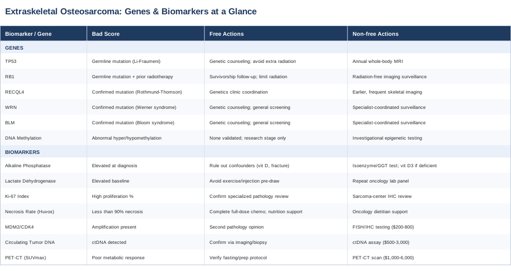
7 Biomarkers Worth Tracking With Extraskeletal Osteosarcoma
Extraskeletal osteosarcoma is a malignant bone-forming tumor that arises in soft tissue rather than in bone itself, most often in the thigh, and it is rare enough that large randomized trials specific to it barely exist. Because of that, oncologists borrow heavily from the biomarker playbook developed for conventional (skeletal) osteosarcoma, adjusting for the fact that soft-tissue tumors do not always behave identically. The StatPearls overview of osteosarcoma and a published case review of extraskeletal osteosarcoma are useful starting points for understanding how closely the two conditions overlap and where they diverge.
Why Tracking Numbers Beats Waiting for the Next Scan
Imaging tells you what a tumor looks like at one moment. Biomarkers, when interpreted correctly and in trend rather than in isolation, can hint at what is happening biologically between scans, flag when a treatment plan needs adjusting, and give a surveillance program more resolution than "wait six months and re-scan." None of the markers below are meant to be self-interpreted outside of an oncology relationship. They are explained here so that when your team mentions them, you already understand the stakes.
Alkaline Phosphatase (ALP)
Alkaline phosphatase is produced by active bone-forming cells, among other tissues, and roughly 30 to 40 percent of people with osteosarcoma have an elevated level at diagnosis. It correlates loosely with tumor burden and bone turnover, and serial measurements are sometimes used alongside imaging to track response and post-treatment surveillance. In extraskeletal disease, elevation is less consistent than in bone-based osteosarcoma, since the tumor is not embedded in the skeleton, so a normal ALP does not rule anything out.
How to measure it: ALP is part of a standard comprehensive metabolic or liver panel, drawn from a routine blood sample. Out of pocket, this typically costs between 10 and 30 US dollars where it is not already bundled into standard oncology labs and insurance coverage.
If the level is elevated, the plan without supplements: the first step is always identifying the source, since liver, biliary, and bone isoenzymes all raise total ALP. Your team may order an isoenzyme fractionation or a gamma-glutamyl transferase (GGT) test to clarify whether the elevation is bone-related. Ruling out confounders like vitamin D deficiency, secondary hyperparathyroidism, a recent fracture, or normal growth-related elevation in children matters more than reacting to a single number.
If the level is elevated, the plan with supplements or equipment: there is no supplement that lowers a cancer-driven ALP elevation, and treating the number directly would be misguided. The one legitimate adjunct is correcting an actual vitamin D deficiency if bloodwork confirms one, since low vitamin D can independently raise ALP through secondary hyperparathyroidism. Typical correction uses 2,000 to 5,000 IU of vitamin D3 daily, retested after 8 to 12 weeks, and stopped or reduced once levels normalize to avoid the rare but real risk of hypercalcemia from over-supplementation.
Frequency and cautions: ALP is usually rechecked at each oncology visit during active treatment, then roughly every three months during the first two years of surveillance, tapering afterward per your team's protocol.
Lactate Dehydrogenase (LDH)
LDH is a nonspecific enzyme released by cells under metabolic stress or turnover, and in osteosarcoma an elevated baseline LDH has been associated with higher tumor burden and, in several cohort studies, with less favorable outcomes. It is one of the more established prognostic blood markers in this disease family, though like ALP it is not specific to cancer and can rise with hemolysis, muscle injury, liver conditions, or strenuous exercise before a blood draw.
How to measure it: LDH is included in most standard metabolic panels, costing roughly 10 to 25 US dollars as a standalone test, and is almost always bundled into routine oncology bloodwork at no extra cost during active treatment.
If the level is elevated, the plan without supplements: avoid strenuous exercise and intramuscular injections in the 48 hours before a blood draw, since both are common non-cancer causes of a falsely elevated result. If elevation persists on a repeat, clean draw, your oncology team will interpret it alongside imaging and other markers rather than in isolation.
If the level is elevated, the plan with supplements or equipment: as with ALP, no supplement meaningfully lowers a cancer-driven LDH elevation, and attempting to "treat the number" with antioxidant megadosing during active chemotherapy is generally discouraged by oncology teams, since some evidence suggests high-dose antioxidants may blunt the oxidative mechanisms some chemotherapy and radiation regimens rely on. Adequate hydration and avoiding unnecessary muscle strain around lab draws is the realistic scope of self-directed action here.
Frequency and cautions: typically drawn at the same intervals as ALP, more frequently during active treatment, spacing out during long-term surveillance.
Ki-67 Proliferation Index
Ki-67 is a protein expressed in actively dividing cells, and pathologists stain tumor tissue for it to estimate what percentage of cells are proliferating at any given time. A higher Ki-67 index generally correlates with a more aggressive, faster-growing tumor and factors into grading and prognosis discussions.
How to measure it: Ki-67 is assessed via immunohistochemistry on biopsy or resected tumor tissue, as part of the standard pathology workup. It is usually bundled into surgical pathology fees, though a standalone stain can run 50 to 150 US dollars if billed separately.
If the score is unfavorable, the plan without supplements: because Ki-67 reflects an existing tumor's intrinsic biology at the moment of biopsy, it is not something lifestyle changes modify after the fact. The practical response is making sure the specimen was reviewed by a pathologist experienced in bone and soft-tissue sarcomas, ideally at a sarcoma referral center, since grading calls can vary between general and specialized pathology labs.
If the score is unfavorable, the plan with supplements or equipment: there is no supplement or device shown to change a tumor's proliferation index. Framing Ki-67 as something to "fix" outside of oncologic treatment would be misleading; its value is in informing treatment intensity and follow-up frequency, not as a personal target.
Frequency and cautions: measured once per tissue specimen, meaning at initial biopsy and again only if recurrent or metastatic tissue is later obtained.
Percentage of Tumor Necrosis After Chemotherapy (Huvos Grade)
For patients who receive neoadjuvant (pre-surgical) chemotherapy, pathologists measure what percentage of the tumor is dead tissue at the time of definitive surgery, graded on the Huvos scale from grade I (minimal necrosis) to grade IV (no viable tumor detected). A necrosis rate of 90 percent or higher is the traditional threshold associated with meaningfully better long-term outcomes in osteosarcoma, and while extraskeletal cases are studied less extensively, the same logic is generally applied.
How to measure it: this requires no separate test; it is generated by the pathologist examining the surgical specimen after chemotherapy and before or during the resection, and is included in standard surgical pathology.
If the necrosis rate is low, the plan without supplements: the most impactful lever is completing chemotherapy at full planned dose and schedule wherever medically tolerable, since dose reductions and delays are a common, modifiable reason for suboptimal response. Proactive management of nausea, mucositis, and nutritional intake during chemotherapy helps patients tolerate full dosing, which indirectly supports the best possible necrosis outcome.
If the necrosis rate is low, the plan with supplements or equipment: no supplement has been shown to increase tumor necrosis in response to chemotherapy, and several oncology teams specifically advise against high-dose antioxidant supplementation during active treatment for the reason noted above. If nutritional status is a concern, working with an oncology dietitian to maintain adequate protein and caloric intake is a more defensible "equipment" than any pill.
Frequency and cautions: this is a one-time measurement tied to a specific surgery; it cannot be retested or cycled.
MDM2/CDK4 Amplification Status
This molecular test looks for amplification of the MDM2 and CDK4 genes in tumor tissue, and it matters because it helps distinguish true extraskeletal osteosarcoma from a dedifferentiated liposarcoma that has developed osteosarcoma-like areas, a distinction that changes both prognosis and treatment approach. Tumors with MDM2/CDK4 amplification generally point toward a liposarcoma lineage rather than pure osteosarcoma.
How to measure it: performed via fluorescence in situ hybridization (FISH) or immunohistochemistry on tumor tissue at diagnosis, typically costing 200 to 800 US dollars when ordered, and generally covered by insurance when clinically indicated for diagnostic clarity rather than ordered as routine screening.
If the result is unfavorable, the plan without supplements: there is no lifestyle lever here at all. The only meaningful action is obtaining a second pathology opinion from a sarcoma referral center if the diagnosis is ambiguous, since misclassification between these two tumor types has real treatment consequences.
If the result is unfavorable, the plan with supplements or equipment: not applicable; this is a fixed molecular characteristic of the tumor's genome, not something influenced by diet, supplements, or external devices.
Frequency and cautions: typically tested once at diagnosis; retesting is only relevant if a new or recurrent tumor is biopsied and classification is again in question.
Circulating Tumor DNA (ctDNA)
ctDNA testing looks for tumor-derived genetic fragments shed into the bloodstream, and in several sarcoma subtypes early research suggests it may detect molecular evidence of recurrence before it becomes visible on imaging. Evidence in osteosarcoma and extraskeletal osteosarcoma specifically is still early-stage and largely investigational rather than a standard of care, so this belongs in your surveillance conversation as an emerging option rather than an expected test.
How to measure it: a blood draw sent to a specialized laboratory, generally costing 500 to 3,000 US dollars depending on the assay and lab, with insurance coverage varying widely and often limited outside of a clinical trial.
If a result is concerning, the plan without supplements: confirm findings with your oncology team through standard imaging and, if needed, tissue biopsy, since ctDNA assays can have false positives and are not yet validated to stand alone as a diagnostic trigger in this tumor type.
If a result is concerning, the plan with supplements or equipment: no supplement or device changes ctDNA shedding; the only "equipment" relevant here is access to a specialized lab and, where available, enrollment in a clinical trial studying ctDNA surveillance in sarcomas.
Frequency and cautions: in research protocols, testing every three months during active surveillance is common; outside of a trial, frequency should be set by your oncology team based on individual risk.
PET-CT Metabolic Response (SUVmax)
A PET-CT scan measures how much glucose tumor tissue is taking up, expressed as a standardized uptake value (SUV). A meaningful drop in SUVmax after a few cycles of chemotherapy is a well-established predictor of good pathological response in bone and soft-tissue sarcomas, and is also used during long-term surveillance to catch metabolically active recurrence.
How to measure it: a PET-CT scan performed at a hospital or imaging center, typically costing 1,000 to 6,000 US dollars without insurance, along with a modest radiation exposure that your team will weigh against the diagnostic benefit.
If the response is poor, the plan without supplements: make sure scan preparation was followed correctly, since fasting status, blood glucose levels, and recent exercise can all distort SUV readings; a poorly prepped scan can look like a poor response when it is actually a measurement artifact. Beyond that, a genuinely poor metabolic response is a signal for your oncology team to reconsider the treatment regimen, not something to self-manage.
If the response is poor, the plan with supplements or equipment: no supplement changes tumor glucose metabolism in a clinically meaningful way. The most useful "equipment" consideration is confirming the scan was read by a radiologist experienced in sarcoma imaging, since interpretation can vary.
Frequency and cautions: commonly performed after 2 to 3 cycles of chemotherapy to assess interim response, then at surveillance intervals of roughly every 3 to 6 months for the first two years, spacing out thereafter.
Building a Practical Tracking Rhythm
No single marker above tells the whole story, and none of them are meant to be tracked outside of an active oncology relationship. What is realistic is keeping your own simple log of dates, values, and scan results so that trends, not single data points, drive the conversation at each visit. A rising ALP alongside a rising LDH is a more meaningful signal than either one alone, and a dip in one value between visits is rarely worth independent action without your care team's context.
The Genes and Inherited Syndromes Behind the Numbers
The biomarkers above describe what a tumor is doing right now. A smaller set of genes explains why some people develop osteosarcoma-family tumors in the first place, and why certain families are placed on more intensive surveillance schedules. None of these genes can be "fixed" with a supplement protocol, and framing them that way would be inaccurate; the realistic plan for each is genetic counseling, informed surveillance, and, where relevant, minimizing avoidable radiation exposure, since several of these genes also impair DNA repair and increase sensitivity to radiation-induced malignancy.
TP53 and Li-Fraumeni Syndrome
TP53 is a tumor-suppressor gene often called the "guardian of the genome" because its protein product helps cells detect DNA damage and either repair it or trigger cell death rather than let a damaged cell keep dividing. Germline (inherited) mutations in TP53 cause Li-Fraumeni syndrome, which carries a markedly elevated lifetime risk across a specific cluster of cancers including osteosarcoma, soft-tissue sarcoma, breast cancer, adrenocortical carcinoma, and certain brain tumors, as detailed in the Li-Fraumeni Syndrome GeneReviews entry. This is one of the more well-established genetic links in this disease family, with decades of human cohort data behind it.
The plan without supplements: genetic counseling and testing for at-risk family members, followed by a structured surveillance protocol, which for confirmed carriers often includes annual whole-body MRI and other imaging designed to avoid ionizing radiation, since TP53 carriers may be more sensitive to radiation-induced tumors.
The plan with supplements or equipment: no supplement restores TP53 function or lowers the associated cancer risk; this is an area where "equipment" genuinely means access to whole-body MRI surveillance rather than any product. Frequency is typically annual for whole-body MRI in confirmed carriers, set and adjusted by a genetics or oncology team, with no meaningful side effects beyond the time and cost of imaging itself.
RB1 and Hereditary Retinoblastoma
RB1 is a tumor-suppressor gene central to cell-cycle regulation, and inherited RB1 mutations cause hereditary retinoblastoma, a childhood eye cancer. Survivors of hereditary retinoblastoma carry a well-documented increased lifetime risk of second cancers, with osteosarcoma being one of the more prominent ones, particularly in radiation fields if radiotherapy was used for the original eye tumor. The Retinoblastoma GeneReviews entry outlines this risk and the surveillance considerations it drives.
The plan without supplements: long-term survivorship follow-up that specifically screens for second malignancies, and avoiding additional unnecessary radiation exposure where clinically possible, given the compounded risk in previously irradiated tissue.
The plan with supplements or equipment: again, no supplement changes RB1 function. Low-dose or radiation-free imaging modalities, used at intervals set by a survivorship clinic, are the realistic "equipment" here rather than any oral intervention.
RECQL4 and Rothmund-Thomson Syndrome
RECQL4 is one of several RecQ helicase genes involved in maintaining genome stability during DNA replication. Mutations cause Rothmund-Thomson syndrome, a rare condition associated with skin changes, skeletal abnormalities, and a substantially elevated risk of osteosarcoma, often at a younger age than sporadic cases. Human evidence here comes mainly from case series and registry data rather than large cohort trials, reflecting how rare the syndrome is.
The plan without supplements: earlier and more frequent skeletal imaging surveillance in known carriers, coordinated through a genetics clinic, since early detection is the main lever available.
The plan with supplements or equipment: no supplement compensates for impaired RecQ helicase function. This is a case where the honest answer is that surveillance, not intervention, is the current standard.
WRN (Werner Syndrome) and BLM (Bloom Syndrome)
WRN and BLM are also RecQ helicase family genes, and mutations in each cause distinct premature-aging and cancer-predisposition syndromes, Werner syndrome and Bloom syndrome respectively, both of which carry elevated risk across several cancer types including sarcomas. Evidence linking these specific genes to osteosarcoma risk comes from smaller case series compared to the TP53 and RB1 literature, so it should be treated as suggestive rather than as strongly established as the Li-Fraumeni association.
The plan without supplements: genetic counseling for affected families, general cancer screening appropriate to the syndrome, and awareness among treating physicians that these patients may need individualized surveillance schedules.
The plan with supplements or equipment: there is no supplement-based intervention with credible evidence for either gene in this context; management is coordinated through a specialist familiar with the syndrome.
DNA Methylation Patterns as an Epigenetic Factor
Beyond inherited gene mutations, osteosarcoma-family tumors frequently show abnormal DNA methylation patterns, including hypomethylation in regions that can activate growth-promoting genes and hypermethylation silencing tumor-suppressor pathways such as the RB and CDKN2A pathways. This is an active area of research rather than a settled clinical tool, and no epigenetic-modifying supplement or intervention has human evidence supporting its use to alter cancer risk or outcomes in this disease. Where this research may eventually matter is in refining which tumors are more likely to respond to specific therapies, an area to watch rather than to act on today.
What Peter Attia's Outlive Gets Right About Catching Cancer Earlier
Peter Attia's book Outlive: The Science and Art of Longevity is not written about extraskeletal osteosarcoma specifically, but its central argument about cancer, one of what he calls the "four horsemen" of premature death, reframes how early detection and risk-tracking should be approached in general. For anyone navigating a rare sarcoma diagnosis or a hereditary predisposition, several of his points translate directly into more useful conversations with an oncology team.
1. Cancer Is Usually Detected Too Late to Matter Most
Attia argues that by the time most cancers are found through symptoms or routine screening, a meaningful fraction have already progressed past the stage where treatment is most effective. This is part of his case for more aggressive, individualized surveillance in higher-risk people rather than waiting for standard population screening intervals.2. Risk Stratification Should Drive Screening Intensity, Not Age Alone
Rather than applying the same screening calendar to everyone, Attia advocates tailoring surveillance intensity to actual risk factors, including family history and known genetic predisposition, which is directly relevant to families carrying TP53 or RB1 mutations.3. A Single Marker Is Rarely Enough
He repeatedly stresses that any one biomarker, imaging result, or genetic finding should be interpreted as part of a pattern, not in isolation, echoing the same principle behind tracking ALP and LDH trends together rather than reacting to one lab value.4. Whole-Body MRI Has a Role for High-Risk Individuals
Attia discusses whole-body MRI as a radiation-free surveillance option worth considering for people at meaningfully elevated cancer risk, a point that aligns with existing surveillance protocols for confirmed Li-Fraumeni carriers.5. Exercise Capacity Is an Underrated Longevity Marker
He places heavy emphasis on cardiorespiratory fitness (VO2 max) as a predictor of all-cause mortality, arguing it belongs alongside traditional biomarkers in a full risk picture, even though it is not a cancer-specific marker.6. Muscle Mass Matters During and After Cancer Treatment
Attia connects preserved muscle mass to better tolerance of aggressive treatment and better long-term outcomes across serious illness, which lines up with the emphasis oncology dietitians place on protein intake during chemotherapy.7. Chronic Low-Grade Inflammation Deserves Attention
He discusses inflammatory markers as part of a broader risk picture, while being careful not to claim that lowering them reverses existing cancer, a distinction worth keeping in mind when reading about inflammation and cancer online.8. Statistical Literacy Changes How You Read a Result
A recurring theme in the book is teaching readers to understand sensitivity, specificity, and false positive rates before reacting emotionally to a screening result, which is directly applicable to interpreting an ambiguous ctDNA or imaging finding.9. Prevention-Minded Medicine Requires an Engaged Patient
Attia is candid that this level of individualized risk management usually requires the patient or family to actively drive conversations with specialists, rather than relying solely on standard-of-care defaults.10. Longevity Strategy Is a Portfolio, Not a Single Bet
His closing framework treats healthspan as the sum of many smaller, evidence-based decisions rather than one dramatic intervention, which is a fair way to think about managing risk around a rare cancer: no single test, gene, or habit does the whole job.Complementary Approaches With Real Evidence in Cancer Care
None of the practices below treat extraskeletal osteosarcoma, and none should replace oncologic care. What they have in common is human clinical evidence, mostly from broader cancer populations rather than this specific rare tumor, showing measurable benefit for the anxiety, pain, fatigue, and quality-of-life burden that comes with cancer diagnosis and treatment.
Mindfulness Meditation
Mindfulness training teaches sustained, nonjudgmental attention to the present moment, and it has become one of the more studied complementary practices in oncology because cancer treatment involves prolonged uncertainty and anticipatory anxiety that standard care often does not directly address. For someone managing a rare diagnosis with an uncertain treatment timeline, it offers a structured way to reduce the mental load of waiting between scans and lab results, without any claim of affecting tumor biology.A randomized controlled trial of a brief mindfulness-based intervention in younger breast cancer survivors found reductions in depressive symptoms and markers of inflammatory activity compared to a control condition, published in this study on mindfulness meditation for younger breast cancer survivors. While this trial was conducted in breast cancer survivors rather than sarcoma patients, the intervention itself is disease-agnostic and the mechanism, reducing psychological distress, generalizes reasonably well.
A realistic starting point is a structured 8-week mindfulness-based stress reduction (MBSR) course, either in person through a hospital's integrative oncology program or through a well-validated app-based program, practiced for 20 to 30 minutes daily. Side effects are minimal, though some people initially find sitting with difficult emotions uncomfortable before it becomes easier, and it works best as a steady practice rather than something used only during acute anxiety spikes.
Yoga
Yoga combines physical movement, breath regulation, and attention training, and its relevance to cancer care comes from consistent evidence of improvements in fatigue, mood, and physical function during and after treatment, which matters for anyone trying to maintain strength and tolerate chemotherapy over months of treatment.A Cochrane systematic review and meta-analysis by Cramer and colleagues, Yoga for improving health-related quality of life, mental health and cancer-related symptoms in women diagnosed with breast cancer, found moderate-quality evidence that yoga improved health-related quality of life and reduced fatigue and sleep disturbance compared to no intervention, with more limited evidence compared to other active interventions like structured exercise.
For someone in active treatment, a gentle, restorative or chair-based yoga class, ideally one specifically designed for cancer patients and taught by an instructor trained in oncology-adapted movement, two to three times per week is a reasonable and low-risk starting point. It should be paused or modified around surgery sites, active bone lesions, or periods of low blood counts, in coordination with the treatment team.
Massage Therapy
Massage therapy is relevant here mainly for pain and anxiety management, both of which are common during the surgical and chemotherapy phases of osteosarcoma-family tumor treatment, and it is one of the more consistently studied complementary approaches for cancer-related pain specifically.A 2023 meta-analysis, Massage therapy can effectively relieve cancer pain, found that massage produced a statistically significant reduction in cancer pain scores compared to control conditions across the pooled trials analyzed.
In practice, this means working with a licensed massage therapist experienced in oncology massage, which uses lighter pressure and avoids direct work over surgical sites, tumor locations, or areas with compromised bone integrity. Sessions of 30 to 60 minutes, once or twice weekly during periods of higher pain or anxiety, are a reasonable frequency, with the main caution being that anyone with active bone lesions should have their oncology team clear specific body areas before treatment to avoid fracture risk.
Music Therapy
Music-based interventions, whether delivered by a trained music therapist or through structured listening protocols, are used in oncology mainly to reduce procedural anxiety and pain perception, which is relevant for the frequent scans, infusions, and procedures that come with sarcoma treatment.A systematic review, Music Therapy: A Noninvasive Treatment to Reduce Anxiety and Pain of Colorectal Cancer Patients, found consistent reductions in self-reported anxiety and pain across the reviewed studies, reinforcing findings seen in other cancer populations.
A practical approach is using a personally chosen playlist of calming, familiar music during infusions, scans, or recovery periods, or working with a hospital-based music therapist if one is available through the treatment center. There is essentially no downside risk beyond personal preference, making it one of the lowest-barrier complementary options on this list.
The Bottom Line
Extraskeletal osteosarcoma is rare enough that no single article, biomarker, or gene will give a complete picture of any one person's situation. What actually helps is understanding what each number on a lab report or pathology result is measuring, which ones are realistically modifiable and which are not, and where the inherited genetic component fits into a family's broader risk picture. The seven biomarkers and six genetic factors covered here are the pieces your oncology team is most likely already tracking; knowing what they mean turns a confusing printout into a real conversation.
The most useful next step is a practical one: bring a simple log of your recent ALP, LDH, and imaging results to your next appointment, ask directly whether genetic counseling or testing is appropriate for your specific case, and ask which of the complementary approaches above your treatment center already offers through its integrative oncology program. Better information does not change the biology of a tumor, but it consistently changes the quality of the decisions made around it.