This article contains AI generated content.
· UpdatedFabry Disease: 4 Genes And 6 Biomarkers To Track
Introduction
Living with Fabry disease — or supporting someone who does — means navigating a condition that most physicians encounter only a handful of times in their careers. The rarity of this diagnosis often translates into delayed care, generic symptom management, and a frustrating gap between what patients experience and what standard protocols address. If you have been through rounds of tests before someone finally connected the dots, this article is for you.
Fabry disease is caused by mutations in the GLA gene, which encodes the enzyme alpha-galactosidase A. When this enzyme is absent or severely reduced, a fatty molecule called globotriaosylceramide (Gb3) accumulates over decades in the kidneys, heart, nerves, and skin. The result is a multi-system disease that progresses silently until it causes strokes, kidney failure, or serious cardiac complications. The problem with generic advice here is that Fabry is not one disease — it is dozens, shaped by which mutation a person carries, which organs are most affected, and how quickly the damage accumulates.
Better information genuinely changes outcomes. Patients who track the right biomarkers catch organ damage earlier, when intervention still matters most. Those who understand how certain modifier genes shape their disease trajectory can have more productive conversations with specialists. And those who know which non-pharmaceutical approaches may support lysosomal function are often better prepared to optimize everything around their enzyme replacement therapy or chaperone medication.
This article takes two angles. First, it covers the 6 most clinically meaningful biomarkers to track in Fabry disease — what each one reveals, how to measure it, what it costs, and what to do when the result is poor. Second, it examines the 4 genes most relevant to Fabry disease, including both the causative mutation and the modifier genes that influence severity through lysosomal biology and cellular stress pathways. Understanding both layers gives you a clearer picture of your disease and practical options that go beyond standard care.
6 Biomarkers to Track in Fabry Disease
Standard Fabry monitoring typically covers kidney function panels and cardiac imaging. But research over the past fifteen years has revealed a richer set of biomarkers that capture disease activity, treatment response, and organ risk with much greater precision. Tracking these six — ideally in collaboration with a metabolic disease specialist — gives a far more complete picture of what is actually happening.
1. Lyso-Gb3 (Globotriaosylsphingosine): The Sharpest Signal Available
Lyso-Gb3 is the deacylated form of Gb3 and currently the most sensitive biomarker for Fabry disease monitoring. Unlike Gb3 itself, lyso-Gb3 is elevated in virtually all patients with classic Fabry disease and, critically, in most female carriers who would otherwise test normal on enzyme activity assays. This makes it uniquely valuable in a condition where standard enzyme testing frequently misses affected women due to X-chromosome inactivation.
Why it matters: Plasma lyso-Gb3 correlates with disease severity, progression rate, and treatment response. Patients on enzyme replacement therapy (ERT) who achieve sustained lyso-Gb3 reduction typically show better preservation of organ function over time. Persistently elevated lyso-Gb3 despite treatment signals inadequate disease control or accelerated progression.
How to measure it: Lyso-Gb3 is measured in plasma via liquid chromatography-tandem mass spectrometry (LC-MS/MS), a specialized test performed at reference laboratories or academic Fabry centers. Cost ranges from approximately $150 to $400 depending on the facility. Coverage varies by insurer; verify in advance. In healthy individuals, lyso-Gb3 is typically below 0.5–1.0 nmol/L. In classic Fabry males, levels commonly exceed 50–200 nmol/L. Female carriers often show values in the 2–20 nmol/L range, which are clearly abnormal.
If the score is bad — the plan without supplements: The primary intervention for elevated lyso-Gb3 is enzyme replacement therapy (agalsidase alfa or agalsidase beta administered every 2 weeks intravenously) or, for patients with amenable GLA mutations, migalastat (an oral pharmacological chaperone). Beyond medical therapy, a diet lower in glycosphingolipid-rich foods — particularly processed red meat and certain full-fat dairy — may modestly reduce substrate load. Adequate hydration supports renal clearance. Avoiding hyperthermia, excessive cold, and infection-driven inflammation reduces symptomatic burden while lyso-Gb3 remains elevated. Monitoring frequency: every 6–12 months minimum.
If the score is bad — the plan with supplements or equipment: No supplement has been shown in human Fabry trials to directly reduce lyso-Gb3. However, compounds that support lysosomal function show preclinical interest. Trehalose (a disaccharide that activates TFEB, the lysosomal biogenesis regulator) has been studied in lysosomal storage disease models at 3–5g/day; generally well tolerated. Curcumin as phytosome or with piperine: 500–1000mg/day; cycle 8 weeks on, 2 weeks off; GI upset is possible at high doses and drug interactions with blood thinners warrant caution. These are not replacements for ERT — discuss any additions with your metabolic specialist. Research on lyso-Gb3 in Fabry disease monitoring.
2. Plasma and Urine Gb3/GL-3: Tracking the Substrate Burden
Globotriaosylceramide (Gb3, also called GL-3) is the primary substrate that accumulates when α-Gal A enzyme is deficient. It can be measured in plasma, urine, or tissue biopsies. Urine Gb3 was historically a cornerstone of Fabry monitoring, though lyso-Gb3 has largely superseded it in sensitivity. Gb3 nonetheless remains useful for tracking renal substrate burden specifically and for longitudinal comparisons in patients established on this marker.
Why it matters: Urinary Gb3 reflects the kidney's substrate load and correlates with podocyte dysfunction — the specialized kidney cells that are among the earliest targets of Gb3 toxicity. Plasma Gb3 gives a systemic view but is more subject to dietary and technical variation than lyso-Gb3.
How to measure it: Both plasma and urine Gb3 are measured by LC-MS/MS at reference laboratories. Cost is similar to lyso-Gb3 ($100–350). Some specialized centers batch both tests simultaneously, which can reduce cost and turnaround time.
If the score is bad — the plan without supplements: ERT or migalastat is the primary intervention. Dietary glycosphingolipid reduction, hydration, and regular monitoring every 6–12 months are the supporting measures. The trend over successive measurements matters as much as any single value.
If the score is bad — the plan with supplements or equipment: The lysosomal support compounds noted for lyso-Gb3 (trehalose, curcumin) apply here as well. N-acetylcysteine (NAC) is also of interest as a general lysosomal stress reducer through its antioxidant replenishment of glutathione. Dose: 600mg twice daily. Cycling at 12 weeks on, 2 weeks off is reasonable. GI upset is the most common initial side effect. Always confirm with your specialist before adding NAC to a regimen that includes ERT, as drug interaction data in Fabry is limited.
3. Alpha-Galactosidase A (α-Gal A) Enzyme Activity: The Diagnostic Anchor
Measuring α-Gal A enzyme activity is the cornerstone of Fabry diagnosis in males. It is affordable, widely available, and definitive in classic presentations. However, it has a critical limitation: up to 30–40% of females with Fabry disease have normal or borderline enzyme activity due to random X-chromosome inactivation (lyonization). Enzyme testing alone is insufficient for female carriers — genetic sequencing and lyso-Gb3 measurement are always required alongside it.
How to measure it: Enzyme activity is measured in leukocytes (blood draw), plasma, or dried blood spots (DBS). The DBS method is the most accessible — samples can be mailed to specialized labs and are used for newborn screening and confirmatory testing. Cost ranges from $50–300 depending on sample type and laboratory. In classic Fabry males, activity is typically less than 1% of normal. Late-onset Fabry males retain 5–30%. Females show a wide and unreliable range.
If the score is bad — the plan without supplements: Low enzyme activity in a confirmed Fabry patient is a key consideration for ERT initiation — guided by the treating metabolic physician and based on overall disease burden and organ involvement. Enzyme assay results also help evaluate migalastat eligibility: if enzyme activity increases on migalastat challenge, the mutation is likely amenable and the chaperone is stabilizing residual protein. Regular enzyme monitoring (every 6–12 months) tracks stability.
If the score is bad — the plan with supplements or equipment: No supplement restores α-Gal A activity in patients with loss-of-function GLA mutations. Migalastat (Galafold) is not a supplement — it is a targeted prescription drug for patients whose specific GLA mutation allows some residual protein folding. Preclinical interest exists in endoplasmic reticulum stress-reducing compounds (such as berberine 500mg twice daily) for potentially stabilizing misfolded proteins with partial activity, but human evidence for this in Fabry disease is absent. This remains a research-stage direction.
4. eGFR (Estimated Glomerular Filtration Rate): The Kidney Clock
Kidney disease is one of the most serious long-term consequences of Fabry disease. eGFR measures kidney filtration capacity, and in untreated Fabry patients it typically declines at approximately 3–5 mL/min/year — substantially faster than the age-related decline of 1–2 mL/min/year seen in healthy adults. ERT initiated before significant damage can slow this trajectory meaningfully, making eGFR trajectory one of the most clinically consequential numbers to watch.
Why it matters: A single normal reading can be misleading. What matters is the slope — a persistent downward trend of even 2–3 mL/min/year beyond the expected rate signals a red flag requiring ERT optimization or nephrology input. eGFR below 60 mL/min/1.73m² indicates chronic kidney disease Stage 3, a threshold associated with significantly higher cardiovascular and renal event risk.
How to measure it: eGFR is calculated from serum creatinine combined with age and sex, available in standard metabolic panels ($10–30). However, cystatin C-based eGFR (CKD-EPI Creat-Cys) is more sensitive for early decline and less affected by muscle mass variation — an important distinction given that Fabry patients may have reduced muscle mass from fatigue or restricted activity. Cystatin C adds $25–75. Peter Attia specifically recommends cystatin C as the more accurate early detection tool.
If the score is bad — the plan without supplements: Blood pressure control is essential (target 130/80 or lower in proteinuric Fabry patients). A low-protein diet (0.8g/kg body weight) reduces hyperfiltration demands on damaged nephrons. Sodium restriction below 2g/day reduces fluid overload and glomerular pressure. Adequate hydration prevents concentration-related nephron stress. SGLT2 inhibitors (e.g., empagliflozin, dapagliflozin) are increasingly used in Fabry-related CKD for their nephroprotective effects, pending Fabry-specific trial data.
If the score is bad — the plan with supplements or equipment: Omega-3 fatty acids (2–4g/day combined EPA+DHA) have evidence for reducing proteinuria and modestly preserving eGFR in CKD populations. CoQ10 (ubiquinol form, 200–400mg/day) is studied in mitochondrial and lysosomal disease contexts for supporting cellular energy production in energy-deprived nephrons. Side effects at standard doses are minimal — mild GI upset occasionally. Always coordinate supplementation with your nephrology team. Research on eGFR progression and ERT in Fabry disease.
5. Urine Albumin-to-Creatinine Ratio (UACR): The Early Kidney Warning
Proteinuria — protein leaking into the urine — frequently appears before eGFR begins to fall in Fabry disease, making UACR one of the earliest measurable signs of renal podocyte damage. The spot urine UACR is convenient (no timed collection required) and reproducible enough for longitudinal tracking.
Why it matters: In Fabry patients, UACR above 30 mg/g (microalbuminuria) signals active glomerular injury. Above 300 mg/g (macroalbuminuria), the risk of progressive renal failure rises sharply and the urgency for intervention increases substantially. UACR is also an independent predictor of cardiovascular events, making it doubly important in a condition already carrying elevated cardiac risk.
How to measure it: Spot urine collection — first morning urine provides the most reliable result. Cost: $15–40. Should be measured every 6–12 months in all Fabry patients, and more frequently if already elevated. Some centers prefer a 24-hour urine protein collection for confirmation of macroalbuminuria.
If the score is bad — the plan without supplements: ACE inhibitors (ramipril, enalapril) and ARBs (losartan, valsartan) are standard-of-care for Fabry-related proteinuria, even in normotensive patients once UACR exceeds 300 mg/g. Sodium restriction (less than 2g/day), blood pressure optimization, ERT initiation or dose optimization, and avoidance of nephrotoxic drugs (NSAIDs, certain antibiotics) all contribute. High-protein diets should be avoided.
If the score is bad — the plan with supplements or equipment: The DASH dietary pattern — high in plant foods, low in sodium and processed meat — has evidence for reducing urinary albumin in CKD populations. Omega-3s (2–4g/day) appear here again, with evidence for reducing urinary protein excretion. Emerging interest surrounds sulforaphane (from broccoli sprouts or standardized supplements, 25–50mg/day) for its Nrf2-activating and renal oxidative stress-reducing properties. 4–6 week cycles are typical in research protocols; side effects are minimal. This is extrapolated from general CKD and Nrf2 research — no Fabry-specific human data exist yet.
6. NT-proBNP and High-Sensitivity Cardiac Troponin T: The Heart's Early Signals
Cardiac involvement in Fabry disease — including hypertrophic cardiomyopathy, arrhythmias, and progressive heart failure — is a leading cause of morbidity and mortality. NT-proBNP (N-terminal pro-B-type natriuretic peptide) reflects cardiac wall stress, while high-sensitivity cardiac troponin T (hs-cTnT) detects subtle ongoing myocardial cell injury. Together they track cardiac disease activity more dynamically than echocardiography alone and can signal whether ERT is adequately protecting the heart.
How to measure it: Both are standard blood tests available at most hospital labs. NT-proBNP cost: $30–100. Hs-cTnT cost: $30–80. Frequency: every 6–12 months as part of standard Fabry monitoring, or more often when cardiac involvement is significant. NT-proBNP above 125 pg/mL is typically flagged. Hs-cTnT above 14 ng/L (males) or 9 ng/L (females) is considered elevated. Trends over time matter as much as absolute values.
If the score is bad — the plan without supplements: Elevated NT-proBNP or troponin requires cardiology evaluation in a center familiar with Fabry disease. Primary interventions include ERT optimization, blood pressure control, avoidance of dehydration, extreme temperatures, and stimulants. Some Fabry patients with cardiac arrhythmia or symptomatic cardiomyopathy benefit from beta-blockers or antiarrhythmic agents — always guided by a Fabry-specialized cardiologist. Moderate, regular aerobic exercise is generally safe and cardioprotective unless contraindicated.
If the score is bad — the plan with supplements or equipment: CoQ10 (ubiquinol, 200–400mg/day) has evidence for supporting cardiac mitochondrial function in cardiomyopathy contexts. Magnesium glycinate (300–400mg/day) may help with arrhythmia susceptibility and cardiac wall stress; it is inexpensive, well-tolerated, and does not require cycling. Omega-3s (EPA+DHA, 2–4g/day) have established evidence for reducing arrhythmia risk and supporting cardiac outcomes in at-risk populations. Blood thinning at these doses is a consideration if anticoagulants are already prescribed. Research on cardiac biomarkers in Fabry disease.
With biomarker tracking established, the next layer of understanding involves the genes driving these abnormalities — and the modifier genes that explain why two people with Fabry disease can have vastly different disease trajectories.
The Genetic Architecture of Fabry Disease
Understanding your biomarkers tells you what is happening right now. Understanding the underlying genetics tells you why — and points toward additional levers beyond standard therapy. The genetics of Fabry disease is more nuanced than most patients are initially told.
1. The GLA Gene: The Root of the Diagnosis
The GLA gene, located on the X chromosome at position Xq22.1, encodes alpha-galactosidase A. Over 1,000 pathogenic and likely pathogenic variants have been reported. These range from single nucleotide changes (missense mutations) to large deletions. The specific mutation type is highly predictive of disease severity.
Mutation type and phenotype: Nonsense mutations, frameshift variants, and large deletions typically cause classic Fabry disease — near-zero enzyme activity, childhood onset, and severe multi-organ involvement. Missense mutations causing partial enzyme dysfunction often lead to late-onset phenotypes that disproportionately affect the heart. The distinction matters enormously for prognosis and treatment eligibility.
Variants of uncertain significance (VUS): A growing challenge in Fabry genetics is the VUS — a DNA change that cannot yet be classified as clearly pathogenic or benign. If your genetic report includes a VUS, functional enzyme assay data and lyso-Gb3 levels become essential tools for clinical interpretation. Do not rely on the VUS classification alone.
If the mutation is confirmed — the plan without supplements: Medical management is the central pillar: ERT (intravenous, every 2 weeks) or migalastat for amenable mutations. Genetic counseling for family members is critical — mothers, sisters, and daughters of affected males should be offered screening. A comprehensive monitoring plan with a Fabry center (kidney, cardiac, neurological) should be established. Newborn screening programs are expanding in many countries.
If the mutation is confirmed — the plan with supplements or equipment: No supplement replaces enzyme function for classic GLA mutations. Migalastat (Galafold) is a targeted prescription pharmacological chaperone for patients whose mutation allows some residual enzyme folding — not a supplement, but a highly relevant option for eligible patients. For late-onset mutations with residual activity, any approach that reduces protein misfolding stress (avoiding extreme heat, infections, and oxidative stressors) may help preserve the remaining enzyme function. GLA gene variant research in Fabry disease.
2. TFEB: The Lysosomal Master Switch
TFEB (Transcription Factor EB) is not mutated in Fabry disease, but it is functionally impaired by the disease's downstream effects. TFEB is the master transcription factor controlling lysosomal biogenesis — it activates hundreds of genes involved in lysosomal production, autophagy, and cellular waste clearance. In healthy cells, TFEB translocates into the nucleus during nutrient deprivation or cellular stress, triggering a lysosomal production program.
In lysosomal storage disorders including Fabry disease, Gb3 accumulation drives dysregulation of the mTORC1-TFEB axis. mTORC1, anchored to the lysosomal surface, phosphorylates TFEB and traps it in the cytoplasm — blocking the very lysosomal response that could help clear substrate. This creates a compounding cycle: more Gb3 accumulates, mTOR is inappropriately activated, TFEB is suppressed further, and lysosomal function progressively deteriorates.
Research status: Multiple preclinical studies have demonstrated that activating TFEB in Fabry disease cell and animal models reduces Gb3 accumulation and improves cellular function. Trehalose and curcumin both act (in part) through TFEB activation pathways. Human clinical data remain limited but the pathway is considered a high-priority adjunct target. TFEB activation research in lysosomal storage disorders.
If TFEB signaling appears compromised — the plan without supplements: TFEB is activated by two powerful non-pharmaceutical stimuli: caloric restriction/fasting and aerobic exercise. Both suppress mTORC1, allowing TFEB to enter the nucleus and activate lysosomal biogenesis. Time-restricted eating (16:8 pattern) and zone 2 aerobic exercise (60–70% max heart rate, 30–45 minutes, 4–5 times per week) are the most evidence-supported approaches. Exercise must be individualized in Fabry disease — avoid hyperthermia and ensure adequate cooling.
If TFEB signaling appears compromised — the plan with supplements or equipment: Trehalose (3–5g/day) is generally well tolerated and has lysosomal storage disease model data. Curcumin (phytosome or piperine-enhanced, 500–1000mg/day): cycle 8 weeks on, 2 weeks off; GI upset possible at higher doses. Resveratrol (250–500mg/day) activates SIRT1 which cross-talks with the mTOR-TFEB axis; 8-week cycling is reasonable. All three are adjunct, supportive approaches — not replacements for ERT. Introduce one at a time to monitor response.
3. MTOR: The Cellular Traffic Controller
mTOR (mechanistic target of rapamycin) sits at the intersection of nutrient sensing, cellular growth, immune response, and lysosomal function. It is one of the most studied signaling nodes in biology. Under healthy conditions, mTOR cycles predictably with nutrient availability — high when calories are abundant, low during fasting or stress. In cells burdened with Gb3, this calibration breaks down: mTOR becomes inappropriately activated, blocking autophagy and preventing TFEB-mediated lysosomal cleanup.
This chronic mTOR dysregulation contributes to the cellular damage seen in Fabry podocytes (leading to proteinuria), cardiomyocytes (contributing to hypertrophy), and dorsal root ganglion neurons (contributing to neuropathic pain). Addressing the mTOR-autophagy axis is therefore relevant to multiple organ systems simultaneously.
Research status: Rapamycin (an mTOR inhibitor used clinically for transplant immunosuppression) has been studied in lysosomal storage disease models with encouraging results. At very low "rapalog" doses, it may enhance autophagy without the immunosuppressive profile seen at therapeutic doses. This remains experimental and is not part of standard Fabry care. mTOR pathway research in lysosomal storage disease.
If mTOR appears dysregulated — the plan without supplements: Fasting, caloric restriction, and aerobic exercise are the principal tools. High-intensity interval training (HIIT) has a particularly strong effect on mTOR modulation through AMPK activation. 2–3 sessions per week (20 minutes each, 4×4 or Tabata format) are well supported. In Fabry disease, HIIT must be practiced cautiously — avoid exercising in hot environments, maintain adequate cooling, and start with shorter intervals before progressing.
If mTOR appears dysregulated — the plan with supplements or equipment: Berberine (500mg twice daily) activates AMPK and modulates mTOR, with established human evidence in metabolic conditions. Often described as having "metformin-like" cellular effects. Cycle 8–10 weeks on, 2 weeks off; GI side effects (bloating, loose stools) are common initially and typically resolve. Metformin (prescription only) also activates AMPK — some longevity researchers have explored low-dose protocols, though not Fabry-specific. Any prescription addition requires physician oversight.
4. ABCB1 (MDR1/P-Glycoprotein): The Transport Variable
ABCB1 encodes P-glycoprotein (P-gp), a membrane efflux transporter that pumps a wide range of substrates — including certain lipids and drugs — out of cells. Common variants in ABCB1, particularly rs1045642 (C3435T) and rs1128503, alter P-gp expression and activity. These polymorphisms have been studied as phenotypic modifiers in various conditions involving lipid accumulation and drug transport.
Research status: The evidence linking ABCB1 variants to Fabry disease outcomes is early and observational. A small number of studies have suggested that ABCB1 genotype may influence intracellular lipid handling and potentially modulate how cells respond to glycosphingolipid accumulation. This is not established at the clinical level. It is included here as a research-stage modifier that may become more actionable as pharmacogenomics advances and Fabry biobank data matures. ABCB1 and lysosomal lipid handling research.
If you carry an ABCB1 variant of concern — the plan without supplements: At this stage, there are no specific clinical interventions targeting ABCB1 variants in Fabry disease. The most reasonable approach is heightened monitoring — more frequent biomarker assessment (lyso-Gb3, UACR, eGFR) — to detect any accelerated progression early.
If you carry an ABCB1 variant of concern — the plan with supplements or equipment: Some natural compounds including quercetin (500–1000mg/day) and piperine (5–20mg, typically paired with curcumin) modulate P-glycoprotein expression. However, modulating P-gp pharmacologically carries complex drug interaction implications. This should not be attempted independently — especially when on ERT or migalastat — without specialist guidance. Consult a clinical pharmacologist or metabolic specialist if this gene variant is confirmed.
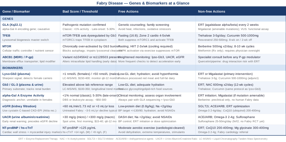
Genes and Biomarkers at a Glance
The table below summarizes all four genes and six biomarkers covered in this article, their concerning thresholds, and the most relevant free and non-free action steps.
Outlive by Peter Attia: 10 Cellular Insights That Reframe Fabry Disease Management
Peter Attia's 2023 book Outlive: The Science and Art of Longevity was not written about Fabry disease — but it may be one of the most useful frameworks a Fabry patient or caregiver can engage with, because it provides a systematic, evidence-grounded approach to cellular health that maps directly onto the mechanisms driving Fabry disease. Attia's central argument — that medicine should focus on preserving cellular and organ function long before disease appears, not after — challenges the default posture of rare disease management and suggests a more proactive toolkit.
1. mTOR Is a Dial, Not a Switch
Attia dedicates significant attention to mTOR as a signaling pathway that needs cycling — high when cellular building is needed, low to enable autophagy and cleanup. In lysosomal storage conditions like Fabry disease, the mTOR dial gets stuck high, preventing the cellular self-cleaning that could reduce substrate burden. The implication: lifestyle interventions that periodically lower mTOR (fasting, exercise) are mechanistically relevant, not just generally healthy.
2. Zone 2 Cardio Is Mitochondrial Medicine
Attia makes a compelling case that sustained low-intensity aerobic exercise — zone 2, approximately 60–70% of maximum heart rate — is one of the most powerful tools available for improving mitochondrial efficiency and lysosomal function. For Fabry patients, zone 2 cardio offers a way to activate TFEB, suppress mTOR, and support cardiac and renal cellular health without the hyperthermia risk associated with high-intensity effort.
3. Fasting Activates the Cellular Cleanup Crew
Caloric restriction and time-restricted eating lower mTOR and activate autophagy — the cellular process by which damaged organelles and protein aggregates are cleared. In Fabry disease, where lysosomal function is already compromised, supporting autophagy through fasting (even a daily 14–16 hour overnight window) provides a non-pharmaceutical contribution to cellular maintenance. Evidence for autophagy induction in humans through these protocols is strong.
4. Muscle Mass Is a Metabolic Buffer
Attia frames muscle as the most underappreciated longevity organ. For Fabry patients dealing with fatigue, neuropathic pain, and exercise intolerance, maintaining muscle through progressive resistance training is both metabolically protective and functional. Resistance training also independently activates AMPK — which modulates mTOR and supports the cellular energy sensing that lysosomal function depends on.
5. Cystatin C-Based eGFR Is More Sensitive for Early Detection
Attia specifically advocates for cystatin C as a more accurate eGFR marker than creatinine-only calculations, particularly in the early stages of kidney function decline. Cystatin C is not affected by muscle mass, diet, or exercise — variables that confound creatinine-based estimates. In Fabry disease, where catching kidney decline before it becomes irreversible is a clinical priority, this is a directly actionable recommendation: ask your nephrologist for the CKD-EPI Creat-Cys equation rather than creatinine alone.
6. ApoB Matters More Than LDL for Cardiovascular Risk
Drawing on the work of lipidologist Thomas Dayspring and cardiologist Allan Sniderman, Attia argues that apolipoprotein B (ApoB) is a more accurate predictor of atherosclerotic cardiovascular event risk than LDL cholesterol. Fabry patients with cardiac involvement should track ApoB alongside standard lipid panels — particularly because Fabry-related cardiomyopathy already creates elevated baseline cardiac risk.
7. Continuous Glucose Monitoring Reveals Hidden Metabolic Stress
Attia describes using continuous glucose monitoring (CGM) not only for diabetes management but as a real-time window into metabolic health — including insulin sensitivity and post-meal glucose spikes. Metabolic stress and chronic hyperglycemia accelerate lysosomal dysfunction and cellular aging. A CGM worn for 2–4 weeks reveals dietary and lifestyle patterns that create unnecessary metabolic load and can guide more precise nutritional adjustments.
8. Sleep Deprivation Is Organ Damage in Slow Motion
Attia treats sleep as a non-negotiable foundation for cellular repair, mTOR cycling, and brain waste clearance through the glymphatic system. In Fabry disease, where neuropathic pain and autonomic dysfunction frequently disrupt sleep architecture, prioritizing sleep quality becomes a therapeutic target. Target: 7–9 hours in a dark, cool (65–68°F / 18–20°C) room. Sleep tracking via a wearable can help identify patterns and improvement over time.
9. Protein Intake Requires Careful Calibration in Kidney Disease
Attia recommends higher protein intake (1.6–2.2g per kg body weight) for muscle preservation in most healthy adults. But Fabry patients with proteinuria or declining eGFR face a genuine tension: higher protein intake increases glomerular filtration demands and can accelerate kidney damage. The practical resolution is to target the lower end of protein adequacy (0.8–1.2g/kg) for those with kidney involvement, while optimizing protein quality — prioritizing leucine-rich sources (fish, eggs, legumes) over quantity.
10. Emotional Health Is Part of the Treatment Stack
Attia is unusually candid about the role of psychological health in longevity outcomes, dedicating a full chapter to what he calls "the missing pillar." For Fabry patients — who frequently experience long diagnostic delays, chronic pain, social isolation, and anxiety about disease progression — addressing emotional health through structured therapy, peer support networks, and stress-reduction practices is not peripheral. It is mechanistically relevant: chronic stress elevates cortisol, which disrupts mTOR regulation, worsens sleep, and impairs immune function.
Complementary Approaches with Evidence for Fabry Disease
Fabry disease involves chronic neuropathic pain, autonomic dysfunction, GI symptoms, and cardiac and renal stress. Several evidence-informed complementary modalities address these dimensions. None replace standard medical care, but the three below have meaningful clinical evidence and practical application.
Mindfulness Meditation and MBSR for Neuropathic Pain
Neuropathic pain — burning extremity pain known as acroparesthesias — is one of the most debilitating features of Fabry disease, particularly in children and young adults. It is poorly responsive to standard analgesics and significantly affects quality of life. Mindfulness-Based Stress Reduction (MBSR) is an 8-week structured program teaching body scanning, breath awareness, and non-reactive attention to pain sensations. It does not eliminate pain but consistently reduces pain catastrophization — the amplifying cognitive response to pain — and improves functional capacity.
A randomized controlled trial in JAMA Internal Medicine found that MBSR significantly reduced pain severity and improved daily functioning in adults with chronic pain compared to standard care. While not Fabry-specific, the mechanism (modulation of central pain processing) is relevant across neuropathic pain conditions sharing similar central sensitization features. The evidence base for MBSR in chronic neuropathic pain is robust enough to warrant consideration as an adjunct.
For practical integration in Fabry disease: begin with 10–15 minutes of daily body scan practice focused on the painful extremities, working up to the full MBSR protocol over 4–8 weeks. The program requires 8 weekly group sessions of approximately 2.5 hours plus a full-day retreat and 30–45 minutes of daily home practice. Accessible options include Palouse Mindfulness (free, online), University of Massachusetts MBSR-certified programs, or clinical MBSR embedded in pain management clinics. Consistent practice across 8 weeks or more appears necessary for lasting neurological benefit.
Biofeedback for Autonomic Dysfunction
Fabry disease causes autonomic nervous system dysfunction — impaired sweating (anhidrosis or hypohidrosis), abnormal heart rate variability, gastrointestinal dysmotility, and temperature dysregulation. These are direct consequences of autonomic nerve fiber involvement by Gb3 accumulation. Biofeedback provides real-time physiological feedback — on heart rate variability, skin conductance, or respiratory rate — that allows individuals to consciously regulate otherwise automatic bodily functions.
Heart rate variability (HRV) biofeedback has the strongest evidence base for autonomic conditions. Clinical studies in dysautonomia-adjacent populations have demonstrated improved vagal tone, reduced symptom burden, and enhanced stress resilience with regular HRV biofeedback practice. Vagal tone improvement is particularly relevant for Fabry patients given the widespread autonomic nerve involvement.
Protocol: HRV biofeedback can be practiced using accessible consumer devices — the Polar H10 chest strap paired with the Elite HRV app, or the Inner Balance sensor, provide reliable HRV feedback. The standard technique involves paced breathing at the individual's resonance frequency (typically around 0.1 Hz, approximately 6 breaths per minute). Sessions of 20 minutes, 4–5 times per week, produce measurable autonomic improvements over 6–8 weeks. Discuss with your cardiologist before beginning if significant arrhythmia is present.
Breathing-Based Therapies for Stress and GI Symptom Management
Beyond structured biofeedback, deliberate slow breathing practices directly address autonomic dysregulation at low cost and with no equipment barrier. Coherent breathing (5–6 breath cycles per minute), diaphragmatic breathing, and the physiological sigh (double nasal inhale followed by a prolonged oral exhale) all shift autonomic balance toward parasympathetic dominance. This matters for Fabry patients because autonomic dysregulation drives GI dysmotility, sleep disruption, temperature intolerance, and cardiovascular instability.
Research from the Huberman Lab at Stanford and other groups has demonstrated that brief physiological sigh protocols (2–5 minutes) significantly reduce subjective stress and measurable physiological arousal markers compared to mindfulness meditation or rest alone. For Fabry patients with autonomic instability, stress-triggered GI symptoms, or sleep disruption, daily breathing practice represents a zero-cost, zero-risk intervention with genuine mechanistic relevance to the autonomic pathology of the disease.
Protocol: 5 minutes of coherent breathing (5 seconds in through the nose, 5 seconds out through the nose) twice daily — morning and before sleep. Progress to 10–15 minutes over 4 weeks. Include 2–3 physiological sighs at moments of acute stress or pain. No equipment is required. Avoid extended breath holds if syncope risk is present or if cardiac arrhythmia is active. This can be started independently and adjusted based on individual tolerance.
Conclusion
Fabry disease demands a level of self-knowledge that goes well beyond most chronic condition management. The six biomarkers covered here — lyso-Gb3, Gb3/GL-3, α-Gal A enzyme activity, eGFR, UACR, and cardiac markers — form a practical monitoring framework that captures disease activity, organ risk, and treatment response with far greater precision than standard annual checkups. The four genes — GLA, TFEB, MTOR, and ABCB1 — explain not just why Fabry occurs, but how its underlying cellular machinery can be influenced through lifestyle, nutrition, and targeted supplements as genuine complements to ERT or migalastat.
The next smart step is not to try everything at once. Start by asking your Fabry specialist about adding lyso-Gb3 and cystatin C-based eGFR to your monitoring panel if they are not already there. Add zone 2 aerobic exercise if your cardiologist clears it. Consider MBSR if neuropathic pain is significantly affecting your quality of life. Approach any supplement mentioned here by introducing one at a time, at conservative doses, with your care team informed. Better information will not replace medical treatment — but it consistently improves the quality of the decisions made around it.
Neurological: Nerve Conditions
Cardiovascular: Heart Conditions Heart Rhythm Conditions
Urological: Kidney Conditions