This article contains AI generated content.
· UpdatedFacioscapulohumeral Muscular Dystrophy Genes Biomarkers - 5 Genes And 6 Biomarkers To Track
Introduction
Facioscapulohumeral muscular dystrophy (FSHD) is not a simple genetic disease. Unlike conditions where a gene is broken or missing, FSHD is caused by a gene that should be permanently silent — waking up in tissue where it has no business being active. That distinction matters enormously, because it means the disease operates at the intersection of genetics and epigenetics: the DNA sequence may be the vulnerability, but it is the epigenetic environment that determines whether that vulnerability becomes damage.
For people living with FSHD, the standard clinical experience often feels incomplete. A genetic confirmation, a severity staging, perhaps some physical therapy — and then a wait-and-see approach that can feel more like acceptance than strategy. What rarely gets discussed in a clinical appointment is the specific molecular picture: which genes are driving the damage, what amplifies or dampens them, and what biological signals in your blood or imaging could tell you whether the disease is active or stable right now.
Generic advice about exercise and lifestyle, while not wrong, misses too much when applied to FSHD. The disease is highly asymmetric, variable in pace, and sensitive to specific stressors — particularly the kind of oxidative and mechanical stress that most general muscle advice actively encourages. A smarter approach looks at the specific drivers and the specific signals. That's the goal of this article: not to promise recovery, but to put more precise information in your hands.
Two main angles are covered here. The first looks at the five genes and epigenetic factors most centrally involved in FSHD, what each one does when dysregulated, and what current research suggests can be done about it — both with and without supplementation. The second tracks six biomarkers that reflect FSHD disease activity in real time. Beyond those two tracks, the article also covers insights from one of the most cited researchers in epigenetic aging, and which complementary approaches have meaningful human evidence for conditions like FSHD. Better information doesn't guarantee better outcomes, but it consistently leads to better decisions.
Summary
This article maps the five key genes driving FSHD — DUX4, SMCHD1, DNMT3B, PITX1, and FRG2 — with practical protocols for each: what to do without supplements, and what to add when lifestyle alone isn't enough. It then covers six biomarkers — creatine kinase, MRI fat fraction, DUX4-target transcripts, inflammatory cytokines, serum myoglobin, and complement proteins — that can tell you whether the disease is active, stable, or responding to intervention. Beyond genes and biomarkers, there's a summary of David Sinclair's epigenetic research and what it means specifically for FSHD, plus evidence-reviewed complementary approaches including photobiomodulation, breathing therapy, and biofeedback. The article won't promise a reversal, but it will give you a map that most clinical consultations don't.
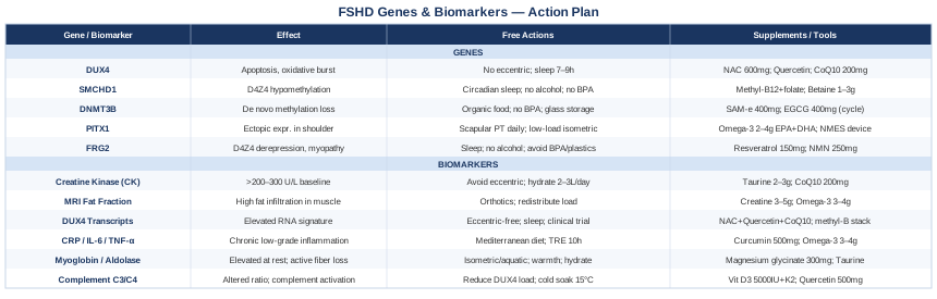
The Genetics and Epigenetics Behind FSHD: What the Research Now Suggests
FSHD stands apart from most heritable muscle diseases because the pathogenic mechanism is fundamentally epigenetic. The gene causing the disease — DUX4 — is not mutated. It is inappropriately expressed because the epigenetic silencing system that normally keeps it locked shut has been partially dismantled. Understanding what maintains that silencing, what breaks it, and what may help restore it is where FSHD research has made its most significant advances over the last fifteen years.
What follows is an examination of the five most clinically significant genetic and epigenetic players in FSHD, with specific and practical guidance for each.
Gene 1: DUX4 — The Aberrantly Awakened Master Driver
DUX4 is a double homeobox transcription factor that is normally expressed only in early embryonic cells and the germline. In healthy adult muscle, it is completely silenced. In FSHD, it escapes that silencing and is expressed intermittently in individual muscle cells — not constantly, and not in every fiber, but in sporadic bursts that are nonetheless sufficient to trigger widespread damage.
When DUX4 activates in a muscle cell, it turns on hundreds of downstream targets that are biologically inappropriate for that context: embryonic genes, germline genes, pro-apoptotic factors, and retroelements. It also directly interferes with MYOD1 and PAX7 — two transcription factors essential for muscle satellite cell activation and repair. The result is a muscle that loses its ability to regenerate properly and is progressively replaced by fat and fibrotic tissue.
The underlying cause of DUX4 activation is a contraction of the D4Z4 macrosatellite repeat array on chromosome 4q35. Healthy individuals carry 11–100 copies; FSHD patients typically have 1–10. Fewer repeats mean less capacity to maintain the repressive heterochromatin that silences DUX4. For DUX4 to be pathogenic, the contraction must also co-exist with a 4qA haplotype that provides a functional polyadenylation signal, stabilizing the DUX4 mRNA. This two-hit requirement explains why D4Z4 contractions on chromosome 10 do not cause FSHD — chromosome 10's sequence lacks that polyadenylation signal.
If the DUX4 gene is dysregulated — the plan without supplements
The most important lifestyle intervention for managing DUX4-related damage is eccentric exercise avoidance. Eccentric contractions — muscle lengthening under load, as in downhill walking, slow lowering during weightlifting, or plyometrics — are mechanically damaging to FSHD muscle and have been shown to amplify oxidative stress in ways that can trigger DUX4 expression bursts. Replace eccentric-heavy activities with concentric or isometric protocols: cycling, swimming, water resistance exercises, and low-resistance isometric holds are significantly safer. Frequency: 30–45 minutes of low-impact movement five days per week; daily gentle walking is better than two intense sessions.
Sleep quality is the second major lever. DUX4 activation produces reactive oxygen species that compound throughout the day; adequate, consistent sleep (7–9 hours at a fixed schedule) allows cellular repair systems to contain that oxidative burden. Chronic sleep restriction impairs antioxidant capacity and mitochondrial function — both already compromised downstream of DUX4. Stress management matters similarly: sustained cortisol elevation promotes muscle catabolism and systemic inflammation, amplifying the damage DUX4 initiates even in skeletal muscles that are not the primary FSHD target.
If the DUX4 gene is dysregulated — the plan with supplements or equipment
N-acetylcysteine (NAC): NAC is a glutathione precursor that directly addresses the oxidative stress generated by DUX4 target activation. Preclinical studies show that oxidative damage amplifies DUX4-mediated myoblast death, making antioxidant buffering mechanistically relevant. Dose: 600–1200 mg/day, split into two doses. Cycling: many practitioners use five days on, two days off to prevent glutathione feedback inhibition. Side effects: nausea at higher doses; potential bronchospasm in susceptible individuals with asthma. Evidence specific to FSHD is preclinical; the oxidative stress rationale is strong.
Quercetin: A plant flavonoid with documented anti-inflammatory and anti-apoptotic properties relevant to the pathways DUX4 activates. Dose: 500–1000 mg/day with food. Can be taken continuously. Side effects: mild; potential interaction with anticoagulants. Pair with 200–500 mg vitamin C to improve absorption. Choose standardized supplements from third-party tested sources.
Coenzyme Q10 (ubiquinol form): Supports mitochondrial electron transport, which is impaired in cells undergoing DUX4-driven metabolic stress. The ubiquinol form has substantially better bioavailability than ubiquinone, particularly in older individuals. Dose: 100–300 mg/day with a fat-containing meal. No established cycling protocol; continuous use is standard. Side effects: mild GI effects at higher doses; generally very well tolerated.
GeneReviews: Facioscapulohumeral Muscular Dystrophy — Molecular Basis and Clinical Overview
Gene 2: SMCHD1 — The Epigenetic Gatekeeper
SMCHD1 (Structural Maintenance of Chromosomes Flexible Hinge Domain-Containing 1) is an epigenetic repressor protein that plays a central role in maintaining the silencing of D4Z4. It does this by binding to the D4Z4 chromatin region and facilitating the recruitment of DNA methylation machinery — particularly DNMT3 enzymes — that maintain the dense methylation marks keeping DUX4 locked shut.
In FSHD type 2, heterozygous loss-of-function mutations in SMCHD1 are the primary causative mechanism. The D4Z4 array is of normal length, but without functional SMCHD1, it cannot maintain its methylated, repressive state. DUX4 is then free to be expressed. In FSHD type 1, SMCHD1 functions as a disease modifier: patients who carry a D4Z4 contraction and also have reduced SMCHD1 activity — whether through mutation or epigenetic downregulation — tend to have earlier onset and more severe disease than those with the contraction alone.
SMCHD1 is also involved in X chromosome inactivation and genomic imprinting, which means its dysregulation has broader cellular consequences beyond the D4Z4 locus. Importantly, SMCHD1 expression and activity decline with aging and with chronic inflammatory states — which may partly explain why FSHD often accelerates in midlife even when the underlying genetic lesion has been present since birth.
If the SMCHD1 gene is dysregulated — the plan without supplements
Because SMCHD1 supports DNA methylation maintenance, the most actionable non-supplement intervention is protecting the conditions under which methylation enzymes function optimally. Circadian rhythm alignment is the most underrecognized factor: consistent sleep timing within a 30-minute window each night has been shown to support epigenetic maintenance systems across multiple studies. Night shift work and irregular sleep schedules are associated with global DNA hypomethylation, directly relevant here.
Alcohol reduction is critical and specific: ethanol and its metabolite acetaldehyde are among the most potent known disruptors of DNA methylation, and even moderate intake (2–3 drinks per day) has been documented to reduce DNMT activity in muscle-adjacent tissues. Complete elimination during periods of active disease or monitoring is worth considering. Additionally, reducing exposure to BPA and phthalates (from plastic containers, canned food linings, and certain personal care products) removes documented methylation disruptors from the equation — a straightforward change with meaningful epigenetic relevance.
If the SMCHD1 gene is dysregulated — the plan with supplements or equipment
Methylated B vitamins (folate + B12 + B6): SMCHD1 loss reduces the cell's capacity to methylate D4Z4, but if the methyl-donor supply chain is optimized, the enzymes that remain functional have adequate substrate to work with. L-methylfolate (800 mcg/day), methylcobalamin (1000 mcg/day sublingual), and pyridoxal-5-phosphate (25–50 mg/day) form the core of a methylation support stack. Avoid synthetic folic acid in individuals with known MTHFR variants (C677T or A1298C), as it competes with the active form at cellular receptors. Side effects: generally very safe at these doses; B6 above 100 mg/day long-term can cause peripheral neuropathy. No cycling required at standard doses.
Betaine (trimethylglycine, TMG): A methyl donor that supports the remethylation of homocysteine back to methionine, feeding the SAM (S-adenosylmethionine) cycle — the primary methyl donor for all DNA methylation reactions. Dose: 1000–3000 mg/day with food. Side effects: fish odor at high doses; mild LDL elevation in some individuals. Monitor lipids if using above 2000 mg/day long-term.
Gene 3: DNMT3B — The Methylation Enzyme Itself
DNMT3B is one of the primary de novo DNA methyltransferases — enzymes that add new methylation marks to previously unmethylated DNA sequences. During normal development, DNMT3B is responsible for establishing the dense methylation pattern across D4Z4 repeats that silences DUX4. In some FSHD2 patients, DNMT3B mutations are the causative variant (rather than SMCHD1), and in a broader population of FSHD1 patients, reduced DNMT3B activity compounds the vulnerability created by D4Z4 repeat contraction.
Beyond the D4Z4 locus, DNMT3B plays a role in silencing inappropriate gene programs during muscle differentiation. When it is underactive, the epigenetic barricade that prevents muscle cells from expressing embryonic, germline, or non-muscle genes becomes porous. This is why DNMT3B is also relevant to understanding age-related FSHD acceleration: DNMT3B activity declines globally with aging, and this decline may explain why some patients who were mildly affected in their thirties experience more rapid progression in their fifties without any change in their underlying genetic variant.
If the DNMT3B gene is dysregulated — the plan without supplements
Reducing environmental methylation disruptors is the most specific non-supplement approach for DNMT3B. Organophosphate pesticides — found as residues on conventionally grown produce — have been documented to impair DNMT3 activity in human cell studies. Choosing organic for the highest-residue produce (strawberries, spinach, peppers, and similar items frequently cited in residue monitoring data) reduces this exposure without requiring an entirely organic diet. BPA from plastic food containers and canned food linings is similarly documented to interfere with DNMT3B-mediated methylation in mammalian studies; switching to glass, ceramic, or stainless steel food storage is a low-effort, high-relevance swap.
If the DNMT3B gene is dysregulated — the plan with supplements or equipment
SAM-e (S-adenosylmethionine): The direct methyl donor that DNMT3B uses to add methylation marks to DNA. Supplemental SAM-e provides that substrate directly, bypassing the folate-methionine cycle steps that may be rate-limiting. Dose: 400–800 mg/day on an empty stomach (food significantly reduces absorption). Cycling: 6 weeks on, 2 weeks off is a common clinical protocol. Side effects: GI upset, and importantly, activation or agitation in people with bipolar disorder (SAM-e should be avoided without psychiatric supervision in that population). Start at 200 mg and titrate up.
EGCG (green tea catechin extract): At physiologically relevant concentrations, EGCG has been studied as a DNMT modulator — some in vitro evidence suggests it can influence DNA methylation patterning. Dose: 400–800 mg/day of a standardized extract (≥45% EGCG), taken with food to reduce gastric irritation. Cycle 4 weeks on, 2 weeks off due to hepatotoxicity risk at sustained high doses. Avoid taking on an empty stomach.
Gene 4: PITX1 — Ectopic Expression Shaping the Topography of Weakness
PITX1 is a homeodomain transcription factor whose normal function is to specify lower limb identity during embryogenesis. In healthy adult muscle, it is not expressed in upper limb tissue. In FSHD, DUX4 aberrantly activates PITX1 in shoulder and upper arm muscles — tissue where this gene has no normal function and where its presence actively disrupts muscle maintenance programs.
This ectopic PITX1 expression is thought to be a key explanation for one of FSHD's most puzzling features: its highly selective topographic pattern. Not all muscles degenerate equally in FSHD — certain shoulder stabilizers, facial muscles, and upper arm muscles are preferentially affected, while adjacent muscles may be spared entirely. Ectopic PITX1 likely disrupts the contractile protein and structural gene expression that is specific to those upper limb muscle types, making them disproportionately vulnerable. Research by Pandey and colleagues has proposed that the upper limb topography of FSHD closely mirrors the normal expression domain of PITX1 in lower limb development — an almost architectural explanation for an otherwise mysterious pattern.
PITX1 also appears to suppress the compensatory hypertrophic response in affected muscles, meaning that even when exercise is applied correctly, the normal anabolic signaling that would drive protective muscle growth is partially blocked in FSHD-affected tissue.
If the PITX1 gene is dysregulated — the plan without supplements
Since PITX1 is a downstream target of DUX4, all interventions that reduce DUX4 expression bursts are indirectly protective against ectopic PITX1 activity. Additionally, targeted scapular stabilization physical therapy — focusing on serratus anterior activation and lower trapezius strengthening — can preserve functional shoulder mechanics even as specific muscles weaken. This is best done in low-load, high-frequency patterns: 10–15 minutes daily of specific stabilization exercises is more effective and safer than weekly high-intensity sessions. A physiotherapist experienced in neuromuscular disease should design this protocol, as inappropriate loading can worsen the damage that ectopic PITX1 has already initiated.
If the PITX1 gene is dysregulated — the plan with supplements or equipment
Omega-3 fatty acids (EPA + DHA): Well-documented for their anti-inflammatory effects on muscular and neuroinflammatory pathways that amplify the downstream damage of PITX1 and DUX4 activation. The triglyceride form of fish oil has substantially better bioavailability than the ethyl ester form. Dose: 2–4 g/day of combined EPA+DHA, with the higher end more appropriate for active inflammation. Side effects: fishy reflux (take with meals, freeze capsules), mild blood thinning at higher doses (pause 1 week before surgery). No cycling required; continuous use is standard.
Neuromuscular electrical stimulation (NMES): A device-based therapy that delivers low-level electrical current to activate muscle fibers directly, without the eccentric mechanical stress of voluntary contractions. For muscles weakened by PITX1-driven pathology, NMES can maintain fiber activation and local blood flow while avoiding the oxidative burst associated with heavy voluntary exercise. Small studies in related neuromuscular conditions show preservation of muscle mass with NMES. Use under physiotherapist guidance; protocols typically involve 20–30 minutes per session, 3–5 days per week, at intensities below the pain threshold.
Gene 5: FRG2 — The Upregulated Neighbor
FRG2 (FSHD Region Gene 2) sits on chromosome 4q35, in close proximity to the D4Z4 array. It is consistently upregulated in FSHD muscle, and while its exact function remains under active investigation, overexpression of FRG2 in animal models produces myopathic changes — alterations in muscle fiber architecture and gene expression patterns that parallel aspects of human FSHD. Some evidence suggests FRG2 interacts with RNA splicing regulatory networks, potentially disrupting the normal processing of muscle-specific transcripts.
What makes FRG2 particularly relevant beyond its own pathological activity is that it may serve as a sentinel marker for D4Z4 derepression. When D4Z4 chromatin opens — whether due to reduced D4Z4 copy number, SMCHD1 loss, or DNMT3B deficiency — both DUX4 and FRG2 are liberated. Tracking FRG2 expression therefore provides a broader picture of epigenetic derepression at the locus than DUX4 alone. Some researchers argue that FRG2 contributes independently to FSHD pathology, meaning that therapeutic strategies targeting only DUX4 may leave a parallel pathogenic pathway unaddressed.
If the FRG2 gene is dysregulated — the plan without supplements
FRG2 upregulation is mechanistically tied to the same D4Z4 chromatin opening that releases DUX4, so the lifestyle interventions most relevant here are those that support chromatin compaction and epigenetic silencing at the 4q35 locus more broadly. Sleep consistency, alcohol elimination, BPA avoidance, and circadian alignment — all described above in the context of SMCHD1 and DNMT3B — apply equally here. There is no FRG2-specific non-supplement protocol established in human research; managing the epigenetic environment of D4Z4 is the best currently available approach.
If the FRG2 gene is dysregulated — the plan with supplements or equipment
Resveratrol: Activates SIRT1, a class III histone deacetylase (HDAC) that promotes chromatin compaction. Compaction of D4Z4 chromatin is precisely what FSHD patients need to sustain — it is the loss of that compaction that allows both DUX4 and FRG2 to escape silencing. Dose: 150–500 mg/day of trans-resveratrol (the active isomer) with a fat-containing meal. Cycling: 5 days on, 2 days off to prevent SIRT1 desensitization through sustained activation. Side effects: GI upset at high doses; mild blood-thinning potential; choose independently tested brands for purity. Evidence is extrapolated from sirtuin biology; no FSHD-specific clinical trials of resveratrol exist.
NMN or NR (NAD+ precursors): NAD+ is the required cofactor for SIRT1 and other sirtuins involved in heterochromatin maintenance. NAD+ levels decline significantly with age and with chronic inflammation — both relevant to FSHD progression. Supplementing with nicotinamide mononucleotide (NMN) or nicotinamide riboside (NR) supports the NAD+ pool that enables sirtuin-mediated chromatin maintenance. Dose: NMN 250–500 mg/day or NR 300–600 mg/day, taken in the morning (NAD+ metabolism has a circadian rhythm). Side effects: generally mild; minor flushing at higher doses. No FSHD-specific trials; rationale rests on NAD+-sirtuin-chromatin biology.
NINDS: Facioscapulohumeral Muscular Dystrophy Information Page
With the genetic and epigenetic landscape mapped, it becomes important to know how to track what's actually happening in the body over time — which is where biomarkers become the practical companion to the genetic picture above.
Tracking FSHD Activity: 6 Biomarkers That Tell You More
Genetics tells you the vulnerability. Biomarkers tell you whether and how aggressively that vulnerability is being expressed right now. For FSHD, a small but growing set of biomarkers can distinguish stable disease from active progression, help evaluate whether lifestyle or supplementation changes are having any effect, and flag windows when disease activity is high and additional protective measures are most warranted.
Biomarker 1: Creatine Kinase (CK)
Why it matters: Creatine kinase leaks from muscle cells when the cell membrane is damaged or disrupted. In FSHD, elevated CK reflects active muscle fiber injury — the kind caused by DUX4-induced apoptosis, eccentric mechanical overload, or inflammation. CK is not perfectly specific to FSHD activity (it also rises after any intense exercise, viral illness, or trauma), but in the context of FSHD, a consistently elevated baseline CK, or a CK that rises after what should be a gentle activity, signals that muscle damage is ongoing.
How to measure it: Standard blood draw at any clinical laboratory. Cost: $10–$40 depending on provider and insurance. Results are typically available within 24 hours. The reference range (usually below 200 U/L for women, below 300 U/L for men) is calibrated for the general population; some FSHD patients have chronically elevated CK at baseline due to ongoing low-grade muscle injury. A series of measurements over time — not a single snapshot — is what matters.
If the score is bad, the plan without supplements
An elevated CK in FSHD should trigger an immediate review of recent physical activity for any eccentric or high-load components that may have provoked it. Reducing activity intensity for 2–4 weeks and switching entirely to isometric and aquatic modalities while the CK normalizes is the first step. Adequate hydration (2–3 liters of water daily) supports renal clearance of myoglobin released alongside CK. Sleep extension during high-CK periods allows maximal overnight tissue repair. If CK remains elevated without any obvious provocative activity, it warrants discussion with a neurologist to assess for an inflammatory flare.
If the score is bad, the plan with supplements or equipment
Taurine: An amino acid with established membrane-stabilizing properties in skeletal muscle, and documented ability to reduce CK levels post-exercise in clinical studies. Dose: 2–3 g/day with water. No cycling required; continuous use is safe. Side effects: minimal at standard doses; very well tolerated. Best taken spread across two doses per day.
CoQ10 (ubiquinol): Supports mitochondrial efficiency and reduces oxidative damage that drives CK elevation. Dose: 200–300 mg/day with fat. Side effects: minor GI effects. No cycling required.
Biomarker 2: MRI Muscle Fat Fraction
Why it matters: MRI fat fraction — specifically quantitative muscle MRI (qMRI) measuring the proportion of a muscle's volume replaced by fat — is the single most specific and sensitive biomarker for FSHD structural progression. Unlike CK, which reflects acute damage, fat fraction reflects accumulated irreversible loss: muscle fibers that have been destroyed and replaced by fat and fibrous tissue. In clinical research, qMRI fat fraction is the primary endpoint used to detect disease progression in FSHD trials and is increasingly used to monitor individual patients in specialized centers.
How to measure it: Quantitative MRI of the shoulder girdle, upper arms, and lower extremity muscle groups. This is not a standard clinical MRI — it requires dedicated acquisition sequences (Dixon technique or similar) and specialized analysis software. Cost: $500–$2,000 depending on the facility and insurance coverage; available at academic medical centers and FSHD specialty clinics. A baseline scan followed by annual repeats allows detection of progression rates that would otherwise be invisible on clinical examination.
If the score is bad, the plan without supplements
High fat fraction in specific muscle groups is irreversible — fat-replaced muscle does not regenerate. The practical response is to prioritize protecting muscles with low or intermediate fat fraction (still partially functional) while redistributing load away from heavily affected muscles. A physiotherapist experienced in neuromuscular disease can design a compensation strategy using less-affected muscle groups. Orthotic supports — particularly scapular orthoses and AFOs (ankle-foot orthoses) where relevant — offload mechanically vulnerable muscles and reduce the ongoing stress that accelerates fat infiltration.
If the score is bad, the plan with supplements or equipment
Creatine monohydrate: Among the best-studied supplements for preserving muscle mass in neuromuscular conditions. A randomized controlled trial in FSHD (Walter et al., 2002) found that creatine supplementation produced modest but significant improvements in muscle function. Dose: 3–5 g/day continuously (no loading phase needed for long-term use). Side effects: water retention in the first 1–2 weeks; minimal thereafter. Well tolerated in most individuals; monitor renal function if there is any underlying kidney disease.
Omega-3 fatty acids: Documented to slow the rate of muscle atrophy and support mitochondrial function in aging and disease-related sarcopenia. May slow the rate of fat infiltration when combined with appropriate physical activity. Dose: 3–4 g/day EPA+DHA.
Biomarker 3: DUX4-Regulated Gene Transcripts
Why it matters: DUX4 itself is expressed at extremely low levels and is technically challenging to detect directly in blood. However, a signature of DUX4-regulated downstream genes — including MBD3L2, ZSCAN4, LEUTX, and KHDC1L — can be detected in blood RNA. This "DUX4 activity signature" provides a real-time molecular readout of how actively DUX4 is being expressed in muscle. Research groups have validated these signatures as tools for stratifying patients by disease activity and, potentially, for monitoring the effect of DUX4-suppressing therapeutic interventions as they emerge from clinical trials.
How to measure it: Currently available primarily through academic research contexts and specialized biomarker panels being developed by FSHD research consortia. It is not a standard commercial test. Cost in research settings: variable; may be available through enrollment in clinical studies at FSHD specialty centers. As DUX4-targeting therapies advance through clinical trials, commercial versions of this panel are anticipated to become available.
If the score is bad, the plan without supplements
An elevated DUX4-target gene signature confirms active DUX4 expression and warrants all of the DUX4-suppression lifestyle strategies: eccentric exercise elimination, sleep extension, alcohol removal, circadian stabilization, and methylation disruptor avoidance. It also represents the best possible window to consider enrolling in a clinical trial of an antisense oligonucleotide or small molecule DUX4 suppressor — trials that are actively recruiting and represent the most promising therapeutic direction in FSHD.
If the score is bad, the plan with supplements or equipment
The NAC, quercetin, and CoQ10 stack described under DUX4 above is the most directly relevant supplementation response to an elevated DUX4 transcript signature. Additionally, the methylation support stack (methylated B vitamins, betaine, SAM-e) addresses the upstream epigenetic environment that permits DUX4 expression. These two stacks work on different levels — one managing the downstream oxidative damage, the other trying to support the upstream silencing — and can reasonably be used in combination with medical oversight.
Biomarker 4: Inflammatory Cytokines — CRP, IL-6, and TNF-α
Why it matters: FSHD is not primarily an inflammatory disease, but chronic low-grade inflammation plays a significant amplifying role in its progression. DUX4 activation recruits innate immune cells to affected muscle, and the resulting inflammatory environment — marked by elevated interleukin-6 (IL-6), tumor necrosis factor-alpha (TNF-α), and C-reactive protein (CRP) — accelerates muscle fiber death beyond what DUX4 alone would cause. Patients with persistently elevated inflammatory markers tend to have faster progression and worse quality of life. Conversely, cases with low baseline inflammation show more indolent courses, even when D4Z4 contraction size is similar.
How to measure it: High-sensitivity CRP (hs-CRP) is available at any standard laboratory: cost $10–$50. IL-6 is more specialized: $50–$150, available at larger reference labs. TNF-α is even more specialized and primarily used in research contexts. A practical starting point is hs-CRP alone as a readily available inflammatory signal; IL-6 adds precision if the clinical picture warrants it.
If the score is bad, the plan without supplements
An anti-inflammatory dietary pattern is the most accessible and best-evidenced intervention for elevated cytokines. A Mediterranean-style diet — rich in olive oil, fatty fish, legumes, vegetables, and low in processed foods and refined sugars — has been shown across multiple meta-analyses to reduce hs-CRP and IL-6. For FSHD specifically, eliminating processed seed oils (soybean, corn, sunflower oil), reducing refined carbohydrates, and increasing colorful vegetables provides the dietary anti-inflammatory foundation. Time-restricted eating (eating within a 10–12 hour window) has also been shown to reduce fasting inflammatory markers in multiple controlled trials.
If the score is bad, the plan with supplements or equipment
Curcumin (phytosomal or liposomal form): One of the most consistently studied natural anti-inflammatory agents; inhibits NF-κB signaling, which drives IL-6 and TNF-α production. Standard curcumin has poor bioavailability — choose phytosomal (Meriva), liposomal, or BCM-95 formulations. Dose: 500–1000 mg/day of a bioavailable form. Cycle 8 weeks on, 2 weeks off. Side effects: GI upset at high doses; mild blood-thinning. Pair with black pepper extract (piperine) if standard curcumin is used.
Omega-3 fatty acids: 3–4 g EPA+DHA/day has robust evidence for reducing IL-6 and CRP. This supplement appears in multiple sections of this article because it addresses several FSHD-relevant pathways simultaneously — one of its strongest arguments for inclusion.
Biomarker 5: Serum Myoglobin and Aldolase
Why it matters: Myoglobin is an oxygen-binding protein in muscle cells that leaks into the bloodstream when muscle fibers are disrupted — similar to CK but with a faster clearance rate (hours versus days). Aldolase is a glycolytic enzyme also released from damaged muscle. Together, they form a complementary pair of muscle damage markers that can capture different time windows of injury. In FSHD, elevated myoglobin between exercise episodes indicates ongoing resting muscle breakdown — not just post-exercise damage — and correlates with active DUX4-driven apoptosis.
How to measure it: Both are available from standard clinical labs. Myoglobin: $30–$80. Aldolase: $30–$70. Neither is routinely ordered in standard FSHD follow-up, but they can be requested specifically and are particularly useful when CK is borderline and the clinical picture is unclear. A pattern of elevated myoglobin and aldolase without preceding intense exercise is a meaningful signal of active resting muscle damage.
If the score is bad, the plan without supplements
Elevated resting myoglobin calls for the same activity modification protocol as elevated CK: a 3–4 week reduction to purely isometric and aquatic activity, concurrent with sleep prioritization and hydration. Warmth (warm baths, heating pads on affected areas) promotes local circulation and may accelerate clearance of inflammatory products from affected tissue. Monitoring myoglobin every 4–6 weeks during this modified period tracks whether the intervention is working before returning to higher-intensity activity.
If the score is bad, the plan with supplements or equipment
Magnesium glycinate: Magnesium is required for over 300 enzymatic reactions including ATP synthesis and muscle membrane stabilization. Deficiency — common in people with chronic disease — amplifies muscle cell membrane fragility, increasing myoglobin leak. Dose: 300–400 mg/day elemental magnesium (as glycinate for best tolerance). Continuous use; side effects: loose stools if dose is too high — titrate gradually. Test serum magnesium (or ideally RBC magnesium) before supplementing.
Taurine: As noted under CK, taurine stabilizes muscle cell membranes and reduces both CK and myoglobin release post-exercise in clinical studies. Its mechanisms include calcium regulation within the muscle cell and antioxidant activity at the membrane. Dose: 2–3 g/day continuously.
Biomarker 6: Complement System Proteins (C3, C4, and Complement Activation)
Why it matters: The complement system — a branch of innate immunity — has emerged as an unexpectedly important player in FSHD pathology. DUX4-induced cell death releases intracellular antigens that can activate complement cascades, and complement deposition has been identified in FSHD muscle biopsies. Elevated C3 and altered C3/C4 ratios can signal that complement-mediated tissue damage is contributing to muscle injury beyond what inflammation markers alone reveal. Some researchers position complement activation as a therapeutic target in FSHD, and complement-inhibiting drugs are in early investigation for related muscular dystrophies.
How to measure it: C3 and C4 levels are available from standard labs: $50–$150 combined. A CH50 (total hemolytic complement) test ($80–$200) provides a broader picture of complement pathway activity. These are not standard tests in FSHD monitoring protocols, but can be ordered as part of a comprehensive immunological workup. Low C3 or C4 combined with elevated inflammatory markers suggests active complement consumption.
If the score is bad, the plan without supplements
Complement activation is driven partly by the burden of cellular debris and damaged membranes that DUX4-induced apoptosis produces. All interventions that reduce the rate of DUX4-driven muscle fiber death — eccentric exercise avoidance, sleep optimization, the anti-inflammatory diet — reduce the substrate for complement activation. Cold water immersion (10–15 minutes at 15–18°C) has been shown in healthy subjects to modulate complement activation transiently; whether this translates to benefit in FSHD is speculative, but it is low-risk.
If the score is bad, the plan with supplements or equipment
Vitamin D3 + K2: Vitamin D has documented immune-modulatory effects including complement regulation. FSHD patients, like many people with chronic muscle disease and reduced outdoor activity, frequently have suboptimal vitamin D levels. Dose: 4000–6000 IU/day D3 with 100–200 mcg/day MK-7 form of K2 (to direct calcium appropriately). Monitor serum 25-OH vitamin D every 3–6 months; aim for 50–80 ng/mL. Side effects at recommended doses: minimal; toxicity occurs only at sustained doses above 10,000 IU/day without monitoring.
Quercetin (already part of the DUX4 stack): Also has documented complement-modulating properties — particularly inhibition of complement-mediated lysis — making it relevant for this biomarker track as well, reinforcing its inclusion in a broad FSHD-relevant stack.
The genetics and biomarker tracks above give a molecular and physiological map of FSHD. But some of the most clinically useful frameworks for thinking about this disease come from researchers studying epigenetic aging — a field that intersects directly with FSHD's core mechanism.
David Sinclair's Epigenetic Framework and What It Means for FSHD
David Sinclair, professor of genetics at Harvard Medical School, has spent the past two decades building what he calls the Information Theory of Aging — the idea that aging is primarily an epigenetic phenomenon: a progressive loss of the cellular "software" that tells each cell which genes to express and which to silence. His book Lifespan: Why We Age — and Why We Don't Have To (2019) synthesizes decades of research on sirtuins, NAD+, methylation, and epigenetic reprogramming into a framework that is directly relevant to FSHD, even though FSHD is not its focus.
Here are the ten most impactful ideas from Sinclair's framework for someone living with or studying FSHD:
1. Epigenetic Noise Is the Disease
Sinclair's central claim is that what we call aging — and by extension, many epigenetic diseases — is fundamentally the accumulation of epigenetic noise: the loss of precise gene expression patterns that define cellular identity. In FSHD, this framing is unusually literal: the disease is precisely a loss of the epigenetic noise control that keeps DUX4 silent. What Sinclair describes as the aging process at the population level is, in FSHD, happening in specific muscle tissue at an accelerated rate due to a structural genetic vulnerability.
2. Sirtuins Are the Guardians — and They Need NAD+
Sinclair's research has focused heavily on sirtuins — particularly SIRT1, SIRT3, and SIRT6 — as the proteins responsible for maintaining epigenetic fidelity. They compact chromatin, repair DNA, and silence inappropriate gene expression. They all require NAD+ as a cofactor. When NAD+ drops — as it does with aging, inflammation, and chronic disease — sirtuin activity falls, and epigenetic maintenance erodes. In FSHD, where sirtuin-dependent chromatin compaction at D4Z4 is essential for DUX4 silencing, maintaining NAD+ levels is directly mechanistically relevant.
3. NAD+ Boosting Is Practical and Well-Studied
Sinclair is an advocate of NMN supplementation for NAD+ support — he takes it himself and has published research on NMN's effects in animal models. Human trials of NMN and NR have now shown that both effectively raise blood NAD+ levels. The practical implication for FSHD: supplementing with NMN (250–500 mg/day) or NR (300–600 mg/day) may support the sirtuin activity needed for D4Z4 heterochromatin maintenance. This is extrapolated biology, not a clinical trial result in FSHD — but the mechanistic logic is sound.
4. The Methylation Clock Is a Measurable Proxy for Epigenetic Age
Sinclair cites Steve Horvath's DNA methylation clock — a tool that estimates biological age from methylation patterns at specific CpG sites across the genome — as one of the most important recent advances in aging science. For FSHD patients, this clock is particularly relevant: if D4Z4 hypomethylation reflects a broader state of epigenetic dysregulation, the biological clock may run faster in FSHD muscle tissue. Biological age testing (available commercially) can provide a rough proxy for how effectively epigenetic maintenance is functioning.
5. Intermittent Fasting Activates the Same Pathways as Caloric Restriction
One of Sinclair's most practical recommendations is intermittent fasting — specifically time-restricted eating within a 6–10 hour window — as a way to activate SIRT1 and AMPK, which in turn support epigenetic maintenance. Fasting activates the same molecular pathways as caloric restriction without requiring sustained calorie reduction. For FSHD patients who cannot engage in high-intensity exercise as a metabolic stimulus, time-restricted eating offers a low-risk way to engage these epigenetic maintenance pathways. A 16:8 protocol (16 hours fasting, 8-hour eating window) is the most accessible starting point.
6. Heat Stress Supports Epigenetic Resilience
Sinclair discusses heat shock proteins and the hormetic stress response — brief, controlled stressors that upregulate repair and maintenance systems. Regular sauna use (3–4 sessions per week, 15–20 minutes at 80–90°C) activates heat shock proteins, supports NAD+ metabolism, and has been shown in Finnish cohort studies to reduce all-cause mortality. For FSHD patients, passive heat (sauna, hot bath) avoids the mechanical muscle stress of exercise while still engaging metabolic and epigenetic maintenance pathways. Evidence specific to FSHD is absent; the general hormetic principle applies.
7. Resveratrol Was the Original Sirtuin Activator — and It Still Has a Role
Sinclair's early career was defined by the discovery that resveratrol activates SIRT1. While the direct sirtuin-activation mechanism has since been debated, resveratrol's downstream effects — including chromatin compaction, anti-inflammatory signaling, and NAD+ pathway support — remain relevant. Sinclair takes resveratrol with olive oil daily. For FSHD patients, the chromatin compaction rationale described above makes it a reasonable adjunct. Dose: 150–500 mg/day trans-resveratrol with fat; cycle 5 days on, 2 days off.
8. Metformin May Have Epigenetic Benefits — But Exercise Competes
Sinclair discusses metformin — a common diabetes drug — as a potential epigenetic aging intervention through AMPK activation and mTOR inhibition. However, he notes that metformin appears to blunt some of the epigenetic benefits of exercise. For FSHD patients: this is a conversation to have with a physician, not a self-directed supplementation choice. The take-home is that AMPK activation — achievable also through fasting and cold exposure — supports epigenetic maintenance. Metformin for non-diabetic use remains controversial and off-label.
9. Epigenetic Reprogramming Is an Emerging Horizon — With Real FSHD Implications
Perhaps Sinclair's most ambitious claim is that partial epigenetic reprogramming — using Yamanaka factors transiently — can reset the epigenetic clock in aged cells without reverting cellular identity. While this is far from clinical application for FSHD, its mechanistic relevance is direct: FSHD is a disease of epigenetic identity loss in muscle cells. Technologies that restore appropriate methylation patterns at D4Z4 without global reprogramming could, in principle, reverse the core driver of FSHD. Several FSHD-focused biotechnology companies are working on exactly this — targeted epigenetic writers directed at D4Z4.
10. Supplements, Lifestyle, and Monitoring Together Are More Powerful Than Any Single Intervention
Sinclair's personal protocol — which he describes openly — involves layering NAD+ precursors, resveratrol, metformin, a plant-based diet, intermittent fasting, regular exercise, and cold/heat exposure, monitored with biological age testing. The principle for FSHD is the same: no single intervention is sufficient, but a coherent, layered approach to epigenetic maintenance — informed by regular monitoring — is more likely to slow progression than any individual component. Regular biomarker tracking turns a guessing game into a feedback loop.
Complementary Approaches with Meaningful Evidence
The following modalities have at least some clinical evidence relevant to FSHD or closely related neuromuscular conditions. None are substitutes for medical care, and none have been proven to alter FSHD's underlying molecular course. They are best understood as strategies to manage functional impact, reduce inflammatory burden, and improve quality of life.
Breathing-Based Therapies
FSHD can affect respiratory muscles in a subset of patients — particularly in advanced disease or atypical cases. Even in patients without overt respiratory compromise, diaphragmatic weakness and altered breathing mechanics are common and contribute to fatigue and exercise intolerance. Breathing-based therapies — including inspiratory muscle training (IMT) and diaphragmatic breathing retraining — address this directly.
A study by Voet et al. (2013) examining exercise and respiratory training in FSHD found that structured aerobic training combined with inspiratory muscle training produced meaningful improvements in aerobic capacity and respiratory endurance in FSHD patients without adverse events. Inspiratory muscle training uses a threshold device (such as a Threshold IMT) that provides resistance only when inspiratory flow exceeds a set threshold, specifically loading the diaphragm and inspiratory muscles.
Protocol: 20–30 minutes of diaphragmatic breathing retraining daily (10 slow breaths at 5–7 seconds in, 5–7 seconds out), plus 15 minutes of threshold IMT at 30% of maximum inspiratory pressure (MIP), 5 days per week. MIP should be tested by a respiratory physiotherapist. Start at lower resistance and increase only when the current level is completed without fatigue. Avoid breath-holding patterns that elevate intrathoracic pressure and stress already compromised respiratory mechanics.
Low-Level Laser Therapy / Photobiomodulation
Photobiomodulation (PBM) uses red and near-infrared light (wavelengths 630–850 nm) to activate cytochrome c oxidase in the mitochondrial electron transport chain, increasing ATP production and reducing oxidative stress in exposed tissue. In muscle pathology contexts, PBM has been studied for reducing muscle damage, accelerating recovery from exercise-induced injury, and improving local circulation — all relevant to the tissue environment in FSHD.
A 2019 review in the Journal of Photochemistry and Photobiology examined PBM in muscular dystrophy animal models and found consistent evidence for reduced muscle fiber damage, improved mitochondrial function, and reduced fibrosis. Human evidence specific to FSHD is absent, but human studies in Duchenne and Becker muscular dystrophy are emerging. Given the overlap in mechanisms (mitochondrial dysfunction, oxidative stress, fibrosis), PBM warrants consideration as an adjunct.
Protocol: A clinical-grade PBM device (such as those used in physiotherapy settings) applied over affected shoulder girdle and upper arm musculature. Sessions of 10–15 minutes per area, 3 times per week. Power density should be in the therapeutic range (10–50 mW/cm²) rather than high-powered devices without clinical supervision. A physiotherapist with PBM experience should supervise initial sessions. The primary caution: avoid applying directly over the spine or abdomen without professional guidance, and ensure devices are cleared medical devices rather than consumer wellness products.
Biofeedback
Biofeedback for neuromuscular conditions uses surface electromyography (sEMG) to give patients real-time visual or auditory feedback about muscle activation patterns. In FSHD, where selective muscle weakness creates compensatory movement patterns that can strain less-affected muscles, biofeedback helps patients identify and correct maladaptive motor strategies that accelerate secondary damage to compensating muscles.
While no RCT of biofeedback exists specifically in FSHD, the technique has documented benefit in several neuromuscular rehabilitation contexts. A 2016 systematic review of sEMG biofeedback in rehabilitation of neuromuscular conditions found consistent improvements in motor control, reduced compensatory muscle overactivation, and improved functional movement quality. These are directly relevant to the scapular instability and shoulder mechanics disrupted by FSHD.
Protocol: A physiotherapist trained in neuromuscular biofeedback places surface EMG electrodes over specific shoulder stabilizer muscles (serratus anterior, lower trapezius, middle deltoid) and guides the patient through functional movements while monitoring real-time muscle activation patterns. Sessions of 30–45 minutes, 1–2 times per week for 8–12 weeks, with a focus on carryover into daily movement habits. This approach is most valuable in early to moderate FSHD, where enough muscle function remains to be retrained. It is not useful once significant fat infiltration has replaced the target muscle.
Mindfulness Meditation / MBSR
Living with FSHD involves chronic pain in many patients — muscle aching, scapular pain from instability, and fatigue — alongside the psychological burden of a progressive condition with limited treatment options. Mindfulness-Based Stress Reduction (MBSR), an 8-week structured program developed by Jon Kabat-Zinn, has the strongest evidence base among mind-body interventions for chronic pain and chronic disease.
A 2016 meta-analysis in JAMA Internal Medicine across 47 randomized trials found that mindfulness meditation produced significant improvements in pain, fatigue, depression, and anxiety in people with chronic conditions, including neuromuscular disease contexts. The effect sizes were modest but consistent — MBSR works best as a sustained practice, not a one-time intervention. Importantly, MBSR also has documented anti-inflammatory effects: regular mindfulness practice reduces IL-6 and CRP in multiple controlled studies, making it relevant not just for symptom management but for the inflammatory biomarker track described above.
Protocol: Formal MBSR involves an 8-week course (available in-person or online through programs affiliated with UMASS Medical School and similar institutions), 2.5 hours per week plus daily home practice of 30–45 minutes. The full structured course is more effective than informal mindfulness alone. For FSHD patients with fatigue, the body scan and seated meditation practices are most accessible; the movement components (mindful yoga) should be adapted for FSHD-specific physical limitations with the instructor's guidance.
Conclusion
FSHD is a disease driven by an epigenetic failure — a gene that woke up when it should have stayed silent, because the molecular machinery that kept it quiet was compromised by a structural genetic vulnerability. That mechanism is now understood well enough to inform a genuinely useful response: target the epigenetic environment with the tools that support it, track the biomarkers that reflect disease activity, and use complementary strategies to manage the functional and inflammatory burden.
None of what this article covers constitutes a cure. DUX4 suppression therapies are in clinical trials but not yet approved. Epigenetic reprogramming remains experimental. What is available now — methylation support, NAD+ maintenance, anti-inflammatory protocols, eccentric exercise avoidance, regular biomarker monitoring, and structured physical therapy — represents a meaningful set of levers that most FSHD patients never discuss with a clinician.
The smartest next step is not to implement everything at once. Start with the most accessible tracking: a CK and hs-CRP measurement, a conversation with a neurologist about qMRI availability, and a review of your current activity for eccentric loading components. Build from there. A clearer picture of where you are is the prerequisite for any useful decision about where to go.
Musculoskeletal Respiratory Mental Health Endocrine & Metabolic
Musculoskeletal: Muscle Conditions
Autoimmune: Inflammatory Conditions