This article contains AI generated content.
· UpdatedFarber Disease - 3 Genes And 6 Biomarkers To Track
Introduction
Living with Farber disease — or caring for someone who has it — means navigating a condition that most physicians have read about once, if at all. The classic clinical triad of painful swollen joints, subcutaneous nodules, and a hoarse or weak cry in infancy can spend years being misdiagnosed as juvenile arthritis or an unclassified connective tissue disorder. By the time the right name is found, the right monitoring questions have rarely been asked.
What makes Farber disease distinct from the inflammatory conditions it mimics is that it has a specific, measurable biochemical mechanism. Ceramide accumulation is not a vague background process — it generates real, trackable signals in blood and tissue. Those signals can be followed over time, compared across therapeutic interventions, and interpreted in relation to which organs are bearing the most burden. Yet most clinical monitoring defaults to general inflammatory markers that capture only the surface of what is actually happening.
Generic advice — "manage inflammation," "support immune function," "reduce stress" — is not wrong, but it is too broad for a lysosomal storage disorder with a known enzymatic root cause. The right data points, measured at the right intervals, allow far more precise conversations with specialists, clearer evaluation of treatment responses, and earlier detection of organ involvement.
This article takes a targeted approach. The primary section covers six biomarkers that directly reflect ceramide metabolism, enzymatic activity, and organ stress — with guidance on how to measure each one and what the evidence supports for improving abnormal values. A second section examines the three key genes in the ceramide metabolism pathway, what their variants predict about disease severity, and how to work with the pathway around each one. A third section distills the most impactful insights from the growing body of ceramide research in metabolic medicine, and a final section covers complementary approaches with genuine human clinical evidence for the symptoms most central to Farber disease. Better information does not cure anything — but it consistently leads to better decisions.
Summary
This article breaks down six biomarkers that matter most for tracking Farber disease — starting with plasma ceramide species (the most direct measure of the core biochemical defect) and moving through acid ceramidase enzyme activity, inflammatory markers, liver function, sphingosine-1-phosphate, and complete blood count. For each biomarker, you will find exactly why it matters for this condition specifically, how to get it measured, what it costs, and what the evidence supports for improving it — both through lifestyle changes and through targeted supplementation or equipment. The genetics section covers the ASAH1 gene with its genotype-phenotype correlations, plus two pathway modifier genes (CERS2 and SMPD1) that can significantly influence the inflammatory character of ceramide accumulation — each with a specific plan for reducing the burden. Beyond laboratory targets, the article synthesizes ten key insights from leading metabolic medicine researchers on ceramide biology that most clinicians do not routinely apply, and reviews three complementary approaches — mindfulness-based stress reduction, massage therapy, and breathing-based therapy — with meaningful clinical evidence for the joint pain, inflammation, and respiratory involvement that define Farber disease.
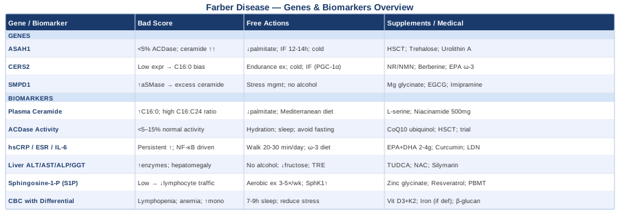
6 Biomarkers to Track in Farber Disease
Farber disease is not a condition where routine blood panels tell the full story. Standard inflammatory markers and basic metabolic panels will often flag problems but cannot explain them. The biomarkers below were chosen because each one directly reflects a specific mechanism of the disease: ceramide accumulation, enzymatic failure, downstream signaling disruption, or organ stress. Together, they create a functional monitoring picture that a standard check-in cannot provide.
1. Plasma Ceramide Species
Why it matters
Ceramide accumulation is the defining biochemical event in Farber disease. The ASAH1 gene encodes acid ceramidase, the lysosomal enzyme responsible for hydrolyzing ceramide into sphingosine and a free fatty acid. When this enzyme is deficient or absent, ceramide builds up in cells throughout the body — most visibly in macrophages and histiocytes within joints, liver, lung, and nervous tissue. Measuring plasma ceramide species by acyl chain length — particularly C16:0, C18:0, C20:0, C22:0, and C24:0 — provides the most direct biochemical confirmation of disease activity and the clearest baseline for tracking whether any intervention is shifting the lipid profile.
Short-chain ceramides, especially C16:0, are strongly pro-apoptotic and pro-inflammatory, activating cell death pathways and NF-κB signaling. Long-chain ceramides (C24:0) have more cytoprotective and membrane-stabilizing properties. The C16:0 to C24:0 ceramide ratio is therefore not merely a number — it reflects the balance between death-signaling and membrane integrity in tissues where the enzyme is absent. A ratio skewed toward C16:0 indicates a more aggressive inflammatory and apoptotic ceramide burden.
How to measure it
Plasma ceramide species profiling is performed via liquid chromatography-tandem mass spectrometry (LC-MS/MS). This is a specialized test, not available at general clinical laboratories, but offered at academic medical centers with metabolic biochemistry programs and through select commercial reference laboratories. Cost range: $200–$600 depending on the number of species quantified and the institution. Insurance coverage is variable; prior authorization under rare metabolic disease codes is often required. Repeat measurement every 6–12 months provides the trend data needed to evaluate interventions.
If the score is bad, the plan without supplements
The most impactful dietary change is reducing palmitic acid (C16:0) intake — the primary substrate for de novo ceramide synthesis via serine palmitoyl transferase. Palmitic acid dominates palm oil, butter, full-fat dairy, and fatty red meat. Replacing these with oleic acid-rich fats — olive oil, avocado, macadamia nuts — shifts the ceramide synthesis substrate pool without requiring extreme fat restriction. The change in ceramide species profile from this dietary shift has been documented in lipid metabolism research within 8–12 weeks.
Eliminating secondary ceramide triggers is equally important. Oxidative stress, high-fructose diets, and excessive omega-6 vegetable oils all activate sphingomyelinase enzymes, generating additional ceramide from sphingomyelin breakdown. A Mediterranean-pattern eating approach addresses most of these factors simultaneously and has the best human evidence base for shifting ceramide species in a favorable direction.
If the score is bad, the plan with supplements or equipment
L-serine (2–4 g/day in divided doses, continuous, no cycling required): Competes with alanine in the first rate-limiting step of de novo ceramide synthesis at serine palmitoyl transferase. This competition shifts production toward less toxic deoxysphingolipid species. Human safety data at these doses are available from ALS and macular degeneration trials. Side effects are minimal. This is among the most mechanistically direct supplementation approaches for modulating ceramide production at its source.
Niacinamide (vitamin B3) (500 mg twice daily; cycle 5 days on / 2 days off for long-term use): Inhibits the neutral sphingomyelinase (SMPD2/3) enzyme family, reducing ceramide generated from sphingomyelin breakdown. The evidence base is primarily cellular but mechanistically coherent. Do not substitute flush niacin — the mechanism and side effect profiles differ substantially. Monitor liver enzymes with extended use at these doses.
Fenretinide (investigational — do not self-administer): A synthetic retinoid shown in preclinical and early human work to reduce ceramide accumulation in lysosomal storage models. Relevant only within a clinical trial framework under specialist supervision.
2. Acid Ceramidase Enzyme Activity
Why it matters
Measuring residual acid ceramidase (ACDase) activity in peripheral blood leukocytes or cultured fibroblasts is both the diagnostic gold standard and a functional indicator of phenotypic severity. Patients with classic Farber disease type 1 typically retain less than 5% of normal ACDase activity. Those with milder forms — particularly type 3, which presents primarily with joint disease without neurological involvement — may retain 5–15% residual activity. Even this narrow difference corresponds to meaningfully different clinical trajectories and different timelines for considering interventions.
Repeat measurements add clinical value because enzyme activity can fluctuate with secondary stressors. Acute infections, significant oxidative insults, and certain medications can transiently suppress lysosomal enzyme function, creating artificially low readings that do not reflect the true genetic baseline. Establishing a pattern across multiple measurements is more informative than a single result.
How to measure it
The assay uses a fluorescent substrate (4-methylumbelliferyl-ceramide) applied to leukocytes isolated from fresh EDTA whole blood. The sample requires transport to a specialized biochemical genetics laboratory with same-day or overnight processing requirements — standard courier arrangements are usually sufficient when coordinated in advance. Available at reference centers including the Kennedy Krieger Institute (Baltimore), Massachusetts General Hospital's Lysosomal Disease Program, and the University of Minnesota's biochemical genetics division. Cost: $150–$400, with variable insurance coverage under rare metabolic disease billing codes.
If the score is bad, the plan without supplements
Residual ACDase activity is determined by genotype — the specific ASAH1 variants present — and cannot be increased through lifestyle changes. What can be changed is the functional burden on the lysosomal system around the deficient enzyme. Maintaining adequate hydration (dehydration impairs lysosomal membrane integrity and acidification), avoiding prolonged caloric restriction that depletes lysosomal cofactors, and maintaining a consistent sleep-wake cycle (lysosomal function and autophagy are regulated in part by circadian biology) all reduce the pressure on an already compromised clearance apparatus. These interventions do not treat the enzyme deficiency, but they reduce the rate at which the system is overwhelmed.
If the score is bad, the plan with supplements or equipment
Hematopoietic stem cell transplantation (HSCT) is currently the only intervention that addresses the enzymatic deficiency in peripheral tissues for patients without significant central nervous system involvement. Early HSCT — ideally before major joint damage or substantial organ infiltration — has been shown in published case series and registry data to stabilize or improve joint disease, voice quality, and subcutaneous nodules in Farber disease types 1 and 3. This is a medical intervention requiring a specialist center and careful selection, not a supplement — but it belongs on the table of every discussion with a metabolic geneticist as soon as diagnosis is confirmed.
Recombinant human acid ceramidase (rhACDase — velcerase alfa) is in active clinical development as an enzyme replacement therapy. Early clinical trials are underway; families and clinicians can monitor enrollment at ClinicalTrials.gov by searching "acid ceramidase deficiency."
Coenzyme Q10 (ubiquinol form) (100–300 mg/day with a fat-containing meal, continuous): Supports mitochondrial membrane integrity and reduces the oxidative stress that destabilizes lysosomal membranes, contributing indirectly to lysosomal system support. Not a direct ACDase activator, but a reasonable adjunct with a strong safety profile and widely available evidence in mitochondrial and lysosomal disease contexts.
3. High-Sensitivity CRP, ESR, and IL-6
Why it matters
Ceramide accumulation in macrophages within synovial tissue drives the granulomatous joint inflammation that is the most visible and debilitating feature of Farber disease. High-sensitivity C-reactive protein (hsCRP) and erythrocyte sedimentation rate (ESR) provide a non-specific but practical measure of systemic inflammation intensity. IL-6 is more mechanistically specific: it is a direct downstream mediator of the ceramide-to-NF-κB inflammatory cascade, and persistently elevated IL-6 is associated with joint, lung, and liver involvement in more severe Farber phenotypes.
One clinically important pattern: in Farber disease, hsCRP may remain persistently elevated even in the absence of acute infection — reflecting metabolic-inflammatory signaling rather than a reactive response. Recognizing this pattern is useful when evaluating treatment effects over time and distinguishing disease activity from intercurrent illness.
How to measure it
All three are available at standard clinical laboratories. hsCRP: $15–$50. ESR: $10–$30. IL-6 (serum or plasma): $50–$150; less routinely covered by insurance and requires advance processing to prevent ex vivo cytokine induction artifacts. For meaningful trend data, measure at consistent intervals (every 3–6 months) in a fasted state, at approximately the same time of day. Note any acute illness within the two weeks before measurement, as it will transiently elevate all three markers independent of disease activity.
If the score is bad, the plan without supplements
Sustained low-intensity aerobic movement — 20–30 minutes of walking daily — meaningfully reduces IL-6 and hsCRP through myokine signaling. Contracting muscle tissue releases IL-10 and soluble IL-6 receptor antagonists, producing a systemic anti-inflammatory effect that is independent of weight change. For patients with significant joint involvement where walking is difficult, warm water hydrotherapy provides an equivalent movement signal with substantially reduced mechanical joint load.
Dietary shifts with strong human evidence: replacing refined carbohydrates with fibrous vegetables, increasing omega-3 fatty acid intake through fatty fish or algae-based DHA, and eliminating trans fats from processed foods reduce hsCRP by a clinically meaningful margin in inflammatory conditions. The cumulative effect of these changes is larger than any single modification in isolation.
If the score is bad, the plan with supplements or equipment
Omega-3 fatty acids (EPA + DHA combined) (2–4 g/day with food, continuous): Inhibit the arachidonic acid cascade and NF-κB activation; reduce circulating IL-6 in multiple meta-analyses of inflammatory conditions. No cycling required. Monitor for minor bleeding risk at the higher end of this range, particularly with concurrent anticoagulant use.
Curcumin (phospholipid complex or BCM-95 formulation) (500–1,000 mg/day with a fat-containing meal; cycle 6 weeks on / 2 weeks off for extended use): Inhibits NF-κB and COX-2 at the pathway level relevant to ceramide-driven inflammation. Formulation matters critically — standard curcumin powder has insufficient bioavailability to produce meaningful blood levels. Avoid with active gallbladder disease.
Low-dose naltrexone (LDN) (1.5–4.5 mg nightly, prescription required, off-label use): Modulates macrophage activation and TLR4-mediated signaling through mechanisms particularly relevant to storage disease-driven macrophage infiltration in joints and liver. A growing body of human data supports LDN in macrophage-driven inflammatory conditions. Requires a physician comfortable with off-label prescribing and appropriate baseline liver function monitoring.
4. Liver Function Panel (AST, ALT, ALP, GGT)
Why it matters
The liver is a primary site of ceramide accumulation in Farber disease, particularly in more severe phenotypic forms. Hepatomegaly is present in approximately half of patients with classic type 1 disease. Elevated ALT and AST indicate hepatocyte stress; elevated ALP and GGT suggest bile duct and Kupffer cell (liver macrophage) involvement — directly relevant given that ceramide-laden histiocytes infiltrating liver tissue are the cellular hallmark of Farber disease. Liver involvement can progress silently, and regular testing creates a longitudinal trend that a single measurement cannot provide.
The liver function panel also serves as a safety monitoring tool for any supplementation protocol or off-label pharmacological intervention being added alongside standard care — making it a doubly useful routine measure.
How to measure it
Standard liver function panel (LFP) or comprehensive metabolic panel (CMP): $20–$60, routinely covered by insurance with clinical indication. Measure every 3–4 months if hepatomegaly is present; every 6 months for patients with mild or absent liver involvement. For metabolic monitoring purposes, a more sensitive ALT target is below 25 U/L for women and below 30 U/L for men — more conservative than standard lab reference ranges, and better calibrated for detecting early hepatocyte stress before overt disease develops.
If the score is bad, the plan without supplements
Complete alcohol elimination is the most impactful single dietary change for hepatic ceramide burden. Even modest alcohol intake activates de novo ceramide synthesis in hepatocytes and stimulates sphingomyelinase activity in liver macrophages — directly compounding an already compromised clearance system. There is no meaningful safe threshold in this context.
Reducing dietary fructose — particularly high-fructose corn syrup from processed foods — decreases de novo hepatic lipogenesis and the associated ceramide burden on hepatocytes. Time-restricted eating (a 12–14 hour overnight fast with consistent meal timing) has been shown in metabolic liver research to reduce hepatic inflammatory markers and improve lysosomal autophagy efficiency without requiring any pharmacological intervention.
If the score is bad, the plan with supplements or equipment
TUDCA (tauroursodeoxycholic acid) (500–1,000 mg/day with food; cycle 8 weeks on / 4 weeks off): A bile acid derivative shown to reduce endoplasmic reticulum stress in hepatocytes and stabilize lysosomal membranes under metabolic stress conditions. The mechanism is directly applicable to ceramide-driven hepatocyte stress in lysosomal storage contexts. Mild gastrointestinal effects are possible. Do not use with active gallstones or bile duct obstruction.
NAC (N-acetylcysteine) (600 mg twice daily; cycle 5 days on / 2 days off): Replenishes intracellular glutathione, the liver's primary antioxidant defense. Well-studied in hepatocyte protection across multiple disease contexts. Generally safe at standard doses; minor blood pressure effects have been reported at higher doses in some individuals.
Silymarin (milk thistle extract) (140 mg three times daily with food; can be used continuously): Hepatoprotective, anti-inflammatory in liver tissue, and modestly antifibrotic through stellate cell modulation. Human evidence is consistent across metabolic liver conditions. Excellent long-term safety profile and suitable as an ongoing adjunct to monitoring.
5. Sphingosine-1-Phosphate (S1P)
Why it matters
Sphingosine-1-phosphate (S1P) sits directly downstream of the ceramide pathway. In normal physiology, ceramide is cleaved by acid ceramidase into sphingosine, which sphingosine kinases (SphK1 and SphK2) then phosphorylate to S1P. S1P carries broadly pro-survival and pro-proliferative signaling properties, functioning as the molecular counterweight to pro-apoptotic ceramide. The ceramide/S1P rheostat is one of the most studied lipid regulatory axes in cell biology.
In Farber disease, because ceramide cannot be efficiently cleaved by deficient ACDase, downstream production of sphingosine — and therefore S1P — is impaired. Low S1P is associated with disrupted lymphocyte trafficking (contributing to the lymphopenia documented in some Farber patients), impaired endothelial barrier function, and reduced tissue repair capacity. Tracking S1P alongside ceramide provides a more complete picture of where this regulatory axis stands, rather than seeing only one side of the balance.
How to measure it
S1P measurement from plasma or whole blood uses LC-MS/MS and is available at academic lipid metabolism laboratories and through specialized metabolomics reference labs that offer full sphingolipid profiling panels. It is not a routine clinical test but is increasingly accessible through research partnerships and metabolomics services. Cost: $150–$400 as part of a sphingolipid panel. Coverage is typically limited outside research protocols, though this is evolving as ceramide testing gains clinical recognition.
If the score is bad, the plan without supplements
Regular aerobic exercise is the most direct non-pharmacological intervention for the ceramide/S1P balance. Moderate aerobic activity increases sphingosine kinase 1 (SphK1) activity in skeletal muscle and vascular endothelial cells, driving more sphingosine toward S1P production. As little as 20–30 minutes of moderate-intensity aerobic exercise three to five times per week has been shown in cardiometabolic research to shift the ceramide/S1P balance favorably — a genuinely pathway-level effect that no supplement fully replicates.
If the score is bad, the plan with supplements or equipment
Zinc (as zinc glycinate or bisglycinate) (15–25 mg elemental zinc/day with food; cycle 3 months on / 1 month off to prevent copper depletion): Activates SphK1 in immune cells and serves as a cofactor for lysosomal enzyme function generally. Pair with 1–2 mg/day of copper during cycling periods to prevent secondary copper deficiency. Zinc excess is more problematic than deficiency — stay within this range.
Resveratrol (high-bioavailability formulation) (500 mg/day with a fat-containing meal; cycle 6 weeks on / 2 weeks off): Activates SphK1 and increases S1P production in cellular models. Standard resveratrol powder has poor bioavailability — micronized or liposomal formulations significantly improve absorption. May interact with anticoagulant medications; note this with prescribing physicians.
Photobiomodulation (red and near-infrared light therapy) (630–850 nm wavelength; 10–20 minutes/day over major joints and the abdomen; continuous use appropriate): Stimulates mitochondrial cytochrome c oxidase, supporting ATP synthesis and reducing the oxidative ceramide generation that suppresses SphK activity. Evidence in lipid signaling specifically is mechanistically coherent but emerging rather than established — use as an adjunct to lifestyle interventions rather than a primary strategy.
6. Complete Blood Count With Differential
Why it matters
The CBC with differential functions as a broad functional surveillance tool in Farber disease for three specific disease-relevant reasons. First, lymphopenia (low absolute lymphocyte count) has been documented in severe Farber phenotypes and directly reflects disrupted S1P-mediated lymphocyte trafficking from lymphoid organs — a consequence of the ceramide/S1P imbalance described above, not an incidental finding. Second, anemia can indicate bone marrow infiltration by ceramide-laden histiocytes, a feature of more severe or systemically advanced disease that warrants escalation of care. Third, elevated monocyte percentage or neutrophilia in the absence of acute infection signals active systemic granulomatous inflammation, often preceding detectable changes on imaging.
Tracking CBC trends over time — even when individual values remain within reference ranges — creates a longitudinal trajectory that point-in-time measurements cannot reveal.
How to measure it
Standard CBC with differential: $15–$50, covered by most insurance plans with clinical indication. Measure every 3–6 months in confirmed Farber patients. Watch specifically: total lymphocyte count below 1,000 cells/μL (concerning for S1P-mediated trafficking failure); monocyte percentage above 12% in a non-infectious context (granulomatous macrophage activation signal); and hemoglobin trend over 12–18 months rather than a single absolute value. Ferritin and transferrin saturation should be checked simultaneously when anemia is detected to distinguish iron-deficiency anemia from inflammatory anemia — a distinction that determines treatment entirely.
If the score is bad, the plan without supplements
For inflammatory anemia — the more likely mechanism in active Farber disease — addressing the underlying inflammatory driver through the CRP/IL-6 protocol above is the appropriate primary step. Iron supplementation is ineffective and potentially harmful when anemia is driven by chronic inflammation rather than iron depletion; do not supplement iron without confirming the mechanism.
For lymphopenia: adequate, consistent sleep (7–9 hours nightly) is one of the most direct non-pharmacological interventions for lymphocyte count — lymphocyte production peaks during slow-wave sleep and is acutely suppressed by sleep disruption. Reducing chronic psychological stress (cortisol chronically suppresses lymphopoiesis) is a parallel lever with the same mechanism and direct applicability given the SMPD1 pathway link discussed in the genetics section.
If the score is bad, the plan with supplements or equipment
Vitamin D3 with K2 (MK-7 form) (2,000–5,000 IU D3 daily with a fat-containing meal; 100–200 mcg K2 MK-7; continuous): Vitamin D deficiency is associated with lymphopenia and impaired monocyte differentiation. Target serum 25-OH vitamin D at 40–60 ng/mL. Safe at standard doses; do not exceed 10,000 IU/day without serum level monitoring. Test before starting to calibrate the dose.
Iron (ferrous bisglycinate) (25–50 mg elemental iron/day away from calcium, tea, and coffee; 3 months on, then recheck CBC and ferritin): Use only if iron-deficiency anemia is confirmed by both low ferritin and low transferrin saturation. Bisglycinate form is significantly better tolerated than sulfate or oxide forms with respect to gastrointestinal side effects.
Beta-glucan-rich mushroom extracts (Reishi, Turkey Tail) (1–3 g/day standardized extract; cycle 6 weeks on / 2 weeks off): Polysaccharide beta-glucans support NK cell activity and monocyte maturation through TLR2/6 receptor signaling. Human immunomodulation evidence is available for these extracts, though not disease-specific to Farber. Avoid during active autoimmune flares.
Genes Driving Ceramide Accumulation: ASAH1, CERS2, and SMPD1
Understanding the genetic architecture of Farber disease moves beyond diagnosis into severity prediction and pathway-level intervention. While ASAH1 is the sole causative gene, two additional genes in the ceramide metabolism pathway — CERS2 and SMPD1 — can substantially modify the ceramide species profile and inflammatory phenotype in ways that influence both prognosis and the prioritization of specific interventions.
ASAH1: The Causative Gene
What the gene does
Located on chromosome 8p22–p21.3, ASAH1 encodes acid ceramidase (ACDase), the lysosomal enzyme that catalyzes ceramide hydrolysis into sphingosine and a free fatty acid. ACDase is expressed as a single precursor protein that is processed into a heterodimeric alpha-beta structure within the lysosome, requiring correct folding, proteolytic processing, and the lipid activator protein saposin D for full function. Pathogenic variants that disrupt any of these steps result in enzyme deficiency and ceramide accumulation in multiple organs.
Over 80 distinct pathogenic variants in ASAH1 have been described, spanning missense mutations (the most common), nonsense mutations, splice-site alterations, and small insertions or deletions. The T222K and P362R missense variants appear repeatedly in published case series and have been used as reference points in genotype-phenotype correlation analyses.
Genotype-phenotype correlations
The Farber disease phenotypic spectrum spans seven recognized subtypes (types 1–7). Type 1 (classic) presents in infancy with all three cardinal features — joint disease, nodules, and hoarseness — plus liver and neurological involvement, typically with very low or absent ACDase activity. Type 3 (mild) presents primarily with joint disease, often without neurological involvement, and correlates with higher residual activity. Compound heterozygotes carrying two different pathogenic variants frequently show intermediate clinical severity. Knowing the specific ASAH1 variants a patient carries allows for prognostication and earlier planning for interventions, particularly the timing of HSCT evaluation.
If the gene is bad: the plan without supplements
Residual ACDase activity is fixed by genotype. What can be modified is the ceramide substrate load entering a system that cannot clear it efficiently. Reducing de novo ceramide synthesis by limiting palmitic acid in the diet directly reduces ceramide production rate at source. Intermittent fasting (12–14 hour overnight fast) activates TFEB, the master transcription factor for lysosomal biogenesis, increasing lysosomal number and functional capacity even when the enzyme complement is genetically compromised. Brief cold exposure (2–3 minute cold showers 4–5 times per week) activates AMPK-mediated autophagy and lysosomal turnover, clearing ceramide-damaged organelles and maintaining organelle health. These are not substitutes for medical intervention, but they are real biological levers with mechanistic coherence across lysosomal storage research.
If the gene is bad: the plan with supplements or equipment
Trehalose (1–3 g/day dissolved in water between meals; continuous): A naturally occurring disaccharide that activates TFEB in cell models and animal studies of lysosomal storage disorders, increasing lysosomal biogenesis and improving substrate clearance. Food-grade and well-tolerated; human safety data are available, though disease-specific trials in Farber disease are limited. The mechanism is shared across lysosomal storage disorders and the safety profile is favorable enough to make it a reasonable adjunct.
Urolithin A (500–1,000 mg/day; continuous): A mitophagy activator that selectively removes dysfunctional mitochondria and supports lysosomal recycling capacity. Phase 2 human data are available in aging and metabolic contexts demonstrating improved cellular clearance function. Supplement form bypasses the gut microbiome variability that makes natural urolithin A production from pomegranate highly inconsistent between individuals.
HSCT and rhACDase enzyme replacement therapy: As discussed in the biomarker section, these are the primary medical interventions for the enzyme deficiency. Specialist referral for HSCT evaluation in non-neurological phenotypes before significant organ damage occurs is the highest-yield clinical decision in the early management of Farber disease.
CERS2: The Ceramide Chain Length Modifier
What the gene does
CERS2 (also called LASS2), located on chromosome 1q21, encodes ceramide synthase 2 — the enzyme responsible for synthesizing very long-chain ceramides, specifically C22:0 and C24:0 species. These longer-chain ceramides tend to be less pro-apoptotic and more membrane-stabilizing than shorter-chain C16:0 ceramide. CERS2 therefore shapes the composition of the ceramide pool that accumulates when ACDase is deficient: a patient with reduced CERS2 expression produces a ceramide accumulation profile biased toward shorter, more pro-inflammatory C16:0 species, potentially worsening both the apoptotic burden on affected tissues and the intensity of the inflammatory phenotype.
CERS2 is not a cause of Farber disease. But common functional variants that reduce its expression act as meaningful modifiers of severity — a genetic context worth knowing, particularly when interpreting a ceramide species profile that is skewed toward short-chain accumulation.
If the gene is bad: the plan without supplements
CERS2 expression is upregulated by PGC-1α activation — the master regulator of mitochondrial biogenesis and metabolic adaptation. Endurance exercise (30 minutes at moderate intensity, five days per week) is the most reliably evidence-based PGC-1α activator in humans. Cold exposure and intermittent fasting both also activate PGC-1α through AMPK and SIRT1 pathways. Together, these lifestyle interventions shift ceramide synthase expression in a direction that favors longer-chain, more cytoprotective ceramide species — a meaningful if modest effect on the overall ceramide composition.
If the gene is bad: the plan with supplements or equipment
Nicotinamide riboside (NR) or NMN (300–500 mg in the morning; cycle 3 months on / 1 month off): Elevates NAD+ levels, activating SIRT1, which then stimulates PGC-1α and downstream CERS2 upregulation. Human data in metabolic aging contexts are available; generally well-tolerated. Take in the morning to align with circadian NAD+ rhythms and avoid sleep disruption.
Berberine (500 mg with meals twice daily; cycle 8 weeks on / 4 weeks off to preserve gut microbiome diversity): AMPK activator that modulates ceramide synthase gene expression in hepatic models, with a directional shift toward longer-chain species. Monitor blood glucose with extended use — berberine can produce clinically significant glucose lowering and is contraindicated with certain diabetes medications without dose adjustment.
EPA-enriched omega-3 supplementation (per the Biomarker 3 dosing above): EPA specifically has been shown in cellular models to influence ceramide synthase gene expression toward longer-chain species, complementing the dietary and lifestyle shift approach without requiring additional supplementation if omega-3s are already being used for inflammation management.
SMPD1: The Upstream Ceramide Generator
What the gene does
SMPD1, located on chromosome 11p15.4, encodes acid sphingomyelinase (aSMase) — the enzyme that generates ceramide from sphingomyelin, upstream of the ACDase step. Biallelic loss-of-function mutations in SMPD1 cause Niemann-Pick disease types A and B, a related lysosomal storage disorder. For Farber disease, SMPD1 acts as a genetic modifier: common functional variants that increase aSMase activity generate more ceramide upstream, directly amplifying the substrate load that the deficient ACDase must attempt to process.
Beyond genetics, aSMase activity is acutely sensitive to environmental and physiological stressors. Psychological stress and elevated cortisol activate aSMase in blood cell membranes, transiently increasing ceramide generation. Alcohol strongly activates hepatic aSMase. Oxidative stress from poor diet, environmental toxins, and chronic systemic inflammation all upregulate aSMase activity. This means that for Farber disease patients, lifestyle factors interact with SMPD1 function in ways that are mechanistically direct and clinically meaningful, independent of the patient's specific SMPD1 genetic variants.
If the gene is bad: the plan without supplements
The clearest non-pharmacological intervention targeting aSMase activity is chronic stress reduction — not as a vague lifestyle recommendation but as a specific mechanism. The HPA axis-ceramide axis is a well-documented pathway in lipid stress biology: chronic psychological stress activates aSMase through cortisol signaling, generating ongoing ceramide from sphingomyelin reserves in multiple tissues. Consistent stress reduction — whether through structured mindfulness practice, regular low-intensity outdoor activity, or maintained social connection — reduces the frequency and intensity of aSMase activation events.
Complete alcohol avoidance is the other highest-yield change in this context: alcohol is among the most potent aSMase activators in hepatic tissue, directly amplifying ceramide generation upstream of an already compromised clearance enzyme.
If the gene is bad: the plan with supplements or equipment
Magnesium glycinate (300–400 mg/day in the evening; continuous): Cofactor for ceramide pathway enzymes; shown to reduce aSMase activity under oxidative stress conditions in cell models. Excellent safety profile. Use glycinate form — magnesium oxide and citrate cause substantially more gastrointestinal effects at equivalent elemental doses.
EGCG (green tea extract) (400–600 mg standardized EGCG/day; cycle 6 weeks on / 2 weeks off): Inhibits aSMase in cell models and has demonstrated anti-ceramide effects in lipid metabolism research. Liver enzyme monitoring is advisable with extended use at the upper end of this dose range, given the hepatotoxicity risk associated with very high EGCG supplementation in susceptible individuals.
Low-dose imipramine (prescription, clinical context only): A tricyclic antidepressant that at sub-antidepressant doses specifically inhibits acid sphingomyelinase activity through a mechanism distinct from its effects on serotonin and norepinephrine reuptake. This has been noted in scientific literature on Niemann-Pick disease and related lysosomal storage conditions with ceramide involvement. Not a self-administered intervention — requires physician involvement and appropriate monitoring given imipramine's drug interaction profile.
What Ceramide Research Reveals That Standard Medicine Often Misses
The scientific literature on ceramides has accelerated significantly over the past decade, driven largely by cardiovascular medicine researchers who discovered that ceramide testing predicts heart disease risk independently of — and often more accurately than — LDL cholesterol. While most of this research targets cardiovascular patients rather than Farber disease specifically, the underlying biology is identical: ceramide as a lipid signaling mediator, not just a structural fat. The insights below challenge several assumptions embedded in standard clinical monitoring and treatment approaches, and they translate directly to the Farber disease context.
Peter Attia, in his book Outlive: The Science and Art of Longevity (2023) and on his podcast The Drive, has synthesized this research more clearly than almost any other clinical voice, working extensively with lipidologist Thomas Dayspring on advanced ceramide testing protocols. What follows are ten of the most impactful insights from this body of work, reframed for monitoring and understanding Farber disease.
1. Standard Cholesterol Panels Are Blind to Ceramide Biology
Total cholesterol, LDL-C, HDL-C, and triglycerides tell you almost nothing about ceramide status. A patient with completely normal lipid panels can have dramatically elevated ceramide levels with active tissue damage in progress. Routine lipid panels will miss the primary biochemical driver of Farber disease entirely — which is one reason why general practitioners following a Farber patient with standard metabolic panels may see "reassuring" results that do not reflect the actual disease state.
2. C16:0 Ceramide Is the Most Damaging Short-Chain Species
Not all ceramide accumulation is equally harmful. C16:0 ceramide, synthesized primarily by CERS5 and CERS6, is the most strongly pro-apoptotic and pro-inflammatory ceramide species. In cardiovascular research, elevated C16:0 specifically predicts major adverse events, independent of total ceramide. In Farber disease, a ceramide accumulation profile skewed toward C16:0 — influenced by CERS2 functional status — represents a qualitatively different and more inflammatory burden than accumulation biased toward C24:0.
3. The C16:0 to C24:0 Ratio Matters More Than Total Ceramide
Attia and Dayspring emphasize ratio interpretation over absolute concentration in ceramide testing. The balance between short-chain pro-apoptotic and long-chain cytoprotective ceramide species is a more functionally meaningful measure than total ceramide alone. Tracking this ratio as interventions are applied — dietary, exercise, supplement-based — provides more actionable data than a total ceramide number that changes slowly and does not distinguish between beneficial and harmful species.
4. Saturated Fat Is the Primary Modifiable Driver of Ceramide Synthesis
Palmitic acid is the immediate substrate for the rate-limiting step in de novo ceramide synthesis. Reducing palmitate intake directly reduces ceramide production rate. This is not a generic anti-fat recommendation — it is a substrate-level biochemical intervention with human evidence. Replacing palm oil and butter with oleic acid-rich fats shifts the de novo synthesis substrate pool away from ceramide precursors in a way that is measurable in ceramide species profiles within weeks.
5. Ceramide and Insulin Resistance Share a Bidirectional Relationship
Ceramide impairs insulin signaling by activating protein phosphatase 2A (PP2A) and PKCζ, which block AKT phosphorylation — a central node in insulin signal transduction. Simultaneously, hyperinsulinemia and metabolic dysfunction upregulate ceramide synthesis. For Farber disease patients, this creates a self-reinforcing cycle: ceramide accumulation generates insulin resistance in peripheral tissues, which in turn drives more ceramide production via de novo synthesis. Managing metabolic health and insulin sensitivity is not peripheral to Farber disease — it is directly relevant to the ceramide feedback loop.
6. Exercise Shifts the Ceramide Landscape More Broadly Than Diet Alone
Aerobic exercise activates sphingosine kinase in skeletal muscle, driving both ceramide catabolism and S1P production simultaneously. It upregulates CERS2, shifting ceramide synthesis toward longer-chain cytoprotective species. It reduces NF-κB-driven inflammation that otherwise stimulates sphingomyelinase. And it activates TFEB, supporting lysosomal biogenesis. No single supplement replicates the breadth of this ceramide-targeted effect — which is why exercise appears in every intervention protocol in this article.
7. Ceramide-Driven Inflammation Is Mechanistically Distinct From Cytokine-Driven Inflammation
Anti-inflammatory medications that target TNF-α, IL-1β, or JAK-STAT pathways address downstream inflammation but do not address ceramide accumulation itself. This is why patients with Farber disease can show persistently elevated ceramide and ongoing tissue damage even when standard inflammatory markers are pharmacologically suppressed. Targeting the lipid pathway directly — through substrate reduction, lysosomal support, or ceramide disposal pathway activation — addresses a different and more upstream level of the mechanism.
8. The Liver Is Both the Primary Producer and a Primary Target of Ceramide Stress
Hepatocytes are the major site of ceramide biosynthesis and simultaneously among the most vulnerable to ceramide-induced apoptosis. This creates an internal feedback loop in Farber disease: as ceramide accumulates in hepatic lysosomes, hepatocyte death accelerates local ceramide release, which triggers further NF-κB activation and macrophage recruitment. The liver function panel is not a secondary monitoring target — it reflects a self-amplifying process that can accelerate if not tracked consistently.
9. TFEB Activation Is One of the Most Promising Pathways in Lysosomal Disease
TFEB (transcription factor EB), the master regulator of lysosomal biogenesis and autophagy gene expression, is an active research target across multiple lysosomal storage disorders. When activated, TFEB increases lysosomal number, improves lysosomal acidification efficiency, and accelerates substrate clearance. Multiple research groups are exploring TFEB activators as therapeutic adjuncts in lysosomal storage disorders. Lifestyle interventions that robustly activate TFEB — intermittent fasting, endurance exercise, and cold exposure — are the only tools currently available outside clinical trials.
10. Serial Ceramide Profiling Is a Functional Outcome Marker, Not Just a Diagnostic Tool
In current clinical practice, ceramide testing is ordered once at diagnosis and rarely repeated. The framework emerging from metabolic medicine research argues for treating it as a serial functional biomarker — measured every 6–12 months to assess whether interventions are actually shifting ceramide species composition over time. For Farber disease, this means ceramide profiling should function like HbA1c in diabetes: a trend biomarker that captures cumulative metabolic state, not a snapshot reserved for initial workup.
Complementary Approaches for Pain, Inflammation, and Respiratory Involvement
The three approaches below address the specific symptoms most prominent in Farber disease — joint pain, systemic inflammation, and respiratory involvement from laryngeal nodules. Each was selected because it has meaningful human clinical evidence, not merely theoretical plausibility, for conditions with overlapping mechanisms.
Mindfulness-Based Stress Reduction (MBSR) for Chronic Pain and Inflammation
Chronic joint pain in Farber disease involves two distinct components: direct ceramide-driven synovial inflammation and central sensitization — the nervous system's adaptive lowering of pain thresholds in response to persistent nociceptive input. MBSR, the 8-week structured mindfulness program developed by Jon Kabat-Zinn, addresses both through separate mechanisms. It reduces psychological amplification of pain through sustained attentional training, and it reduces cortisol-mediated aSMase activation — creating a direct link between the stress reduction practice and the upstream ceramide generation pathway described in the SMPD1 section.
A 2016 randomized controlled trial comparing MBSR to cognitive behavioral therapy and usual care for chronic musculoskeletal pain found significant improvements in pain, functional limitation, and quality of life in the MBSR group at 26-week follow-up. Evidence is not disease-specific to Farber disease, but the pain mechanisms addressed — central sensitization and inflammatory contribution — apply directly.
Practical application: the standard MBSR program runs 8 weeks with 45–60 minutes of daily home practice and one weekly group session. Online adaptations are available through several certified centers for patients with limited mobility. Begin with 10-minute body scan practices before progressing to full-length sessions. The anti-inflammatory HPA axis effect requires consistency over weeks — outcomes measured at 8 weeks are substantially better than at 2. This is not a one-session intervention; sustained practice is what produces the physiological signal.
Massage Therapy for Joint Mobility and Nodule Management
The subcutaneous nodules in Farber disease are composed of ceramide-laden macrophages (histiocytes) and can cause direct discomfort and secondary restriction of joint mobility in affected areas. Therapeutic massage — particularly manual lymphatic drainage and gentle connective tissue techniques — does not dissolve the nodules, but it can improve local tissue circulation, reduce secondary perilesional edema, maintain range of motion in affected joints, and reduce the muscle guarding that develops around chronically painful areas. For pediatric patients, pressure and frequency require careful individual calibration.
Systematic evidence for massage therapy in inflammatory joint conditions — including juvenile idiopathic arthritis and related disorders with joint inflammation — shows consistent short-term reductions in pain and stiffness with minimal adverse effects across multiple clinical trials. Evidence is not specific to Farber disease, given its extreme rarity, but the joint inflammation mechanism shares enough overlap to make massage a clinically reasonable adjunct to standard care.
In practice: 30–60 minute sessions one to two times per week with a therapist trained in gentle joint work and lymphatic drainage techniques. Communication between the massage therapist and the treating metabolic specialist is important — certain nodule locations may be anatomically sensitive to pressure, and treatment should progress at the patient's reported comfort level. Aggressive pressure directly over nodule formations is contraindicated.
Breathing-Based Therapy for Laryngeal and Respiratory Involvement
Hoarseness and voice weakness caused by ceramide-laden nodules on the laryngeal mucosa are among the most distinctive and earliest features of Farber disease. In older patients, progressive laryngeal involvement can affect voice quality, swallowing coordination, and respiratory effort during exertion. Breathing-based therapies — specifically those used in speech-language pathology and respiratory physiotherapy — can help maintain respiratory muscle coordination, optimize the use of available airway space, and reduce the functional burden of laryngeal restriction on voice production and swallowing.
Diaphragmatic breathing retraining and vocal tract coordination exercises are established approaches in laryngeal conditions involving inflammation and mucosal restriction, including recurrent respiratory papillomatosis and laryngeal amyloidosis. Direct evidence in Farber disease is absent given its rarity, but the anatomical and functional overlap with these conditions is sufficient to justify early referral to a speech-language pathologist experienced in laryngeal disorders, ideally at the time of diagnosis before significant voice change has occurred.
Application protocol: referral to a speech-language pathologist with laryngeal specialization is the starting point. Breathing exercises should be performed in a supported lying or seated position, focusing on lower thoracic and diaphragmatic expansion, 10–15 minutes daily. Avoid techniques requiring forced phonation or high-effort vocalization against restricted airways — these can irritate already inflamed laryngeal tissue and are counterproductive. Progress should be guided by the therapist with input from the metabolic team managing the underlying disease activity.
Conclusion
Farber disease has a known enzyme deficiency, a known lipid substrate that accumulates when that enzyme fails, and measurable downstream consequences that can be tracked with the right panel of tests. The six biomarkers covered in this article — plasma ceramide species, acid ceramidase enzyme activity, inflammatory markers, liver function, sphingosine-1-phosphate, and CBC with differential — create a functional monitoring picture that standard clinical check-ins cannot provide. The genetic context from ASAH1 genotype, CERS2 expression, and SMPD1 activity helps explain why inflammatory severity varies between patients sharing the same primary mutation and points toward specific pathway-level interventions.
None of this replaces a specialist metabolic disease team — Farber disease is serious, and its medical management requires expert oversight. But the gap between "wait for a cure" and "actively monitoring with the right data" is large enough to be worth closing. The next step is practical: review which of these biomarkers are already being tracked, identify the gaps, and bring specific questions about ceramide species profiling and lysosomal function to the next appointment with a metabolic geneticist. More precise information is the first tool available to anyone navigating a condition this complex.
Respiratory Skin Endocrine & Metabolic
Musculoskeletal: Joint Conditions
Digestive: Liver & Gallbladder Conditions
Autoimmune: Inflammatory Conditions