This article contains AI generated content.
· UpdatedFemoral Anteversion: 5 Genes And 7 Biomarkers To Track
Introduction
If you or your child has been diagnosed with femoral anteversion — or if you've spent years wondering why your hips look rotated, your knees tend to cave inward, or your gait simply feels off — you've probably encountered advice that ranges from "wait and see" to generic stretching or, in more severe cases, surgery. That advice has its place, but it rarely addresses the biological environment underneath the geometry.
Femoral anteversion is the forward twist of the femoral neck relative to the knee axis. Some rotation is normal — roughly 10 to 15 degrees in adults. When it exceeds that range, it alters how forces travel through the entire lower kinetic chain, concentrating stress on specific cartilage surfaces, straining medial knee structures, and creating compensatory tension patterns that can extend to the lower back. It is more prevalent in adults than commonly recognized, and its consequences accumulate quietly over years.
What often goes unaddressed is that the degree to which this condition progresses, stagnates, or improves is not determined solely by the original geometry. Bone quality, inflammatory load, connective tissue resilience, cartilage turnover rates, and the hormonal environment governing tissue repair all shape what your hip is doing right now — and what it will be doing in five years. Several of these factors are measurable. Several are modifiable.
This article takes two complementary lines of inquiry. The first — and most immediately actionable — examines seven specific biomarkers that reflect the metabolic landscape shaping your hip's trajectory. Each can be tested, tracked, and improved with targeted intervention. The second examines five genes that influence skeletal architecture and connective tissue quality, with practical frameworks for supporting or compensating for each variant. Together, they offer what a standard orthopedic consultation rarely provides: a biology-grounded roadmap rather than a population-average protocol.
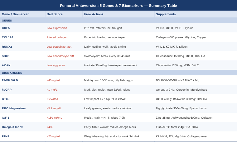
7 Biomarkers to Monitor for Femoral Anteversion
The body leaves measurable traces of what is happening at the tissue level — in cartilage turnover rates, bone formation activity, inflammatory state, and nutrient availability. Tracking these markers does not change the angle of your femoral neck overnight, but it identifies exactly where the biological terrain is weakest, and where targeted intervention will have the most traction. The following seven biomarkers are the most informative for anyone navigating femoral anteversion or its downstream effects.
1. 25-OH Vitamin D (Calcidiol)
Why it matters for femoral anteversion. Vitamin D is essential for calcium absorption, bone mineralization, and the neuromuscular control that buffers hip joint load during movement. In children, severe deficiency causes rickets — a condition that directly exaggerates rotational lower limb deformities, including femoral anteversion. In adults, suboptimal levels (below 40 ng/mL) are associated with reduced cortical bone density, impaired hip muscle activation, and slower periarticular tissue repair. Since femoral anteversion chronically concentrates load on specific joint surfaces, bone and muscle quality become significant determinants of whether the condition remains tolerable or worsens.
How to measure it. A standard 25-hydroxyvitamin D blood test is available at any clinical laboratory. Cost: $30–$80. Testing twice yearly — late winter and late summer — captures meaningful seasonal variation. Target range for musculoskeletal optimization: 40–60 ng/mL. Levels above 100 ng/mL may indicate toxicity risk and warrant clinical review. For a detailed evidence overview, the NIH Office of Dietary Supplements Vitamin D Fact Sheet provides a thorough clinical reference.
If the score is low: the plan without supplements
Safe midday sun exposure is the most physiologically natural route. Exposing large skin surfaces — arms and legs — for 15 to 30 minutes between 10 AM and 2 PM, four to five days per week from spring through early fall, raises levels substantially in most individuals. Oily fish (wild salmon, sardines, mackerel), egg yolks, and liver provide modest but consistent dietary contributions. Scheduling outdoor physical activity during peak solar hours simultaneously addresses the vitamin D deficit and provides the mechanical loading the hip needs.
If the score is low: the plan with supplements
Vitamin D3 (cholecalciferol, not D2) at 2,000–5,000 IU daily is the standard repletion protocol. It must be co-administered with Vitamin K2 (MK-7 form, 100–200 mcg/day) to direct calcium into bone matrix rather than soft tissue. Magnesium is required to convert 25-OH D to its active form (calcitriol), so supplementing without addressing magnesium status limits results — see biomarker 4. Retest after 90 days and adjust dose accordingly. Standard doses carry no clinically meaningful side effects; doses above 5,000 IU daily warrant laboratory monitoring every 6 months. No cycling required at standard doses.
2. High-Sensitivity C-Reactive Protein (hsCRP)
Why it matters for femoral anteversion. hsCRP is the most accessible marker of systemic low-grade inflammation. Femoral anteversion creates abnormal joint contact patterns — focal cartilage compression, repetitive periarticular ligament strain — and inflammation is the biological amplifier of that chronic mechanical irritation. Elevated hsCRP (above 1 mg/L) signals a state that accelerates cartilage matrix degradation, impairs soft tissue repair, and reduces the threshold for symptomatic flare-ups. Peter Attia consistently ranks hsCRP among the highest-value routine markers in preventive medicine precisely because it reflects risk across multiple organ systems simultaneously — including the joint tissues under chronic mechanical stress from femoral anteversion.
How to measure it. Included in many standard panels or ordered separately. Cost: $20–$50. Optimal target: below 0.5 mg/L. Values between 1–3 mg/L warrant lifestyle intervention. Above 3 mg/L requires clinical evaluation to rule out acute infection or autoimmune activity before interpreting as chronic background inflammation.
If the score is high: the plan without supplements
Dietary pattern has the most consistent clinical evidence for lowering hsCRP. Eliminating refined seed oils (soybean, sunflower, corn), ultra-processed foods, and excess refined carbohydrate — and replacing them with a Mediterranean-pattern diet rich in olive oil, vegetables, legumes, and fatty fish — consistently lowers hsCRP within 4–8 weeks across multiple clinical studies. Progressive resistance training three times per week reduces hsCRP independently of weight change. Improving sleep quality — even one chronically short or disrupted night transiently elevates hsCRP — provides an additive effect and is often the fastest variable to address.
If the score is high: the plan with supplements
EPA and DHA omega-3 fatty acids (2–4 g/day from fish oil or algae oil) have the strongest clinical evidence base for hsCRP reduction among joint-relevant supplements. Curcumin with piperine (500–1,000 mg curcumin daily) has demonstrated statistically significant hsCRP reductions in multiple randomized controlled trials. Magnesium glycinate (300–400 mg/day) independently lowers CRP in deficient individuals. These three can be combined safely and their effects are additive. Cycle curcumin every 8–12 weeks with a 2-week break. Fish oil and magnesium can be taken continuously without cycling.
3. CTX-II (Urine C-Terminal Telopeptide of Type II Collagen)
Why it matters for femoral anteversion. CTX-II is a direct biochemical marker of articular cartilage degradation — it measures fragments of type II collagen released when cartilage matrix is broken down by enzymatic activity. Because femoral anteversion alters the normal distribution of load across the hip joint, it creates focal zones of elevated cartilage stress that accelerate breakdown even in people who are currently pain-free. Tracking CTX-II reveals what is happening at the cartilage surface before structural damage becomes visible on imaging. In the framework used by Allan Sniderman and others in precision medicine — organ damage before symptoms — a rising CTX-II is a measurable early warning that justifies preemptive intervention.
How to measure it. Urine test using the second morning void, which gives the most consistent results. Cost: $50–$150 through specialty laboratories such as DoctorsData or ZRT. Not included in standard panels; must be specifically requested. Results should be interpreted against age-adjusted reference ranges and retested every 6 months during active intervention.
If the score is high: the plan without supplements
Load modification is the most important non-supplemental intervention. Reducing high-impact, repetitive loading — distance running on hard surfaces, plyometric training, heavy compressive squats with poor alignment — lowers cartilage contact stress directly. Replacing these with swimming, cycling, and walking on natural terrain distributes load more evenly across the joint surface. Physical therapy targeting hip external rotator and abductor strengthening is essential: the gluteus medius, piriformis, and deep hip rotators offset the abnormal internal rotation torque created by elevated anteversion, reducing the focal cartilage compression that drives CTX-II elevation. Three to four targeted sessions per week over 12–16 weeks produces measurable reductions.
If the score is high: the plan with supplements
Undenatured type II collagen (UC-II, 40 mg/day) has shown statistically significant reductions in cartilage degradation markers in human clinical trials and is among the most evidence-backed cartilage support supplements available. Boswellia serrata standardized to AKBA (300–500 mg/day) reduces matrix metalloproteinase activity, slowing the enzymatic breakdown of cartilage matrix. Oral hyaluronic acid (80–200 mg/day) supports synovial fluid viscosity, reducing friction-related wear on cartilage surfaces. Begin with UC-II alone for 8 weeks to establish a baseline response before adding others. No significant adverse effects have been reported at standard doses for any of these compounds.
4. RBC Magnesium
Why it matters for femoral anteversion. Standard serum magnesium — the version included in routine metabolic panels — misses the majority of deficiencies because the body maintains serum levels by extracting magnesium from bone and muscle. RBC magnesium measures intracellular status, which is where the mineral actually functions. Magnesium participates in over 300 enzymatic processes, including the activation of vitamin D, calcium regulation in bone matrix, collagen cross-linking, and neuromuscular control of the hip rotators. Low intracellular magnesium impairs all of these — creating a biological environment that is poorly suited to either maintaining bone quality or developing the precise hip muscle coordination that compensates for rotational structural imbalance.
How to measure it. A specific RBC magnesium panel, distinct from the serum magnesium included in standard metabolic panels. Cost: $30–$80. Optimal range: 5.6–6.8 mg/dL. Below 5.2 mg/dL warrants prompt intervention. Retest after 90 days of supplementation. The NIH Magnesium Health Professional Fact Sheet provides comprehensive reference ranges and evidence for skeletal and neuromuscular applications.
If the score is low: the plan without supplements
Dark leafy greens (spinach, Swiss chard), pumpkin seeds, black beans, avocado, and dark chocolate are the most magnesium-dense dietary sources. Light cooking rather than boiling preserves mineral content. Reducing alcohol — which dramatically increases renal magnesium excretion — is often the single most impactful change for people who consume it regularly. Addressing gut health matters as well: magnesium absorption depends on intestinal mucosal integrity, and conditions causing intestinal inflammation substantially reduce it.
If the score is low: the plan with supplements
Magnesium glycinate or malate are the preferred forms for musculoskeletal applications — significantly better absorbed and far less laxative than magnesium oxide. Dose: 300–400 mg elemental magnesium daily, ideally in the evening. Epsom salt baths (magnesium sulfate, 2 cups per 20-minute soak, three times per week) provide transdermal magnesium alongside direct hip rotator muscle relaxation — addressing both the systemic deficiency and the local tissue tension that compounds femoral anteversion symptoms. No cycling required at standard doses; reduce if loose stools occur.
5. IGF-1 (Insulin-Like Growth Factor 1)
Why it matters for femoral anteversion. IGF-1 is the primary mediator of growth hormone's anabolic effects on bone and connective tissue. It stimulates osteoblast proliferation, promotes collagen synthesis, and drives periosteal bone remodeling — the adaptive process by which bone architecture changes in response to mechanical signals. In growing children with femoral anteversion, IGF-1 levels significantly influence whether the femoral neck undergoes the spontaneous derotation that normally occurs between ages three and eight. In adults with residual anteversion, adequate IGF-1 is what makes physical therapy and movement retraining biologically viable: without it, the remodeling response to targeted loading is blunted, and gains are slow and limited.
How to measure it. Standard blood test, typically included in growth hormone panels. Cost: $50–$150. Age-adjusted optimal range for adults aged 30–50: 150–300 ng/mL. Values naturally decline with age, which partially explains why tissue repair slows over time. Retesting every 6 months during active intervention is appropriate.
If the score is low: the plan without supplements
Compound resistance training involving large lower-body muscle groups is the most potent physiological stimulus for IGF-1 production. Hip hinge variations, squat patterns, and single-leg press work are particularly relevant because they load the most mechanically important structures for femoral anteversion management. High-intensity interval training additionally stimulates growth hormone in the 24 hours post-exercise. Adequate protein intake (1.6–2.2 g/kg body weight per day) provides the amino acid substrate for the anabolic response. Sleep optimization — 7–9 hours with consistent timing — is non-negotiable because 70–80% of growth hormone secretion occurs in the first several hours of deep sleep.
If the score is low: the plan with supplements
Zinc (25–40 mg/day with food, balanced with 2 mg copper to prevent displacement) directly supports IGF-1 synthesis and is frequently depleted in people with high training loads. Ashwagandha (KSM-66 extract, 600 mg/day) has demonstrated significant IGF-1 increases in peer-reviewed human randomized trials. Collagen peptides (10–15 g/day, taken 30–60 minutes before exercise with 500 mg vitamin C) enhance connective tissue synthesis in the context of adequate IGF-1 levels, supporting the ligament and cartilage structures strained by abnormal hip rotation. Cycle ashwagandha: 8 weeks on, 2 weeks off; some individuals experience mild sedation. Collagen peptides are safe long-term.
6. Omega-3 Index
Why it matters for femoral anteversion. The Omega-3 Index measures EPA and DHA as a percentage of total fatty acids in red blood cell membranes — a stable, representative indicator of anti-inflammatory fatty acid status throughout the body, including joint tissues. Below 4% indicates a strongly pro-inflammatory state; the research-supported target is 8–12%. EPA and DHA reduce the activity of matrix metalloproteinases (enzymes that degrade collagen and cartilage matrix), promote the resolution of periarticular inflammation, and support the structural integrity of the ligaments chronically stressed by abnormal hip rotation patterns. Peter Attia consistently cites the Omega-3 Index as among the most underutilized markers in routine medicine, particularly for people with musculoskeletal burden.
How to measure it. Finger-prick home test via OmegaQuant, the laboratory used as the standard reference in most published research on this marker. Cost: $50–$75. Retest every 6 months when actively adjusting fatty acid intake to track response. The NIH Omega-3 Fatty Acids Fact Sheet provides a detailed overview of tissue-level mechanisms and evidence.
If the score is low: the plan without supplements
Increasing fatty fish consumption to three to four servings per week — sardines, wild salmon, mackerel, and anchovies are the most concentrated sources — is the most direct dietary approach. Reducing competing omega-6 linoleic acid from refined vegetable oils (sunflower, corn, soybean) improves the omega-6/omega-3 ratio and reduces net inflammatory signaling even without additional omega-3 intake. This ratio adjustment is often as important as the absolute omega-3 number.
If the score is low: the plan with supplements
Triglyceride-form fish oil — significantly better absorbed than ethyl ester forms — at 2–4 g combined EPA + DHA per day is the standard intervention. Algae-based DHA and EPA is equally effective and appropriate for those avoiding fish products. Take with the largest meal of the day for optimal absorption. At 3–4 g per day, the index typically reaches the 8–12% target range within 90–120 days. Enteric-coated formulations minimize fishy reflux. No cycling needed; fish oil is safe long-term at these doses. Above 4 g daily, discuss with a physician if taking anticoagulant medications.
7. P1NP (Procollagen Type 1 N-Terminal Propeptide)
Why it matters for femoral anteversion. P1NP is the most sensitive available marker of active bone formation — it directly reflects osteoblast production of new type I collagen, the first step in building new bone matrix. When paired with a bone resorption marker such as CTX, it provides a real-time picture of the remodeling balance: is bone being built faster than it is being broken down? This matters directly for femoral anteversion management because any strategy aimed at gradual mechanical adaptation — targeted loading, gait retraining, physical therapy — depends on active osteoblast responsiveness. A suppressed P1NP signals that bone formation capacity is insufficient to respond meaningfully to mechanical stimuli, limiting the adaptive potential of any rehabilitation program.
How to measure it. Fasting morning blood draw — bone turnover markers are diurnal, and morning consistency matters for valid comparisons over time. Cost: $50–$150. Should ideally be paired with serum CTX for a complete remodeling picture. Reference ranges are sex and age-dependent; for premenopausal women and adult men, values below 20 ng/mL suggest suppressed formation. Retest every 3–6 months during active intervention.
If the score is low: the plan without supplements
Weight-bearing mechanical loading is the strongest physiological driver of P1NP. Bone formation markers rise within 24–48 hours of a loading session — the response is rapid even when structural change takes months. For femoral anteversion specifically, the most relevant loading protocols include hip abductor strengthening (lateral band walks, clamshells), single-leg stance training (which loads the hip in the positions most affected by excess anteversion), and progressive lower body resistance work with full hip extension. Three to four sessions per week, with at least one rest day between sessions, allows anabolic signaling consolidation. Avoiding prolonged sedentary periods between sessions matters as much as the sessions themselves.
If the score is low: the plan with supplements
Vitamin K2 (MK-7, 100–200 mcg/day) is essential for carboxylating osteocalcin — the protein that mineralizes the new collagen matrix built by active osteoblasts. Without adequate K2, new bone matrix forms but mineralizes poorly. Vitamin D3 (biomarker 1) and magnesium (biomarker 4) are prerequisites for the full bone formation cascade, and this trio — D3, K2, magnesium — should be considered foundational before any more targeted bone agents are added. Collagen peptides (10–15 g/day, taken pre-exercise with vitamin C) provide the glycine and proline substrate that P1NP reflects being deposited. All three are safe long-term at standard doses with no significant interactions.
The Genetics of Femoral Anteversion: 5 Genes Worth Knowing
The biomarkers above reveal what is happening now in your joint tissue. What they do not fully explain is why your hip developed this geometry in the first place — or why two people with the same anteversion angle can have dramatically different tissue quality and symptom trajectories. That is where genetics enters the picture.
Femoral anteversion is substantially heritable. Twin studies and family aggregation data suggest that 50–70% of variation in femoral neck geometry is explained by genetic factors. The genes below influence either the developmental blueprint for skeletal geometry, the quality of bone and cartilage matrix across a lifetime, or both. Genetic testing through consumer services such as 23andMe or clinically oriented panels (GeneDx, Genomind, or direct functional genomics consultation) can identify your status for most of these variants. The frameworks below apply whether you have confirmed variants or are using them as a general risk-stratification guide.
1. GDF5 (Growth Differentiation Factor 5)
What it does. GDF5 is a member of the TGF-β superfamily that plays a central role in limb joint formation during embryonic development, specifically in establishing joint cavity geometry, cartilage morphology, and the spatial positioning of the femoral head relative to the acetabulum. The single nucleotide polymorphism rs143384 — the A allele, commonly referred to as the risk allele — results in reduced GDF5 expression in joint tissues compared to the G allele. Carriers of the AA genotype show measurable differences in joint architecture, including shallower acetabular coverage and altered femoral head-neck geometry, which directly influences the persistence and degree of femoral anteversion. A landmark study published in Nature Genetics (Miyamoto et al.) established this association across large multi-ethnic cohorts. This is among the best-characterized skeletal geometry genes identified in human genome-wide association studies.
What it may affect. Reduced GDF5 signaling during development influences the trajectory of femoral head-neck geometry and may predispose to less favorable spontaneous correction during childhood, as well as accelerated cartilage wear in adulthood under abnormal mechanical conditions.
If the gene variant is present: the plan without supplements
The primary compensatory strategy is optimizing the mechanical environment around the joint morphology you have. This means: physical therapy focused on hip external rotator and abductor strength to offset the rotational torque from altered geometry; movement retraining toward a neutral or slightly externally rotated gait pattern; and avoidance of prolonged W-sitting (heels out, knees in) — the position that chronically reinforces hip internal rotation in people with elevated anteversion. Daily mobility work (10–15 minutes), strength training three times per week, and gait awareness during daily walking are the three pillars. These directly address the mechanical consequences of altered GDF5-driven joint morphology without requiring any supplementation.
If the gene variant is present: the plan with supplements
Vitamin D3 has been shown in preclinical studies to upregulate GDF5-related signaling in cartilage progenitor cells — ensuring optimal D3 status (50–60 ng/mL) is the most biologically supported supplement-based intervention for GDF5 variants. Undenatured type II collagen (UC-II, 40 mg/day) supports cartilage integrity in altered joint geometries. Vitamin C (500–1,000 mg/day) and lysine (500–1,000 mg/day) provide substrate for cartilage collagen matrix maintenance. These do not change your genotype but may partially compensate for reduced endogenous GDF5-driven cartilage regeneration capacity. All are safe long-term at stated doses. Preclinical data on GDF5 modulation is promising; human-specific evidence remains an active research area.
2. COL1A1 (Collagen Type 1 Alpha 1)
What it does. COL1A1 encodes the alpha-1 chain of type I collagen — the primary structural protein of bone matrix, tendons, and ligaments. The Sp1 polymorphism (rs1800012) produces an overexpression of the alpha-1 chain relative to the alpha-2 chain, altering the mechanical properties of the resulting collagen: less resilient, more prone to fatigue failure under repetitive loading. In the context of femoral anteversion, this matters significantly because the periarticular ligaments stabilizing the hip — the iliofemoral and pubofemoral ligaments — are primarily type I collagen structures. Reduced collagen resilience means these stabilizers fatigue faster under the atypical loading patterns created by excess femoral rotation, contributing to joint instability and accelerated wear.
What it may affect. Reduced bone matrix fracture toughness, ligament laxity around the hip, slower connective tissue recovery from mechanical stress, and potentially increased susceptibility to stress-related hip pathology over time.
If the gene variant is present: the plan without supplements
Progressive loading protocols are essential — the goal is to increase connective tissue mechanical capacity gradually rather than applying sudden high loads that exceed the tolerance of structurally reduced-quality collagen. Eccentric exercise protocols (controlled, slow lowering phases in hip abductor and external rotator work) are particularly effective at remodeling tendon and ligament structure. Extended warm-up periods before hip-loading activities allow tissue temperature and viscoelasticity to optimize. Reduce impact loading frequency and progress loads conservatively. Three to four structured sessions per week at moderate intensity, with a conservative 5–10% load progression per 2-week period.
If the gene variant is present: the plan with supplements
Collagen peptides (10–15 g/day taken 30–60 minutes before exercise) with 500 mg vitamin C have demonstrated significantly upregulated collagen synthesis in tendon and ligament tissue in randomized trials — the evidence from Shaw, Baar, and colleagues at UC Davis is among the most mechanistically coherent in the connective tissue field. Glycine (5 g/day) provides additional synthesis substrate. Copper (2 mg/day) is required for lysyl oxidase activity — the enzyme that cross-links collagen fibers and determines their mechanical quality. This is the step most directly affected by COL1A1 variants: supporting the cross-linking enzymatic pathway helps compensate for altered chain composition. Balance copper with zinc to prevent displacement. Collagen peptides and vitamin C are safe long-term.
3. RUNX2 (Runt-Related Transcription Factor 2)
What it does. RUNX2 is the master transcriptional regulator of osteoblast differentiation — the gene that instructs undifferentiated mesenchymal stem cells to become bone-forming cells. Variants that reduce RUNX2 activity produce fewer, less active osteoblasts, resulting in impaired bone mineralization, reduced cortical bone density, and altered skeletal patterning. In clinical extremes, RUNX2 haploinsufficiency causes cleidocranial dysplasia — a multi-skeletal developmental condition. More commonly, subclinical RUNX2 variants reduce the body's adaptive bone remodeling response to mechanical stimulation. For femoral anteversion management, this is directly limiting: the entire premise of mechanotherapy (loading the hip to drive gradual geometric adaptation) requires robust RUNX2-mediated osteoblast activity to function.
What it may affect. Reduced bone formation rate, lower cortical bone density, impaired response to mechanical loading stimuli, and potentially a less favorable spontaneous correction trajectory in children with femoral anteversion.
If the gene variant is present: the plan without supplements
Mechanical loading remains the strongest physiological activator of RUNX2 expression in osteoprogenitor cells, even when baseline transcriptional activity is reduced. High-impact bone-specific loading — progressive resistance training, brief jumping protocols on appropriate surfaces, stair climbing — directly upregulates RUNX2 in bone cells and partially compensates for reduced baseline expression. Consistency matters more than intensity: daily low-level mechanical stimulation (walking, standing on uneven surfaces, active standing workstation use) maintains basal RUNX2 activation, while three sessions per week of higher-load work drives larger remodeling signals. Avoiding prolonged sedentary periods is essential since RUNX2 expression drops substantially with mechanical unloading.
If the gene variant is present: the plan with supplements
Vitamin D3 directly induces RUNX2 expression in osteoblast precursors — this is one of the most mechanistically well-documented nutrient-gene interactions in bone biology, with consistent evidence across in vitro, animal, and human studies. Maintaining vitamin D status at the upper optimal range (50–60 ng/mL) is the single most important supplement-supported intervention for RUNX2 variants. Vitamin K2 (MK-7, 100–200 mcg/day) supports the downstream osteocalcin carboxylation that RUNX2-activated osteoblasts depend on for effective mineralization. Silicon (from orthosilicic acid or bamboo-derived extract, 10–25 mg/day) has emerging evidence for supporting bone matrix initiation, the step immediately downstream of RUNX2 activation. D3 and K2 can be taken long-term without cycling; silicon is well tolerated at standard doses.
4. SOX9
What it does. SOX9 is the master regulator of chondrocyte differentiation — the gene that directs progenitor cells to become the cartilage-forming cells that build and maintain articular cartilage. Variants that reduce SOX9 activity impair the formation of the hyaline cartilage lining the hip joint, and more fundamentally, alter the cartilaginous developmental template (the anlage) that guides bone geometry before ossification. In the developmental context, SOX9 activity shapes the geometry of the cartilaginous femoral neck before it mineralizes into bone — meaning variants in this gene may contribute directly to the degree and persistence of femoral anteversion. In the adult context, reduced SOX9 activity translates to slower cartilage repair and reduced resilience of existing articular cartilage under the chronic stress of abnormal rotation patterns.
What it may affect. Altered developmental cartilage geometry contributing to femoral anteversion persistence, reduced articular cartilage thickness and quality, and impaired chondrocyte repair response to joint stress in adulthood.
If the gene variant is present: the plan without supplements
Low-impact cyclical movement is the most protective regime for people with reduced intrinsic cartilage-forming capacity. Articular cartilage lacks direct blood supply — it is nourished through compression and release cycles (the mechanical pumping of movement). Swimming, cycling, and consistent daily walking maintain cartilage nutrition most effectively. Prolonged static loading, particularly sitting with sustained hip flexion, reduces cartilage fluid exchange and should be interrupted every 30–45 minutes with brief movement. High-impact repetitive loading (hard-surface running, impact sports) should be minimized and replaced with equivalent cardiovascular work at lower joint stress.
If the gene variant is present: the plan with supplements
Glucosamine sulfate (1,500 mg/day) and chondroitin sulfate (1,200 mg/day) are the most studied cartilage matrix support supplements. Evidence for their efficacy in the general population is mixed, but they are mechanistically most relevant to people with reduced endogenous cartilage matrix production — which SOX9 variants may represent. Undenatured type II collagen (UC-II, 40 mg/day) has shown benefits in hip and knee cartilage integrity studies. Oral hyaluronic acid (80–200 mg/day) supports synovial fluid viscosity, reducing compressive friction on already-vulnerable cartilage surfaces. These can be combined. Cycle glucosamine and chondroitin every 6 months with a 4-week break to assess ongoing need. No significant adverse effects at standard doses.
5. ACAN (Aggrecan)
What it does. ACAN encodes aggrecan — the large aggregating proteoglycan that gives articular cartilage its water-retaining capacity and, by extension, its ability to absorb and distribute compressive load. The hip joint withstands forces several times body weight during gait precisely because of the water trapped within aggrecan's negatively charged glycosaminoglycan chains. Variants in ACAN that reduce proteoglycan production result in cartilage with lower water content, decreased compressive stiffness, and faster wear under repetitive loading. These variants have been associated with early intervertebral disc degeneration and early hip and knee osteoarthritis — both of which are downstream risks in individuals whose femoral anteversion chronically shifts joint load to focal cartilage zones.
What it may affect. Reduced cartilage hydration and load-bearing capacity, accelerated cartilage thinning under the focal stress created by femoral anteversion, and increased susceptibility to hip osteoarthritis at younger ages than genetic peers without this variant.
If the gene variant is present: the plan without supplements
Systemic hydration is foundational: aggrecan function is hydration-dependent, and even mild chronic dehydration concentrates joint load on a smaller effective cartilage area. A practical target is 35–40 ml per kg body weight of water daily. Consistent low-impact cyclical movement maintains glycosaminoglycan saturation through mechanical fluid exchange. Reducing cumulative compressive load — avoiding heavy squats below 90 degrees with poor alignment, reducing jump landing impact, managing body weight to lower per-step joint force — protects cartilage that has reduced intrinsic capacity to recover from compressive episodes.
If the gene variant is present: the plan with supplements
Chondroitin sulfate (1,200 mg/day) provides the glycosaminoglycan substrate that ACAN variants may produce in insufficient quantities — a published MRI study in Annals of the Rheumatic Diseases (Ludin et al., 2016) demonstrated significant reduction in cartilage volume loss with chondroitin supplementation. MSM (methylsulfonylmethane, 1,500–3,000 mg/day) provides sulfur required for glycosaminoglycan sulfation, directly relevant to aggrecan proteoglycan function. Vitamin C (500–1,000 mg/day) supports proteoglycan synthesis as a cofactor and is foundational to connective tissue health broadly. Cycle chondroitin sulfate every 6 months with a 4-week break. MSM and vitamin C are safe for continuous long-term use.
"Move Your DNA" by Katy Bowman: The Movement Framework That Reframes Hip Problems
The biomarker and genetic frameworks above give you a measurable, actionable picture of your biological terrain. But they do not fully address the question of why so many people with femoral anteversion develop secondary pain and dysfunction while others manage for decades without significant pathology. The answer often points to something more fundamental: whether the body is receiving the movement signals it was designed for, at the frequency those signals need to arrive.
Move Your DNA: Restore Your Health Through Natural Movement by biomechanist Katy Bowman is one of the most evidence-grounded popular books on how modern movement deprivation shapes — and reshapes — skeletal geometry and joint health. While not written specifically for femoral anteversion, its central thesis is directly applicable: bone geometry is not fixed, it is dynamically maintained and altered by the mechanical signals it receives throughout the day. For anyone managing rotational hip issues, the framework Bowman builds may be more clinically valuable over the long term than any single supplement protocol. Below are ten of the most impactful ideas from the book.
1. Bone Is a Living Load-Bearing Antenna
Bowman opens with what bone biologists call Wolff's Law: bone architecture continuously remodels in response to the mechanical loads placed upon it throughout life. This is observable in every population study comparing habitual loading patterns to skeletal geometry. For femoral anteversion, this means that how you load your hip today is actively influencing its architecture — for better or worse. This is the most foundational principle in the book: your bones are registering every movement pattern, every sustained posture, every habitual position, and responding over months and years.
2. Exercise Is Not the Same as Movement
One of Bowman's central arguments is that 60 minutes of daily structured exercise cannot undo the mechanical consequences of 16 hours of sedentary loading. The total movement input distributed across the day — what she calls the movement diet — matters more than any single workout session. For people with femoral anteversion, this translates to a clear practical directive: the hip needs frequent, varied positional input throughout the day, not just a physical therapy appointment three times per week. Changing how you sit, stand, and move during ordinary hours is the lever that structured sessions cannot replace.
3. Modern Footwear Distorts the Entire Lower Chain
Heeled footwear — including most conventional athletic shoes with elevated heels relative to the toe box — shifts the center of mass anteriorly and internally rotates the femur as a compensatory response. Bowman provides a detailed mechanical analysis of how even a small heel elevation (common in running and cross-training shoes) alters hip loading patterns and can reinforce rather than reduce the rotational stress from femoral anteversion. Transitioning gradually to zero-drop or minimal footwear changes the mechanical input at the femoral level over thousands of daily steps.
4. Sitting in Chairs Is a Full-Day Hip Internal Rotation Session
The standard chair-seated posture — hips and knees at 90 degrees, feet flat on the floor — places the femur in sustained moderate internal rotation and hip flexion. Prolonged daily exposure to this position progressively shortens the hip internal rotators and anterior hip capsule, and chronically trains the femoral head into an anteriorly biased position. For people who sit eight or more hours daily, this represents a constant reinforcement of the exact pattern femoral anteversion already predisposes to.
5. The Floor Is the Most Underused Movement Tool
Bowman strongly advocates for habitual floor-level activity — ground sitting in varied positions such as cross-legged, side-sitting, and long-sitting — as a way to introduce diverse joint positional inputs throughout the day. Each floor position loads the hip capsule differently, maintaining the full rotational range that chair culture progressively eliminates. For people with femoral anteversion, floor sitting naturally incorporates more external rotation positioning than chair sitting, providing a low-cost mechanical counter-stimulus throughout the day.
6. The Deep Squat Resets Hip Mechanics
The full, unassisted deep squat — heels flat, maximum hip flexion, toes forward or slightly out — applies a specific combination of joint distraction and circumferential capsular loading that Bowman describes as one of the most complete mechanical reset positions available. This position requires and simultaneously develops full hip capsule mobility in all directions, including the external rotation range typically reduced in people with elevated femoral anteversion. Daily practice, even for three to five minutes, provides mechanical inputs that no machine-based or table-based exercise replicates.
7. Foot Progression Angle Directly Shapes Femoral Load
The angle at which the foot contacts the ground determines the direction of ground reaction forces entering the femur. A habitual toe-in pattern — common in people with elevated femoral anteversion as an adaptive gait strategy — directs forces in ways that compound the internal rotation position through the hip joint. Bowman explains that consciously working toward a neutral foot progression angle changes mechanical input to the femoral head-neck junction over thousands of steps daily. Even modest adjustments in foot angle, compounded across walking distance, accumulate into significant cumulative mechanical change over months.
8. Barefoot Walking Restores the Proprioceptive Hip Signal
Modern footwear dampens the sensory input from ground contact that normally triggers automatic postural adjustments — including hip rotation micro-corrections during the gait cycle. Barefoot or minimal-shoe walking on varied natural terrain restores these afferent signals, allowing the nervous system to make real-time adjustments to hip positioning during each step. Studies on barefoot walking in children have shown measurable effects on lower limb rotational mechanics. Bowman recommends progressive barefoot exposure beginning on softer surfaces to avoid abrupt calf and plantar loading issues.
9. Hip Rotator Tightness Is Often Protective, Not Pathological
Bowman makes a nuanced point that most therapeutic protocols miss: when the hip external rotators are persistently tight, that tightness frequently reflects the nervous system's attempt to stabilize a mechanically compromised joint — not a primary structural problem to be aggressively stretched away. Stretching external rotators without simultaneously building their strength, and without addressing the underlying loading pattern driving the protective tension, can transiently worsen joint stability. Strengthening and repositioning must accompany — and in some cases precede — flexibility work.
10. Environmental Redesign Beats Adding More Interventions
The overarching recommendation that runs through the book is environmental redesign rather than intervention addition. A standing or variable workstation, consistent floor-level sitting, barefoot time, and daily brief outdoor walking contribute more cumulative mechanical signal when integrated into ordinary life than any isolated therapeutic protocol added on top of an otherwise unchanged sedentary environment. The compounding logic is key: small daily inputs sustained over months accumulate into measurable structural change, while intensive weekly sessions applied to an otherwise static loading environment produce diminishing returns.
Complementary Approaches with Clinical Support
The movement and biology-based strategies above are meaningfully strengthened by evidence-supported complementary modalities that address femoral anteversion from different angles — from joint tissue preparation to neuromuscular repatterning. The following four modalities were selected for having at least moderate human clinical evidence in musculoskeletal hip conditions, movement retraining, or connective tissue health.
Yoga
Yoga addresses hip mobility, external rotator flexibility, and kinesthetic body awareness — three capacities directly relevant to femoral anteversion management. Classic hip-opening yoga poses (pigeon, lizard, reclined figure-four, warrior II) selectively stretch the hip internal rotators and anterior capsule while simultaneously developing the external rotator engagement that counteracts excess internal rotation torque. The proprioceptive component of sustained poses also trains positional awareness that transfers to gait and daily movement.
A 2015 randomized controlled trial published in the Journal of Pain Research found that a 12-week yoga intervention produced significant improvements in hip range of motion, pain scores, and functional outcomes in participants with hip-related musculoskeletal complaints. While large trials specific to femoral anteversion have not been conducted, the mechanical and neuromuscular targets of yoga are directly applicable to the biomechanical demands of this condition.
Practice three to four times per week for 20–45 minutes, with emphasis on hip external rotation and abductor engagement rather than deep passive hip flexion. For femoral anteversion specifically, the most useful poses are those that load the hip in external rotation under mild muscular engagement — pigeon (modified with props if hip impingement is present), warrior II, and lateral angle pose. Avoid hypermobile end-range positions if ligament laxity is part of your presentation — passive overstretching of already-lax periarticular structures is counterproductive for joint stability.
Biofeedback
Biofeedback provides real-time sensory information about a body process that normally operates below conscious awareness. For femoral anteversion, the most clinically relevant application is gait biofeedback — using visual, auditory, or vibratory feedback cues to monitor foot progression angle, knee tracking, and hip rotation patterns during walking and running. The in-toeing gait pattern in femoral anteversion is typically automated and habitual; people cannot reliably correct it through voluntary intention alone because they cannot perceive the error while it is happening. Biofeedback creates the sensory loop required for long-term repatterning.
A 2017 randomized controlled trial in the Journal of Biomechanics (Crowell and Davis) demonstrated that real-time kinematic biofeedback during running significantly modified gait mechanics and that these changes persisted at follow-up after feedback was withdrawn. Biofeedback-driven gait retraining has also shown clinical benefit in patellofemoral pain syndrome, which shares significant mechanical drivers with femoral anteversion-related lower limb malalignment.
Clinically, gait biofeedback is pursued through: a physiotherapist using motion analysis equipment or pressure-sensitive insoles; instrumented footwear available through sports medicine clinics; or structured mirror-based and video-feedback gait retraining at lower cost. Sessions of 20–30 minutes, two to three times per week, over 8–12 weeks have been the typical protocol in published trials. Supervision by a physiotherapist specializing in gait retraining is important to ensure feedback targets are calibrated to your specific rotational pattern — overcorrection into excessive external rotation creates a different set of downstream problems.
Massage Therapy
Deep tissue massage and myofascial release targeting the hip external rotators, piriformis, iliotibial band, and anterior hip flexors is directly relevant to femoral anteversion because excessive femoral internal rotation chronically shortens and sensitizes these tissues. The piriformis — the primary hip external rotator — develops trigger points and adaptive shortening in response to the chronic rotational load from anteversion. Paradoxically, this adaptive shortening progressively reduces the external rotation capacity that is needed most for mechanical compensation, creating a self-reinforcing cycle. Targeted massage interrupts this cycle by releasing adaptive tissue restrictions and allowing physical therapy to access the available functional range.
A 2016 systematic review in the Journal of Clinical and Diagnostic Research found that deep tissue massage significantly improved hip range of motion and reduced pain scores in participants with hip-related musculoskeletal dysfunction. Piriformis-specific myofascial protocols have also shown benefit in piriformis syndrome — a condition with high co-occurrence in people with femoral anteversion due to the chronic rotational loading placed on the muscle.
A practical series of 6–10 sessions targeting the piriformis, deep external rotators, tensor fascia latae, and anterior hip flexors — performed by a therapist with sports or orthopedic massage training — provides the best starting framework. One to two sessions per week in the first four weeks, then once weekly for maintenance. Between sessions, self-massage using a lacrosse ball placed below the posterior iliac crest (side-lying position) targets the piriformis directly and maintains the range gains achieved in professional sessions. Results compound substantially when this soft tissue preparation is combined with the strengthening and gait work described in earlier sections.
Tai Chi
Tai chi is a practice of slow, deliberate movement sequences that develops weight transfer precision, lower limb proprioception, and rotational body awareness — capacities specifically challenged by femoral anteversion's altered gait and loading mechanics. The controlled single-leg weight-bearing transitions that characterize tai chi directly train hip abductor and deep rotator control in the functional range where people with femoral anteversion most commonly show deficits. Unlike isolated hip strengthening exercises, tai chi practices these capacities in the context of whole-body coordination and weight shift, which is closer to how they are actually demanded in daily movement.
A 2018 systematic review and meta-analysis published in JAMA Internal Medicine found that tai chi practice significantly reduced fall risk, improved standing balance, and enhanced proprioceptive accuracy in older adults with lower limb musculoskeletal conditions. Research specific to femoral anteversion is limited, but the balance, proprioception, and lower limb neuromuscular control benefits are directly translatable to the functional demands of managing this condition.
A beginner tai chi program — 30 minutes, three times per week — maintained for a minimum of 12 weeks produces measurable proprioceptive and balance improvements in most published studies. For femoral anteversion, forms emphasizing deliberate hip rotation and single-leg weight transfer are the most relevant. In-person instruction with a qualified teacher is strongly preferred over video-only practice in the first 8 weeks, particularly to ensure that knee and hip alignment during rotational movements is correct — poor alignment in tai chi practice can reinforce rather than correct lower limb rotation problems if unsupervised.
Conclusion
Femoral anteversion is not a condition that resolves on its own in adults, nor does it need to progress inevitably toward hip pathology. The trajectory depends significantly on the quality of the biological environment around the joint, the mechanical inputs the hip receives daily, and how consistently those variables are monitored and adjusted.
The seven biomarkers covered here — vitamin D, hsCRP, CTX-II, RBC magnesium, IGF-1, Omega-3 Index, and P1NP — offer a measurable, modifiable picture of that biological environment. The five genes — GDF5, COL1A1, RUNX2, SOX9, and ACAN — provide context for why your particular tissue quality and joint geometry may be more or less responsive to intervention. Katy Bowman's movement framework and the complementary modalities above complete the picture with the behavioral and mechanical layer that no supplement program can replace.
The most practical next step is to begin with the most accessible and high-yield tests: vitamin D, hsCRP, and RBC magnesium are available through routine blood work at modest cost and give an immediate, actionable picture. Review results with a physician or functional medicine practitioner who can contextualize them against your clinical presentation. Layer in movement changes and targeted physical therapy with clearly defined goals, and track biomarker progress at 90-day intervals. This is not about finding a single solution — it is about building a protocol grounded in your actual biology, adjusted over time as the data evolves.
Musculoskeletal: Bone Conditions Joint Conditions Muscle Conditions Tendon & Ligament Conditions
Autoimmune: Inflammatory Conditions Connective Tissue Conditions