This article contains AI generated content.
Femoral Condyle Subchondral Edema: 5 Genes and 7 Biomarkers to Track
Introduction
If an MRI report has ever handed you the phrase "subchondral bone marrow edema of the medial femoral condyle," you already know how little that sentence actually explains. It names a finding, not a cause. It doesn't tell you why it happened, whether it will resolve, or what you should do differently starting tomorrow. Most people leave that appointment with a brace, a rest recommendation, and a vague sense that their knee has become unpredictable.
Generic advice fills that gap badly. "Lose weight, strengthen your quads, avoid impact" is not wrong, but it treats every case of subchondral edema as identical, when the drivers behind it are not. One person's bone marrow lesion is downstream of years of undiagnosed vitamin D insufficiency and low-grade inflammation. Another's is tied to a genetic variant that quietly weakens how cartilage and subchondral bone remodel under load. A third case is mostly mechanical, following a meniscus tear that changed how force travels through the joint. The same three sentences of advice cannot serve all three people well.
This article takes the more specific route. Instead of repeating what you likely already know, it looks at the measurable biology behind subchondral edema: the blood-based biomarkers that reflect what is actually happening in bone and cartilage turnover, and the genetic variants that help explain why some joints are more vulnerable than others. Both lenses are imperfect on their own, but together they turn a vague MRI finding into a shortlist of things you can actually test, track, and act on.
None of this replaces an orthopedic workup, and nothing here promises to reverse existing edema on a fixed timeline — bone marrow lesions are notoriously variable, and biology doesn't move on command. But better information changes the quality of the decisions you can make: which labs to ask your doctor for, which findings deserve real concern, and which levers are actually yours to pull. The sections below cover a practical biomarker panel first, then the genetic variants worth knowing about, a research-backed perspective on joint-protective training, and a set of complementary approaches with real clinical evidence behind them.
Summary
Subchondral bone marrow edema in the femoral condyle sits at the intersection of mechanical stress, bone metabolism, inflammation, and — for a meaningful subset of people — inherited variation in how cartilage and bone are built and maintained. Blood work can tell you more than most people realize: markers of bone resorption and formation, cartilage breakdown, systemic inflammation, vitamin D status, metabolic health, and even uric acid can each point toward a different mechanism driving the lesion. Genetic variants in genes like GDF5, VDR, COL1A1, IL6, and ESR1 don't determine your fate, but they do shift the odds and, more usefully, suggest which compensations matter most for you specifically. Later sections translate a well-known longevity book's exercise framework into joint-specific terms, and review which mind-body and manual therapies have actual trial data in knee osteoarthritis rather than borrowed reputation. The goal throughout is the same: replace "just rest it" with a short list of numbers and variants worth knowing.
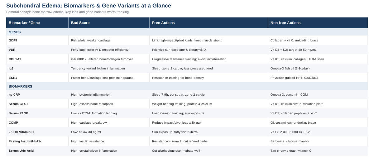
The Seven Biomarkers Worth Tracking
Subchondral bone marrow edema is not a single disease — it's a shared final pathway that shows up on MRI when the bone underneath cartilage is under more mechanical, inflammatory, or metabolic stress than it can quietly absorb. That's exactly why blood biomarkers are useful here: they let you see which of those stresses is actually elevated in your case, rather than guessing. The seven markers below cover the four biological systems most consistently implicated in bone marrow lesions of the knee — systemic inflammation, bone remodeling, cartilage turnover, and metabolic health — with an eye toward what's affordable and what's worth reserving for a more thorough workup.
High-Sensitivity C-Reactive Protein (hs-CRP)
Hs-CRP is a general marker of systemic low-grade inflammation, and it is one of the more direct links to bone marrow lesions specifically. A cohort study following patients with knee osteoarthritis found that higher baseline hs-CRP was associated with both the presence and the progression of bone marrow lesions on MRI (hs-CRP and knee bone marrow lesions study). Elevated hs-CRP suggests that the joint environment is being driven, at least partly, by systemic inflammation rather than pure mechanics — which changes the intervention priority from "protect the joint" to "cool down the whole system."
How to measure it
A standard hs-CRP blood draw costs roughly 15 to 40 dollars out of pocket in the US, or is often bundled into a basic metabolic panel through insurance. It requires no special preparation beyond avoiding acute illness, which transiently spikes the number regardless of joint status.If the score is bad — the plan without supplements
Prioritize sleep consistency (7 to 9 hours, stable wake time), since poor sleep alone raises CRP measurably within days. Reduce ultra-processed food and added sugar intake, which are two of the more reproducible dietary drivers of low-grade inflammation. Add two to three weekly sessions of zone 2 cardio (a pace where conversation is still possible), which lowers CRP over 8 to 12 weeks in most people. Retest at 3 months, not sooner — CRP is noisy week to week.If the score is bad — the plan with supplements or equipment
Omega-3 fish oil (2 to 3 grams combined EPA/DHA daily, with food) has the most consistent evidence for lowering CRP over 8 to 12 weeks; cycle it continuously rather than on and off, and watch for mild GI upset or, at high doses, easy bruising — flag it to a physician if you're on blood thinners. Curcumin with piperine (500 to 1,000 mg daily) has modest supporting evidence and is generally well tolerated, though it can upset the stomach on an empty one. A continuous glucose monitor (two-week sensor, roughly 50 to 100 dollars) can help identify post-meal glucose spikes that correlate with inflammatory load, useful for people whose CRP doesn't respond to the basics above.Serum CTX-I (Bone Resorption Marker)
CTX-I is a breakdown product of type I collagen released when bone is being resorbed. Data from the Osteoarthritis Initiative found that short-term increases in serum CTX-I were associated with a higher likelihood of worsening bone marrow lesions on MRI over the following two years (bone turnover markers and bone marrow lesion changes). In plain terms: if your bone is being broken down faster than it's rebuilt, the subchondral plate under the femoral condyle is one of the first places that shows up as edema.
How to measure it
A serum CTX-I test runs about 40 to 90 dollars through direct-to-consumer lab services, and should ideally be drawn fasted in the morning, since CTX-I has a strong diurnal rhythm and is highest overnight.If the score is bad — the plan without supplements
Add weight-bearing resistance training two to three times a week — bone only remodels favorably in response to mechanical load it hasn't adapted to yet, so progressive loading (not just walking) matters. Ensure adequate dietary protein (roughly 1.2 to 1.6 g/kg body weight daily) and calcium-rich foods, since bone rebuilding needs raw material.If the score is bad — the plan with supplements or equipment
Vitamin K2 (MK-7 form, 90 to 180 mcg daily) supports directing calcium into bone rather than soft tissue; take it with a fat-containing meal, and avoid combining with warfarin without medical guidance since K2 affects clotting. Calcium citrate or carbonate (500 to 600 mg per dose, not exceeding 1,000 to 1,200 mg daily from all sources) if dietary intake is low — excess supplemental calcium has been linked to cardiovascular concerns in some studies, so food sources are preferred where possible. A vibration training platform (10 minutes, 3 times weekly) has some evidence for stimulating bone remodeling in lower-impact-tolerant individuals; side effects are minimal but avoid if you have retinal issues or a pacemaker.Serum P1NP (Bone Formation Marker)
P1NP reflects the rate of new bone matrix formation — the counterpart to CTX-I. Measured together, the two markers reveal whether your bone remodeling is balanced, resorption-dominant (a red flag for lesion progression), or formation-dominant (generally more favorable). The same Osteoarthritis Initiative analysis that linked CTX-I to lesion progression evaluated bone formation markers alongside it, underscoring that the ratio between the two matters more than either number alone (OAI bone turnover marker analysis).
How to measure it
P1NP is typically ordered alongside CTX-I as a "bone turnover panel," adding roughly 40 to 80 dollars. Same fasted, morning-draw protocol applies.If the score is bad — the plan without supplements
Low P1NP relative to CTX-I usually means resorption is outpacing formation — the resistance training and load progression described above is the single most reliable non-supplement lever, since bone formation responds specifically to mechanical strain above a habitual threshold. Sun exposure (10 to 20 minutes several times weekly, skin-tone and season dependent) supports the vitamin D synthesis that formation depends on.If the score is bad — the plan with supplements or equipment
Vitamin D3 (see the dedicated section below) is foundational here, since formation is vitamin-D dependent. Collagen peptides (10 to 15 g daily, ideally with a vitamin C source) provide amino acid substrate for matrix formation; cycle isn't strictly necessary but taking it 30 to 60 minutes before training may modestly enhance the local response. These are low-risk, though collagen supplements are occasionally poorly tolerated by sensitive stomachs.Cartilage Oligomeric Matrix Protein (COMP)
COMP is released when cartilage matrix is being broken down, and it correlates with both radiographic severity and disease progression in knee osteoarthritis, making it one of the more direct proxies for what's happening at the cartilage-bone interface where edema forms (COMP as a biomarker for knee osteoarthritis). It's not knee-specific — any joint or connective tissue stress can raise it slightly — but a persistently elevated COMP alongside imaging findings adds real weight to a mechanical or degenerative process.
How to measure it
COMP is a specialty test, generally 80 to 150 dollars, and not part of routine panels — you'll likely need to request it specifically or go through a sports medicine or rheumatology clinic.If the score is bad — the plan without supplements
Reduce high-impact and pivoting loads temporarily (running, jumping sports) while maintaining low-impact strength work — cartilage stress responds to load type, not just load volume. Address any gait or alignment issue (leg-length discrepancy, valgus/varus alignment) with a physical therapist, since asymmetric loading concentrates cartilage stress on one condyle.If the score is bad — the plan with supplements or equipment
Evidence for glucosamine/chondroitin (1,500 mg / 1,200 mg daily) is mixed but some trials show modest symptomatic benefit with a good safety profile over months of continuous use; avoid if you have a shellfish allergy (glucosamine is often shellfish-derived) and check with a physician if on blood thinners, since chondroitin has a mild anticoagulant effect. Unloading knee braces are a non-drug equipment option that can reduce mechanical load on a specific condyle during the highest-risk activities; they don't need to be worn continuously and are best reserved for higher-load days.25-Hydroxyvitamin D
Vitamin D status is one of the most actionable markers here. A randomized trial in men with symptomatic knee osteoarthritis found that vitamin D supplementation reduced synovial tissue volume and subchondral bone marrow lesion volume compared to placebo (vitamin D and knee bone marrow lesion volume trial). Vitamin D governs calcium handling and bone mineralization, and low levels are common enough that it's worth checking before assuming a more exotic cause.
How to measure it
A 25-OH-D blood test costs 20 to 50 dollars and requires no fasting. Target range for joint and bone purposes is generally considered 30 to 50 ng/mL, though optimal ranges are debated.If the score is bad — the plan without supplements
Regular midday sun exposure on bare skin (arms and legs, 15 to 20 minutes, several times a week, adjusted for skin tone and latitude) can meaningfully raise levels in months with adequate sun. Fatty fish (salmon, mackerel, sardines) two to three times weekly contributes modestly.If the score is bad — the plan with supplements or equipment
Vitamin D3, 2,000 to 5,000 IU daily depending on baseline deficiency severity, taken with a fat-containing meal for absorption; retest at 8 to 12 weeks and adjust the dose rather than guessing. Pair with vitamin K2 (as above) since D increases calcium absorption that K2 helps direct appropriately. Side effects are rare at these doses but very high, sustained intake (above 10,000 IU daily without monitoring) can cause hypercalcemia — this is a marker worth retesting, not a "more is better" supplement.Fasting Insulin and HbA1c
Metabolic health is more entangled with subchondral bone than most people expect. A comprehensive review of metabolic syndrome and subchondral bone alterations describes how obesity, dyslipidemia, and insulin resistance drive changes in subchondral bone independent of simple mechanical overload from body weight — meaning metabolic dysfunction can worsen bone marrow lesions even accounting for weight alone (metabolic syndrome and subchondral bone review). This is a case where addressing "the knee" really means addressing the metabolic system feeding it.
How to measure it
Fasting insulin (15 to 40 dollars) plus HbA1c (10 to 30 dollars) are both widely available, fasted-morning tests. Peter Attia and other metabolically-focused clinicians favor fasting insulin as an earlier warning sign than HbA1c, since insulin resistance often precedes measurable blood sugar changes by years.If the score is bad — the plan without supplements
Resistance training and zone 2 cardio (combined, 4 to 5 sessions weekly) are the most evidence-dense interventions for insulin sensitivity, often improving fasting insulin within 8 to 12 weeks. Reducing refined carbohydrate load and prioritizing protein and fiber at meals blunts the glucose and insulin response that drives this marker.If the score is bad — the plan with supplements or equipment
Berberine (500 mg, two to three times daily with meals) has trial evidence comparable to metformin for improving insulin sensitivity in some populations; cycle in 8 to 12 week blocks with breaks, and watch for GI side effects, which are the most common complaint. A continuous glucose monitor is genuinely useful here — it turns an abstract number into visible feedback on which foods and habits move your glucose curve, which tends to improve adherence more than the number alone.Serum Uric Acid
Uric acid is best known for gout, but its relevance extends further: a study on serum uric acid and knee osteoarthritis found that people with higher uric acid levels showed more frequent bone marrow lesions and more severe radiographic changes than those with lower levels (serum uric acid and knee osteoarthritis severity). The proposed mechanism involves uric acid crystals activating inflammatory pathways in the joint even below the threshold that causes classic gout attacks.
How to measure it
Serum uric acid is inexpensive (10 to 25 dollars) and is often already included in comprehensive metabolic panels — check before ordering it separately.If the score is bad — the plan without supplements
Reduce alcohol (particularly beer) and high-fructose beverage intake, both of which are strong, well-documented drivers of elevated uric acid. Moderate purine-dense food intake (organ meats, certain shellfish) without eliminating them unnecessarily, and maintain good hydration, which supports renal uric acid clearance.If the score is bad — the plan with supplements or equipment
Tart cherry extract (480 to 960 mg daily, or roughly 8 to 12 oz of tart cherry juice) has modest evidence for lowering uric acid and is low-risk for continuous use. Vitamin C (500 mg daily) has a mild uric-acid-lowering effect in some studies; it's inexpensive and safe at this dose, though very high doses can cause GI upset or kidney stone risk in susceptible individuals — this is a marker where a normal number after lifestyle changes should prompt a conversation with a physician before adding gout-specific medication.Taken together, this panel costs a few hundred dollars if run in full, and can reasonably be phased — starting with hs-CRP, vitamin D, and fasting insulin as the cheapest, highest-yield first pass, then adding the bone and cartilage turnover markers if the picture is still unclear or if imaging shows a persistent or worsening lesion.
What Your Genes May Be Telling You
Biomarkers describe your current state; genetics helps explain why that state formed in the first place, and how much room you have to compensate. This is the territory researchers like Ali Torkamani, whose work on genomic risk prediction has shaped how common variants are interpreted in everyday health contexts, and Gary Brecka, known for popularizing accessible biomarker-and-genetic panels, have pushed into the mainstream. The evidence for most joint-related variants below comes from population-level association studies rather than large randomized trials — a genetic variant shifts probability, it does not determine outcome, and lifestyle factors reliably move the needle more than any single gene.
GDF5
GDF5 (growth differentiation factor 5) is involved in cartilage and joint formation, and the rs143383 variant in its regulatory region is one of the most consistently replicated genetic associations with knee osteoarthritis, reaching genome-wide significance in a large study (GDF5 rs143383 and knee osteoarthritis). The risk variant reduces GDF5 expression in joint tissue, which is thought to result in somewhat thinner or less resilient cartilage over a lifetime of loading.
If the gene is unfavorable — the plan without supplements
Since the mechanism is reduced cartilage-supportive signaling rather than active damage, the highest-leverage response is minimizing unnecessary high-impact joint stress (competitive contact sports, excessive running volume on hard surfaces) while still loading the joint enough to maintain muscular support — cartilage itself has minimal capacity to strengthen, but the muscles and tendons around it can absorb more force on its behalf.If the gene is unfavorable — the plan with supplements or equipment
Collagen peptides and vitamin C (as described above) provide raw material for whatever matrix turnover capacity remains. Knee sleeves or unloading braces during high-load activity are a reasonable equipment-based compensation, used situationally rather than continuously to avoid unnecessary dependence.VDR (Vitamin D Receptor)
Variants in the VDR gene, particularly the FokI and TaqI polymorphisms, have been associated with increased knee osteoarthritis susceptibility in multiple populations, including a study in a Latin American cohort (VDR polymorphisms and knee osteoarthritis susceptibility). These variants affect how efficiently cells respond to circulating vitamin D, meaning someone with an unfavorable VDR variant may need a higher blood level of vitamin D to achieve the same cellular effect as someone without it.
If the gene is unfavorable — the plan without supplements
Prioritize consistent sun exposure and dietary vitamin D sources more deliberately than someone without this variant would need to, since the goal is compensating for reduced receptor efficiency, not just reduced supply.If the gene is unfavorable — the plan with supplements or equipment
The same vitamin D3 and K2 protocol described in the biomarker section applies, but with a lower threshold for retesting and a willingness to target the upper-normal range (40 to 50 ng/mL) under physician guidance, since receptor-level resistance may blunt the effect of a merely adequate blood level.COL1A1
COL1A1 encodes a component of type I collagen, the primary structural protein of subchondral bone. The rs1800012 polymorphism has been linked to altered bone turnover and susceptibility to musculoskeletal degenerative conditions, though a meta-analysis found the evidence somewhat inconsistent across studies (COL1A1 rs1800012 and musculoskeletal degenerative disease).
If the gene is unfavorable — the plan without supplements
Progressive resistance training remains the primary lever, since mechanical loading is the strongest known stimulus for favorable bone remodeling regardless of underlying collagen genetics. Avoid prolonged immobilization after minor injuries, since bone with a less resilient collagen matrix may lose density faster during disuse.If the gene is unfavorable — the plan with supplements or equipment
The CTX-I/P1NP-guided bone-support protocol above (vitamin K2, adequate calcium, collagen peptides) is directly relevant here, since it targets the same bone matrix this gene helps build. A DEXA bone density scan (roughly 100 to 250 dollars if not covered by insurance) is a reasonable equipment-based addition to track whether these interventions are working at the skeletal level, not just the joint level.IL6
Interleukin-6 is a central inflammatory signaling molecule, and while the evidence for specific IL6 promoter variants in knee osteoarthritis is mixed — a meta-analysis of the well-studied rs1800795 variant found no consistent association across pooled studies — other IL6 variants have shown positive associations in specific populations (IL-6 promoter polymorphism meta-analysis in knee osteoarthritis). This is a case worth flagging honestly: the genetic signal here is weaker and less consistent than for GDF5 or VDR, so it's more useful as a nudge toward checking hs-CRP than as a standalone concern.
If the gene is unfavorable — the plan without supplements
Treat this as a reason to be more disciplined about the anti-inflammatory basics — sleep, zone 2 cardio, minimizing ultra-processed food — described in the hs-CRP section, since a genetic tendency toward higher inflammatory signaling raises the value of controlling every other inflammatory input you can.If the gene is unfavorable — the plan with supplements or equipment
Omega-3 fatty acids (as dosed above) are again the best-supported option, since they act on the same inflammatory pathways IL-6 participates in.ESR1
Estrogen receptor alpha, encoded by ESR1, influences bone density and cartilage maintenance, which is part of why osteoarthritis risk changes around menopause. A meta-analysis of ESR1 polymorphisms found associations with osteoarthritis risk, though the strength varied by specific variant and population (ESR1 polymorphisms and osteoarthritis meta-analysis). This variant matters most for postmenopausal women, when estrogen's protective effect on bone and cartilage declines regardless of genotype, but declines faster in those with less favorable ESR1 variants.
If the gene is unfavorable — the plan without supplements
Resistance training becomes especially important around and after menopause, since it's one of the few reliably effective non-hormonal levers for maintaining bone density when estrogen's protective signaling declines.If the gene is unfavorable — the plan with supplements or equipment
This is a conversation for a physician rather than a supplement aisle — hormone replacement therapy, where appropriate and medically supervised, has documented effects on bone density that dwarf what any over-the-counter option can offer. Calcium, vitamin D3, and K2 as above remain a reasonable supportive foundation regardless of that decision.The Longevity Framework Worth Borrowing
Genes and biomarkers tell you where you stand; training philosophy tells you what to actually do with your remaining decades of joint use. Peter Attia's book Outlive isn't written about subchondral bone edema specifically, but its central argument — that most people under-invest in strength, stability, and aerobic base until a joint or organ forces the issue — maps directly onto how bone marrow lesions tend to develop: slowly, silently, and usually only addressed once they're symptomatic. Here are the ten ideas from that framework most relevant to protecting a femoral condyle that's already shown signs of stress.
1. Exercise is the most potent intervention you have
Attia is blunt that no supplement, medication, or biomarker optimization comes close to the effect size of consistent exercise on healthspan — and joint-specific outcomes are no exception, since the tissue that supports a stressed condyle is trained, not supplemented, into resilience.2. Stability is a prerequisite, not an afterthought
Poor joint stability concentrates force unevenly across the femoral condyles instead of distributing it. Attia frames stability training (single-leg work, controlled eccentric movement) as the foundation that makes strength and cardio training safe, not an optional add-on.3. The "marginal decade" mindset changes what you train for now
Attia's framing is to train today for the physical demands of your last decade of life — getting up off the floor, climbing stairs, carrying groceries. Applied to a stressed knee, this reframes rehab goals away from "pain-free today" and toward "durable function for decades."4. Zone 2 cardio builds the metabolic base that protects joints indirectly
Given the metabolic-syndrome link to subchondral bone changes covered earlier, zone 2 training's effect on insulin sensitivity and mitochondrial efficiency is directly relevant, not just a cardiovascular side note.5. VO2 max is a stronger longevity predictor than most lab tests
A meta-analysis of cardiorespiratory fitness found it to be one of the strongest predictors of all-cause mortality across large cohort studies (cardiorespiratory fitness and all-cause mortality meta-analysis) — a reminder that joint-friendly aerobic capacity (cycling, swimming, incline walking) is worth building even while a knee is recovering.6. Strength training is protective for knee osteoarthritis specifically, not just generally
A systematic review and meta-analysis of resistance exercise in knee osteoarthritis found consistent improvements in pain and function across trials (resistance exercise and knee osteoarthritis meta-analysis), reinforcing that the "strength first" philosophy has direct condition-specific support, not just general longevity logic behind it.7. Protein intake is a lever most people underuse
Attia advocates for higher protein intake than conventional guidelines suggest, particularly for people actively rebuilding tissue — directly relevant during any recovery phase from a bone marrow lesion, when both bone and surrounding muscle need building material.8. Movement quality prevents the next injury, not just this one
Attia repeatedly returns to correcting movement patterns before they cause the next breakdown, rather than only treating the current one — a preventive framing that maps directly onto avoiding a second, contralateral bone marrow lesion once one knee has already shown vulnerability.9. Centenarian decathlon thinking forces specificity
Attia's exercise for readers to define the specific physical tasks they want to still perform in old age turns abstract "get fit" advice into concrete training targets — for a knee-affected reader, this might mean training for hiking, carrying grandchildren, or getting up from the floor unassisted.10. Waiting for symptoms is the most common and costly mistake
The book's recurring critique of reactive medicine — treating disease only once it's symptomatic — applies precisely to subchondral edema, which often develops gradually under a threshold of noticeable pain. Tracking biomarkers and training preventively, as this article recommends, is the practical antidote.Complementary Approaches With Real Evidence
Beyond bloodwork, genetics, and structured training, a handful of mind-body and manual therapies have actual clinical trial data in knee osteoarthritis — the closest evidence base available for femoral condyle-related joint stress, since bone marrow lesions are rarely studied in complete isolation from the broader osteoarthritic process. The five below were chosen because they have condition-relevant human trials behind them, not because they're popular.
Tai Chi
Tai chi combines slow, controlled weight-shifting movements with breath awareness and balance training, which makes it a low-impact way to build the joint stability and proprioception that a stressed femoral condyle benefits from without adding high mechanical load. Because it trains single-leg stability and controlled deceleration, it directly addresses the uneven-loading mechanism thought to contribute to some bone marrow lesions.
A large randomized comparative-effectiveness trial found that a 12-week tai chi program was as effective as standard physical therapy for reducing pain and improving physical function in people with knee osteoarthritis (tai chi versus physical therapy for knee osteoarthritis), a notably strong result given physical therapy is a first-line, guideline-recommended treatment.
Realistically, this means two sessions a week for at least 12 weeks, ideally with an instructor familiar with modifying stances for people with existing knee sensitivity — deep stances should be avoided or shallowed until pain and swelling are well controlled.
Yoga
Yoga's relevance here comes from its combination of controlled range-of-motion work, isometric strength holds, and flexibility training around the hip and ankle — joints that, when stiff or weak, shift additional load onto the knee and can contribute to the very loading patterns that stress a femoral condyle.
A pilot study of modified Iyengar yoga in people with knee osteoarthritis found meaningful improvements in pain and disability after eight weeks of once-weekly classes (Iyengar yoga for knee osteoarthritis pilot study), though it was a small trial and larger confirmatory studies are still limited.
For someone managing an active bone marrow lesion, poses should be modified to avoid deep knee flexion and any pose that loads the knee in a twisted position; a physical-therapy-informed instructor or a therapeutic yoga class, rather than a general studio class, is the more cautious starting point.
Massage Therapy
Massage therapy is relevant less for the joint itself and more for the surrounding musculature — chronically tight quadriceps, hamstrings, and IT band can alter how force is transmitted across the knee, and manual therapy is one way to address that tension without adding joint load.
A randomized dose-finding trial found that weekly Swedish massage sessions over eight weeks improved knee pain and function in people with osteoarthritis, though the benefit did not persist as strongly without ongoing maintenance sessions at one year (massage therapy dose-finding trial for knee osteoarthritis).
This suggests massage is best used as an ongoing adjunct — perhaps biweekly to monthly — rather than a short course expected to produce lasting change on its own, and it pairs naturally with, rather than replaces, the strength and stability work described above.
Low-Level Laser Therapy (Photobiomodulation)
Low-level laser therapy applies specific wavelengths of light to tissue with the aim of reducing local inflammation and pain, and is typically delivered in a physical therapy or sports medicine setting rather than at home.
A systematic review and meta-analysis of placebo-controlled trials found some evidence for pain and disability improvement in knee osteoarthritis, though the effect appears strongest when combined with exercise therapy rather than used as a standalone treatment, and it is notably absent from most major osteoarthritis treatment guidelines (low-level laser therapy meta-analysis for knee osteoarthritis).
Given the mixed standalone evidence, it's more reasonable to view this as a possible add-on during a structured rehab program than a primary strategy — worth discussing with a physical therapist rather than pursuing in isolation.
Mindfulness Meditation / MBSR
Chronic joint pain and the stress, sleep disruption, and inflammatory signaling that accompany it are tightly linked — mindfulness-based stress reduction targets that psychological and nervous-system layer rather than the joint tissue directly, which matters because both hs-CRP and pain perception respond to chronic stress.
A pilot randomized controlled trial of an eight-week MBSR program in people with knee or hip osteoarthritis found no significant difference in the primary pain scale between groups, but did find a significant reduction in a secondary pain measure at six months in the MBSR group (MBSR pilot randomized controlled trial in knee and hip osteoarthritis) — a genuinely mixed result worth stating plainly rather than overselling.
Given that mixed evidence, MBSR is reasonable as a low-risk complement to the biomarker and training work above, particularly for people whose sleep or stress load is clearly elevated, but it shouldn't be relied on as a primary intervention for the joint itself.
Conclusion
Subchondral bone marrow edema of the femoral condyle is a finding, not a diagnosis in itself — and the difference matters, because it means the real work is in identifying which of several overlapping mechanisms is driving it in your specific case. A focused biomarker panel — hs-CRP, bone turnover markers, COMP, vitamin D, metabolic markers, and uric acid — can turn a vague MRI note into a concrete list of what to address first. Genetic variants add useful context about why your joint may be more or less resilient, though they shift probabilities rather than dictate outcomes, and lifestyle interventions remain the more powerful lever in nearly every case. Structured strength and stability training, informed by a longevity-focused framework, does more for long-term joint durability than any single supplement, and a handful of complementary therapies — tai chi and yoga especially — have real trial evidence behind them when used as part of a broader plan rather than a substitute for one.
The next useful step is a concrete one: ask your physician about ordering a first-pass panel (hs-CRP, 25-OH-D, and fasting insulin are the cheapest and highest-yield place to start), track how your knee responds to a structured strength and stability program over eight to twelve weeks, and bring both the numbers and the pattern of symptoms back to whoever is managing your care. Better information doesn't guarantee a faster resolution, but it consistently produces better decisions along the way.
Musculoskeletal: Bone Conditions Joint Conditions Sports Injuries