This article contains AI generated content.
· UpdatedFemoral Nerve Entrapment: 7 Biomarkers And 5 Genes To Track
Introduction
Living with femoral nerve entrapment is not straightforward pain. It tends to appear as a burning or aching sensation running down the front of the thigh, sometimes paired with quadriceps weakness that makes stairs feel unreliable and standing from a chair feel like a negotiation. You may have been told to rest, avoid hip flexion, and take anti-inflammatories. That advice is not wrong, but it is rarely enough — and for many people, it leaves the underlying drivers completely untouched.
The problem with most generic protocols is that they treat every case of femoral nerve entrapment as though the mechanism were identical: compression, inflammation, rest, recover. In reality, two people with the same imaging finding can have profoundly different healing trajectories depending on their metabolic health, nutritional status, inflammatory load, and even how their nervous system is genetically wired to perceive and amplify pain. One person recovers in six weeks; another is still symptomatic two years later. The difference is rarely just posture or physical therapy compliance.
A more useful approach asks what is making your nerves vulnerable and what is slowing their repair. That means looking at measurable biological data — blood markers that reflect nerve health, metabolic function, inflammation, and nutritional sufficiency — alongside genetic tendencies that shape how your body handles all of the above. None of this replaces a proper clinical evaluation. But it gives you and your practitioners far more to work with than a standard X-ray and a prescription for ibuprofen.
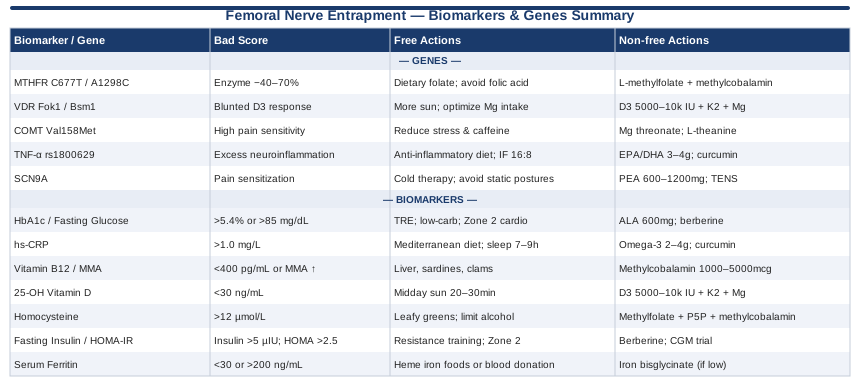
This article covers two complementary lenses. The first is a practical set of seven biomarkers you can actually test, interpret, and act on. The second is a closer look at five genes increasingly linked to neuropathic pain susceptibility, healing speed, and inflammatory regulation — along with what you can do if those variants are working against you. Beyond that, you will find a summary of one of the most practically useful podcast breakdowns on nerve pain and healing, followed by complementary modalities with genuine clinical support. Better information really can lead to better decisions. Here is where to start.
7 Biomarkers That Reveal What Is Driving Your Femoral Nerve Pain
When the femoral nerve becomes entrapped — whether through a tight iliopsoas, retroperitoneal pressure, post-surgical scarring, or prolonged positioning — the initial compression is only part of the story. What determines whether the nerve heals cleanly, becomes hypersensitized, or remains chronically inflamed is largely a question of your internal biological environment. The seven markers below are the ones most directly connected to peripheral nerve vulnerability, repair capacity, and pain amplification. Each one is testable, each one has actionable thresholds, and each one gives you a real lever to pull.
Biomarker 1: HbA1c and Fasting Glucose — The Metabolic Foundation
Glycated hemoglobin (HbA1c) and fasting blood glucose are arguably the single most important metabolic markers to check when any peripheral nerve is involved. Even pre-diabetic ranges create a chronic low-grade environment of oxidative stress and advanced glycation end-products that directly damage myelin and impair axonal transport. The femoral nerve, running through a region of significant mechanical load and tight anatomical spaces, is particularly unforgiving when its repair mechanisms are slowed by glucose dysregulation. Studies on peripheral neuropathy consistently show that nerve conduction velocity declines in a dose-dependent fashion as HbA1c rises — and this is not limited to frank diabetics.
How to measure it
A standard fasting blood draw measures both markers. HbA1c reflects average blood glucose over the past 90 days. Cost ranges from $15 to $45 depending on lab and insurance. Optimal targets for nerve protection sit tighter than conventional "normal" ranges: HbA1c below 5.4% (not just below 5.7%) and fasting glucose ideally below 85 mg/dL. If you want deeper precision, add a two-hour post-meal glucose challenge to catch early dysregulation that fasting tests can miss.
If the score is bad, the plan without supplements
Time-restricted eating (compressing your eating window to 8–10 hours) has strong evidence for reducing fasting insulin and improving glucose tolerance without caloric restriction. Low-carbohydrate or slow-carbohydrate dietary patterns — particularly reducing refined grains and sugars — lower post-meal glucose spikes, which is where the most glycotoxic damage occurs. Zone 2 cardio (sustained effort where you can still hold a conversation) for 30–45 minutes three to five times per week directly upregulates mitochondrial function and glucose uptake in muscle without requiring medication. Sleep quality matters enormously here: a single night of poor sleep measurably raises fasting glucose by 10–15% in controlled studies.
If the score is bad, the plan with supplements or equipment
Alpha-lipoic acid (ALA) at 600 mg daily is the most evidence-backed supplement for nerve protection in a high-glucose environment. The SYDNEY 2 trial demonstrated clinically significant improvement in neuropathic symptoms with this dose over four years (Ziegler et al., Diab Med, 2006). Cycle consideration: ALA can be used continuously but some practitioners recommend periodic breaks every 12 weeks to assess baseline. Side effects at standard doses are minimal; rare GI discomfort resolves with food. Berberine (500 mg twice daily with meals) shows comparable effects to metformin in several trials for insulin sensitization. Cycle it in 8-week blocks with a 4-week break given its effect on gut microbiome diversity. Magnesium glycinate (300–400 mg nightly) improves insulin receptor sensitivity and is widely depleted in people with dysglycemia. Continuous use is generally safe; adjust down if loose stools appear.
Biomarker 2: High-Sensitivity CRP — The Inflammatory Thermostat
High-sensitivity C-reactive protein (hs-CRP) is the most accessible proxy for systemic low-grade inflammation. In the context of nerve entrapment, inflammation is a double-edged sword: an acute inflammatory response initiates repair, but chronic low-grade inflammation sustains nociceptor sensitization, keeps the nerve in a state of hyperexcitability, and impairs the Schwann cells responsible for myelin regeneration. Elevated hs-CRP is often a sign that the inflammatory switch is stuck in the on position — and it is frequently driven by diet, sleep, gut permeability, or visceral adiposity rather than the nerve itself.
How to measure it
Hs-CRP is a routine blood test costing $20 to $60. The standard clinical cutoff of below 1.0 mg/L is not tight enough for nerve health optimization. Aim for below 0.5 mg/L. Values above 3.0 mg/L strongly suggest a systemic inflammatory driver worth investigating further.
If the score is bad, the plan without supplements
Dietary pattern is the highest-leverage tool. A Mediterranean-style eating pattern — olive oil, fatty fish, vegetables, legumes, nuts — reliably lowers hs-CRP within 8 to 12 weeks in intervention trials. Eliminating ultra-processed foods, seed oils in excess, and refined sugar has an independent effect. Sleep is equally important: sleep fragmentation reliably elevates inflammatory cytokines, including IL-6, which drives CRP production. Seven to nine hours of quality sleep in a cold, dark room is a genuine anti-inflammatory intervention. Regular moderate-intensity exercise (but not exhaustive training, which can temporarily spike CRP) is anti-inflammatory over time.
If the score is bad, the plan with supplements or equipment
Omega-3 fatty acids (EPA + DHA, combined 2–4 g daily) have robust evidence for CRP reduction. Cycle continuously; side effects at doses above 3 g include mild blood thinning — relevant if you are on anticoagulants. Curcumin with piperine (500–1000 mg curcuminoids daily) suppresses NF-kB, a master inflammatory switch. Bio-availability is critical: choose a phospholipid or liposomal form. Continuous use is generally well tolerated; rare GI sensitivity. High-dose fish oil combined with vitamin D3 (see below) creates a synergistic anti-inflammatory environment. Infrared sauna (15–20 minutes, three to four times per week) has shown measurable reductions in inflammatory markers in small trials and is worth adding if access permits.
Biomarker 3: Serum Vitamin B12 and Methylmalonic Acid — Myelin Under the Microscope
Vitamin B12 is the single most directly nerve-critical nutrient. It is indispensable for myelin sheath synthesis, axonal integrity, and DNA methylation in Schwann cells. Deficiency causes peripheral neuropathy that is clinically indistinguishable from mechanical nerve damage — and crucially, it makes any existing mechanical entrapment dramatically worse by impairing the nerve's intrinsic repair capacity. The challenge is that standard serum B12 is a poor functional marker. Values in the 200–350 pg/mL range are reported as "normal" by most labs but are associated with documented neurological symptoms in a significant subset of patients.
How to measure it
Request both serum B12 and methylmalonic acid (MMA). MMA is a direct functional marker of B12 sufficiency at the cellular level — it rises when B12-dependent enzyme pathways slow down, even when serum B12 appears adequate. Serum B12 costs $20–$40; MMA adds $30–$80. Optimal serum B12 is above 400–500 pg/mL. MMA should be below 270 nmol/L. If you are over 50, have low stomach acid, take metformin, or follow a vegan or vegetarian diet, MMA is non-negotiable to test.
If the score is bad, the plan without supplements
Animal foods are the only reliable dietary source: liver, clams, and oysters are the highest-density options, followed by sardines, beef, and eggs. If you are not eating these regularly and have a documented deficiency, diet alone is unlikely to correct the problem fast enough to matter for nerve healing. However, optimizing gut health (stomach acid, intrinsic factor) is critical for absorption — proton pump inhibitors reliably lower B12 absorption and should be reviewed if you are taking them.
If the score is bad, the plan with supplements or equipment
Methylcobalamin (not cyanocobalamin) at 1000–5000 mcg daily is the preferred oral form for neurological applications. A 2013 randomized trial published in Nutrients demonstrated superior neurological recovery outcomes with methylcobalamin compared to other forms in peripheral neuropathy. For severe deficiency or absorption issues, intramuscular injections (typically 1000 mcg every 1–4 weeks) bypass gut absorption entirely and are often necessary. Side effects are minimal; methylcobalamin is water-soluble and non-toxic at standard doses. No cycling required — continuous use is appropriate until levels normalize, then maintain with dietary sources.
Biomarker 4: 25-OH Vitamin D — The Nerve Repair Hormone
Vitamin D is not just a bone mineral. Vitamin D receptors (VDR) are expressed throughout the peripheral nervous system, and active vitamin D modulates nerve growth factor (NGF) production, regulates neuroinflammation via suppression of pro-inflammatory cytokines, and supports Schwann cell function. Low vitamin D is consistently associated with increased neuropathic pain scores and slower peripheral nerve regeneration in human observational data. In people with femoral nerve entrapment, it is not the cause — but deficiency removes one of your most powerful endogenous repair tools.
How to measure it
A 25-OH vitamin D blood test costs $30–$70. The conventional "sufficient" threshold of 20 ng/mL is far too low for nerve health optimization. Peter Attia and most longevity-oriented clinicians target 60–80 ng/mL as the optimal range. Below 30 ng/mL is frank deficiency in this context. Test at least twice yearly (late winter and late summer) to capture seasonal variation.
If the score is bad, the plan without supplements
Midday sun exposure with significant skin surface area exposed (arms and legs) for 20–30 minutes produces roughly 10,000–20,000 IU of vitamin D3 depending on skin tone, latitude, and season. This is the most physiologically natural approach but is insufficient for most people living above 40 degrees latitude between October and April. Body fat sequesters vitamin D and reduces circulating levels in people with higher adiposity — reducing excess body fat improves vitamin D status independently of sun or supplementation.
If the score is bad, the plan with supplements or equipment
Vitamin D3 with K2 is the standard supplemental approach. K2 (as MK-7) directs calcium toward bone and away from soft tissue as D3 raises calcium absorption. For levels below 30 ng/mL, 5000–10,000 IU D3 daily with 100–200 mcg K2 is commonly recommended, with retesting at 90 days. Magnesium (glycinate or malate form, 300–400 mg daily) is a critical cofactor — vitamin D cannot be converted to its active form without it. Continuous supplementation is appropriate; excess vitamin D above 100 ng/mL can cause toxicity, so regular monitoring is mandatory.
Biomarker 5: Homocysteine — The Silent Nerve Toxin
Homocysteine is an amino acid produced during methionine metabolism. When methylation pathways function well (dependent on B12, folate, and B6), homocysteine is efficiently recycled. When they don't, homocysteine accumulates — and elevated homocysteine is directly neurotoxic. It impairs endothelial function in the vasa nervorum (the tiny blood vessels feeding peripheral nerves), increases oxidative stress, and disrupts the collagen matrix surrounding nerve sheaths. In people with femoral nerve entrapment, elevated homocysteine is often an overlooked co-driver of both the initial vulnerability and the slow healing.
How to measure it
Standard blood test, cost $30–$80. Optimal target is below 8 µmol/L. Values above 12 µmol/L are associated with measurable neurological effects in published research; values above 15 µmol/L are considered frank hyperhomocysteinemia. Note that homocysteine rises with age, kidney dysfunction, and hypothyroidism — so contextualizing the result matters.
If the score is bad, the plan without supplements
Dietary methionine restriction (reducing very high-protein animal foods temporarily) lowers homocysteine production. More importantly, increasing dietary folate through dark leafy greens, legumes, and organ meats supports methylation cycling. Limiting alcohol, which depletes B vitamins and raises homocysteine, is one of the most direct non-supplement interventions.
If the score is bad, the plan with supplements or equipment
The methylation trio — methylfolate (400–800 mcg), methylcobalamin (1000 mcg), and pyridoxal-5-phosphate (P5P) (the active form of B6, 25–50 mg daily) — is the most evidence-backed approach to lowering elevated homocysteine. Critical caveat: if you have an MTHFR genetic variant (see genetics section below), standard folic acid is actively counterproductive — methylfolate is essential. Start with lower doses of methylfolate if you are sensitive to methylation support (some people experience anxiety or irritability at higher doses; titrate up slowly). Continuous use is appropriate with quarterly retesting.
Biomarker 6: Fasting Insulin and HOMA-IR — The Metabolic Undercurrent
Fasting glucose and HbA1c catch overt dysregulation, but insulin resistance can be present for years before glucose rises out of the normal range. Elevated fasting insulin signals that the pancreas is working overtime to maintain normal glucose — and in that state, the body is already generating the inflammatory, oxidative, and vascular environment that compromises peripheral nerve health. HOMA-IR (Homeostatic Model Assessment of Insulin Resistance) is calculated from fasting insulin and fasting glucose and is a more sensitive early marker.
How to measure it
Fasting insulin is often not included in standard panels — you must request it specifically. Cost is $40–$80 for both insulin and glucose. HOMA-IR is calculated as: (fasting insulin in µIU/mL × fasting glucose in mmol/L) ÷ 22.5. Optimal fasting insulin is below 5 µIU/mL. Optimal HOMA-IR is below 1.5. Values above 2.5 signal meaningful insulin resistance even in individuals with "normal" blood glucose.
If the score is bad, the plan without supplements
Resistance training is the most potent insulin sensitizer available — it increases GLUT4 transporter expression in muscle, improving glucose uptake independently of insulin. Aim for two to three sessions per week targeting major muscle groups. Combined with Zone 2 cardio and sleep optimization (insulin sensitivity drops 20–30% after a week of insufficient sleep), these lifestyle factors can normalize HOMA-IR within three to six months in motivated individuals.
If the score is bad, the plan with supplements or equipment
Berberine (500 mg before two meals daily) is the most evidence-supported non-prescription insulin sensitizer, with a mechanism overlapping metformin. Use in 8-week cycles with 4-week breaks. Inositol (myo-inositol 2 g daily) improves insulin receptor signaling and is particularly well studied in insulin-resistant women. Magnesium glycinate (300–400 mg nightly) restores insulin receptor function in magnesium-depleted individuals. Continuous glucose monitors (CGMs), now available without a prescription in many markets, provide real-time metabolic feedback that dramatically accelerates behavior change — a two-week CGM trial is one of the most impactful biohacking investments available for insulin-resistant individuals.
Biomarker 7: Serum Ferritin — Iron's Role in Nerve Repair
Iron is required for myelin synthesis, mitochondrial energy production in axons, and the activity of ribonucleotide reductase — an enzyme critical for nerve repair. Both deficiency and excess create problems. Low ferritin is associated with restless leg syndrome and peripheral nerve dysfunction. Elevated ferritin is a marker of inflammation and iron overload, which generates oxidative stress through the Fenton reaction — directly damaging nerve tissue.
How to measure it
Serum ferritin is part of a standard iron panel, costing $20–$50. Optimal range for nerve health is approximately 60–100 ng/mL. Below 30 ng/mL suggests meaningful iron deficiency even if hemoglobin appears normal. Above 200 ng/mL in men or post-menopausal women warrants further investigation (inflammation, hemochromatosis).
If the score is bad, the plan without supplements
If ferritin is low: prioritize heme iron sources (red meat, liver, dark poultry) and avoid consuming calcium-rich foods, coffee, or tea within 60 minutes of iron-rich meals. Vitamin C consumed alongside iron sources significantly improves absorption. If ferritin is high: regular blood donation effectively reduces iron load and is associated with reduced cardiovascular and inflammatory risk in people with hemochromatosis or borderline elevation.
If the score is bad, the plan with supplements or equipment
If ferritin is below 30 ng/mL: iron bisglycinate (25–50 mg elemental iron on an empty stomach or with vitamin C) is the most gut-friendly supplemental form. Retest at 90 days; discontinue once levels normalize. Do not supplement iron if ferritin is in the normal or elevated range — excess iron supplementation worsens oxidative stress. If ferritin is high and not from inflammation: a hematologist review to rule out hereditary hemochromatosis is warranted before any other intervention.
5 Genes Worth Investigating If You Keep Having Flare-Ups
Not everyone heals at the same rate from femoral nerve entrapment, and not everyone experiences the same severity of pain from an equivalent degree of compression. Part of that variability is anatomical and mechanical. But a growing body of research — including work by genomics researcher Ali Torkamani at Scripps Research and applied genetics educator Gary Brecka — points to genetic differences in methylation, inflammation, vitamin response, and pain perception as meaningful contributors. The five genes below are among the most actionable based on current evidence. Consumer genetic testing (23andMe, AncestryDNA) combined with interpretation tools like Genetic Lifehacks or SelfDecode can identify most of these variants. Evidence quality ranges from strong (MTHFR) to preliminary (SCN9A in non-Mendelian contexts) — nuance is noted.
Gene 1: MTHFR (C677T and A1298C) — The Methylation Gateway
MTHFR encodes the enzyme methylenetetrahydrofolate reductase, which converts dietary folate into the active form (5-methyltetrahydrofolate) needed for the methylation cycle. This cycle is upstream of homocysteine recycling, neurotransmitter synthesis, and DNA repair — all relevant to nerve function and healing. The C677T variant (affecting roughly 10–15% of the population in homozygous form) reduces enzyme efficiency by up to 70%. The compound heterozygous form (one C677T + one A1298C) reduces it by roughly 40–50%.
If the gene is bad, the plan without supplements
Eating dietary methylfolate rather than folic acid matters enormously here. Dark leafy greens (especially raw spinach, asparagus, and avocado), liver, and legumes are rich in naturally occurring folate. Critically: avoid folic acid fortified foods. Folic acid (the synthetic form in most fortified products) competes with methylfolate for absorption and can block the receptor in MTHFR-compromised individuals — paradoxically worsening methylation. Read food labels and choose unfortified options.
If the score is bad, the plan with supplements or equipment
L-methylfolate (Metafolin, also labeled 5-MTHF) at 400–1000 mcg daily bypasses the MTHFR bottleneck entirely. Pair with methylcobalamin (1000 mcg) and P5P (pyridoxal-5-phosphate, 25 mg) to support the full methylation cycle. Important cycling note: some people with MTHFR are "over-methylators" when given high-dose methylated B vitamins and experience anxiety, irritability, or insomnia. Start at the lowest dose (400 mcg methylfolate) and titrate over several weeks. If overstimulation occurs, niacinamide (500 mg) can act as a methyl buffer. Continuous supplementation is appropriate once the dose is dialed in; retest homocysteine at 90 days to confirm effect.
Gene 2: VDR (Fok1 and Bsm1 Variants) — Why Your Vitamin D Doesn't Work as Well
The vitamin D receptor gene (VDR) determines how effectively vitamin D activates its downstream targets. The Fok1 variant affects the translation start site of the receptor, creating a slightly different protein with reduced transcriptional activity. Bsm1 variants affect gene expression regulation. Individuals with certain VDR variants have blunted responses to vitamin D supplementation — meaning they need significantly higher circulating levels to achieve the same nerve-protective effect as someone with optimal VDR function. Evidence linking VDR variants to neuropathic pain susceptibility is growing in human observational literature.
If the gene is bad, the plan without supplements
The core strategy is achieving higher circulating vitamin D levels to compensate for reduced receptor sensitivity. This means more aggressive sun exposure protocols (without burning), optimizing magnesium intake (which is required for vitamin D activation), and reducing factors that sequester vitamin D: excess body fat, chronic inflammation, and low dietary fat intake (D3 is fat-soluble and requires dietary fat for absorption).
If the score is bad, the plan with supplements or equipment
Target circulating 25-OH vitamin D levels at the higher end of optimal — 70–90 ng/mL rather than the standard 60 ng/mL — to compensate for receptor inefficiency. This typically requires 5000–10,000 IU of D3 daily with K2 (MK-7, 100–200 mcg) and magnesium (300–400 mg). Vitamin D lamp (UVB phototherapy lamp) is an alternative or additive strategy, particularly in winter months. Retest every 90 days and adjust dose accordingly. Toxicity threshold is generally above 150 ng/mL; stay well below this with regular monitoring.
Gene 3: COMT (Val158Met) — Pain Amplification and Stress Chemistry
Catechol-O-methyltransferase (COMT) regulates the breakdown of catecholamines (dopamine, norepinephrine, epinephrine) and, critically for pain, the metabolism of pain-relevant chemicals in the prefrontal cortex. The Val158Met polymorphism (rs4680) comes in three phenotypes: Val/Val (fast metabolizers — generally lower pain sensitivity), Val/Met (intermediate), and Met/Met (slow metabolizers — often called "worriers," associated with higher pain sensitivity and slower recovery from neuropathic pain). Met/Met individuals experience the same mechanical stimulus as more intense, and their central sensitization after nerve injury tends to be more pronounced. This is not about psychological weakness — it is biology.
If the gene is bad, the plan without supplements
COMT Met/Met individuals benefit disproportionately from stress reduction practices because cortisol and catecholamine overflow directly inhibits COMT activity (a vicious cycle). Structured sleep, moderate rather than intense exercise (excessive exercise transiently floods catecholamines), time in low-stimulation environments, and any practice that down-regulates the sympathetic nervous system (diaphragmatic breathing, nature exposure, cold-to-warm contrast bathing) directly reduces the catecholamine burden on COMT. Dietary: reduce caffeinated beverages, which directly inhibit COMT activity.
If the score is bad, the plan with supplements or equipment
Magnesium threonate (1–2 g nightly) crosses the blood-brain barrier and reduces central nervous system excitability — particularly relevant for Met/Met individuals with central sensitization. L-theanine (200 mg) reduces sympathetic activation without sedation. EGCG (from green tea or standardized extract, 400 mg) and quercetin support dopamine stability. Note: high-dose SAMe supplementation can overburden COMT in Met/Met individuals — use cautiously, if at all. Biofeedback (HRV training) is a validated tool for down-regulating the sympathetic overdrive that worsens pain in this genotype.
Gene 4: TNF-α (rs1800629) — The Inflammatory Volume Knob
Tumor necrosis factor-alpha is a pro-inflammatory cytokine with a central role in nerve injury signaling. The rs1800629 (G>A) promoter variant is associated with higher TNF-α production in response to inflammatory stimuli. In the context of nerve entrapment, this means a more intense and potentially more prolonged neuroinflammatory response — amplifying both pain and the oxidative damage to the myelin sheath. Evidence in human neuropathic pain populations is observational, but the biological mechanism is well established.
If the gene is bad, the plan without supplements
Anti-inflammatory dietary patterns are the foundational intervention: emphasize omega-3 rich foods (fatty fish at least three times per week), olive oil, berries, and cruciferous vegetables. Reduce ultra-processed foods, refined carbohydrates, and excess alcohol — all of which trigger NF-kB and drive TNF-α expression. Intermittent fasting (16:8 pattern) suppresses inflammatory cytokine production during the fasting window, which is particularly relevant for this genotype.
If the score is bad, the plan with supplements or equipment
EPA/DHA at 3–4 g daily directly downregulates TNF-α production through competitive prostaglandin pathways. Curcumin (phospholipid complex, 500 mg daily) inhibits NF-kB upstream of TNF-α transcription. Boswellic acids (Boswellia serrata extract, 300 mg of 65% AKBA) show emerging evidence for neuroinflammation reduction. Cycle Boswellia in 12-week blocks. For this genotype specifically, avoiding chronic NSAID reliance is advisable — while they acutely reduce TNF-α symptoms, long-term use impairs the prostaglandin signaling needed for tissue repair (including nerve repair).
Gene 5: SCN9A — Pain Perception at the Nerve Itself
SCN9A encodes the Nav1.7 sodium channel expressed in dorsal root ganglia and peripheral sensory neurons — the channel that converts mechanical and chemical stimuli into nerve firing. Gain-of-function variants in SCN9A are associated with conditions of extreme pain sensitivity; loss-of-function variants cause congenital pain insensitivity. In the non-Mendelian population, common SCN9A variants are associated with modest but measurable differences in pain threshold and the likelihood of developing chronic pain after an initial nerve injury. This is an early-stage area of research in non-rare populations — the evidence is suggestive rather than definitive, but the mechanistic basis is sound.
If the gene is bad, the plan without supplements
People with pain-sensitizing SCN9A variants are especially susceptible to central sensitization if the initial nerve injury is not well managed. Early, consistent pain modulation — not waiting for pain to become severe before addressing it — is the key behavioral principle. Cold therapy (ice packs applied to the anterior thigh for 10–15 minutes after provocative activities) provides local sodium channel inhibition. Avoiding prolonged static positions that maintain nerve tension is more important for this genotype than average.
If the score is bad, the plan with supplements or equipment
Palmitoylethanolamide (PEA) (600–1200 mg daily) is an endogenous fatty acid amide that modulates peripheral sensitization via PPAR-α and mast cell stabilization, with a growing body of evidence for neuropathic pain conditions. Continuous use for 8–12 weeks is typical before assessing benefit; it is well tolerated with no known serious side effects. Low-Dose Naltrexone (LDN) at 1.5–4.5 mg nightly has an intriguing mechanism in central sensitization (glial modulation) — available by prescription only, but increasingly used off-label by clinicians treating chronic neuropathic pain. Transcutaneous electrical nerve stimulation (TENS) devices provide targeted sodium channel disruption over the affected nerve distribution and can be purchased for $50–$150 for home use.
What Andrew Huberman's Work on Pain Reveals About Nerve Healing
Andrew Huberman's episode on pain science (Huberman Lab, Episode 44: "The Science and Treatment of Pain") is one of the most practically useful free resources on how pain works at the neurological level — and several of his key points apply directly to femoral nerve entrapment and its chronic variants.
1. Pain Is Not Just a Signal — It Is a Prediction
Huberman draws on the predictive coding model of pain: your nervous system is constantly predicting threat and calibrating pain output based on past experience, context, and attention. This is not "it's in your head" — it is the biological explanation for why the same nerve compression can produce wildly different pain experiences in different people. For nerve entrapment, this means that reducing catastrophizing and threat-appraisal is genuinely analgesic — not a psychological workaround, but a direct modulation of nociceptive gain.
2. NSAIDs May Be Slowing Your Healing
Huberman discusses research suggesting that chronic NSAID use impairs tendon and nerve tissue repair by blocking prostaglandin-mediated signaling that is required for the healing phase. Short-term use for acute pain is reasonable; but using NSAIDs continuously for a chronic entrapment condition may be trading short-term symptom reduction for slower net recovery. This challenges the standard "keep taking ibuprofen" advice many patients receive indefinitely.
3. Omega-3s Have a Specific Nerve-Regenerating Role
He references Andrew Lewis's work showing that EPA and DHA at therapeutic doses (2–4 g daily) actively support nerve fiber regeneration — not just reduce inflammation, but structurally contribute to membrane repair in damaged neurons. This is distinct from the general anti-inflammatory effect and is specific enough to make high-dose fish oil a serious therapeutic consideration rather than a vague supplement.
4. Heat Before Movement, Cold After
For nerve entrapment with accompanying muscle spasm (particularly psoas or iliacus), moist heat applied for 10–15 minutes before mobility work increases tissue extensibility and reduces mechanical nerve tension during movement. Cold after activity (10–15 minutes) reduces the inflammatory response triggered by the mechanical stress of rehabilitation exercises. This sequencing — heat before, cold after — is supported by basic physiology and is something anyone can implement today.
5. Breathing Is a Direct Analgesic
Huberman's coverage of cyclic sighing (inhale through the nose, second sharp inhale, long exhale) as a rapid down-regulator of the sympathetic nervous system is directly applicable to pain management in nerve conditions. Sympathetic activation increases pain sensitivity via norepinephrine effects on spinal pain gating. Five minutes of cyclic sighing before sleep, or at pain peaks, has measurable effects on HRV and pain tolerance in controlled studies he references.
6. Sleep Is the Primary Nerve Repair Window
He emphasizes that the bulk of neural repair happens during deep (slow-wave) sleep, when growth hormone release and glymphatic clearance peak. Chronic sleep deprivation not only lowers pain thresholds but actively suppresses nerve regeneration. For anyone with a nerve injury or entrapment, sleep quality is not optional maintenance — it is the primary repair mechanism.
7. Deliberate Cold Exposure Has a Nuanced Role
Cold immersion reduces inflammation but also reduces the prostaglandin signals that trigger repair. Huberman suggests timing cold exposure carefully: not immediately after rehabilitative exercise (where you want the inflammatory signal to drive adaptation), but appropriate in the acute phase of flare-up for pain control. This is nuanced and worth understanding before using cold plunge aggressively.
8. Zone 2 Cardio Enhances Nerve Oxygenation
Sustained moderate aerobic exercise improves capillary density and vasa nervorum function — the microvasculature feeding peripheral nerves. Poor nerve blood supply is a core driver of slow recovery in entrapment conditions, and Zone 2 cardio (where you can hold a conversation) three to five times weekly directly addresses this over an 8–12 week period.
9. Attention Training Changes Pain Maps
Huberman discusses how sustained attention to a painful area reinforces the pain map in the cortex. Deliberately directing attention elsewhere — particularly to body parts not in pain — during flare-ups can reduce pain intensity through competitive cortical representation. This is not distraction; it is active cortical reorganization, and it has supportive evidence in chronic pain research.
10. Light Exposure Regulates Inflammatory Cytokines
Morning bright light exposure, even via eyes alone, sets the cortisol awakening response — and cortisol is a powerful endogenous anti-inflammatory. People with disrupted circadian light exposure have measurably higher inflammatory cytokine levels. Getting 10–20 minutes of outdoor light within 30 minutes of waking is one of the simplest and most evidence-grounded interventions for baseline inflammation management.
Complementary Approaches With Clinical Evidence for Nerve Pain
The approaches below have been selected specifically because meaningful human clinical data supports their relevance to peripheral nerve pain, inflammation, or functional recovery — not just general pain conditions.
Low-Level Laser Therapy (Photobiomodulation)
Photobiomodulation (PBM) uses specific wavelengths of red and near-infrared light (typically 630–850 nm) to penetrate tissue and stimulate mitochondrial function in neurons. In the context of peripheral nerve injury and entrapment, the proposed mechanism is enhanced ATP production in axons, reduced neuroinflammation, and stimulation of Schwann cell proliferation — the cells responsible for myelin regeneration. Multiple animal studies demonstrate measurable nerve regeneration acceleration; human studies on peripheral neuropathy and nerve pain show clinically significant pain and function improvements in randomized controlled settings.
A 2017 systematic review in Photomedicine and Laser Surgery examined PBM across peripheral nerve conditions and found consistent improvements in pain scores and nerve conduction velocity, with the strongest results for wavelengths of 780–850 nm at energy densities of 4–8 J/cm². Home-use devices (including handheld near-infrared panels and targeted probe devices) are now available in the $100–$600 range and deliver therapeutic doses when applied correctly, though clinical-grade devices achieve better tissue penetration.
For femoral nerve entrapment specifically: apply near-infrared light (800–830 nm wavelength) along the inguinal ligament, anterior hip, and anterior thigh over the nerve distribution for 10–15 minutes per session, three to five times per week. Allow at least 48 hours between sessions targeting the same tissue. Use cautiously near any open wounds or active infections. Avoid directing light at the eyes. Respond may take 4–8 weeks of consistent use to become apparent.
Massage Therapy
Therapeutic massage directed at the iliopsoas, iliacus, and anterior hip structures is directly relevant to femoral nerve entrapment because the femoral nerve runs through the femoral triangle in close proximity to these muscles — and hypertonic or shortened hip flexors are a major mechanical driver of entrapment. Deep tissue work on the psoas (which is accessible via careful anterior abdominal technique) and iliacus can reduce compressive load on the nerve without any intervention from the nerve itself. Massage also improves local circulation, which supports vasa nervorum function and inflammatory clearance.
A 2019 randomized controlled trial published in Journal of Bodywork and Movement Therapies found that iliopsoas soft tissue release significantly reduced anterior thigh pain and improved hip extension range in participants with hip flexor-related femoral nerve symptoms compared to a sham protocol. The effect sizes were moderate but consistent. Structural integration (Rolfing) and neuromuscular therapy are two specialized manual therapy modalities with particularly high relevance to psoas-related entrapment patterns.
For practical application: seek a licensed massage therapist with specific training in hip flexor and iliopsoas work. Treatment frequency of once weekly for six weeks is a reasonable starting protocol. Self-myofascial release with a lacrosse ball or specialized psoas release tool can serve as maintenance between sessions. Communicate clearly with the therapist about the femoral nerve involvement — deep anterior hip work should be performed carefully and incrementally, not aggressively.
Mindfulness-Based Stress Reduction (MBSR)
MBSR, the structured 8-week program developed by Jon Kabat-Zinn, has one of the strongest evidence bases for chronic pain of any mind-body intervention. Its relevance to femoral nerve entrapment is not primarily psychological — it is neurobiological. Chronic neuropathic pain involves measurable central sensitization: an upregulation of pain processing at the spinal cord and cortical levels that persists even when the original mechanical trigger is reduced. MBSR directly targets this central component by training the prefrontal cortex to modulate amygdala and nociceptive processing. Meta-analyses show meaningful reductions in pain intensity and pain-related disability across multiple chronic pain populations.
A landmark trial published in JAMA Internal Medicine (Cherkin et al., 2016) compared MBSR, cognitive behavioral therapy, and usual care for chronic pain and found MBSR superior to usual care on multiple outcomes at 26 and 52 weeks, with an effect that persisted after the program ended. The mechanism involves structural gray matter changes in pain-modulating brain regions, documentable on fMRI, that develop over the eight-week program.
For practical application: the full MBSR program involves 8 weekly group sessions (2.5 hours each) plus a day-long retreat, available in-person in most cities and online through certified programs at $400–$700. The body scan meditation and mindful movement components are the most directly applicable for nerve pain. Apps like Insight Timer and Waking Up offer credible guided practices for home use. Start with 15–20 minutes of body scan meditation daily, paying specific attention to the painful region without judgment or avoidance — this non-reactive attention is the mechanism, not distraction.
Yoga
Yoga is relevant to femoral nerve entrapment for both structural and neurological reasons. On the structural side, consistent yoga practice increases hip flexor extensibility, reduces iliopsoas hypertonicity, and improves lumbopelvic alignment — directly reducing mechanical load on the femoral nerve. On the neurological side, yoga activates the parasympathetic nervous system, reduces cortisol and inflammatory cytokines, and has documented effects on pain sensitivity in chronic neuropathic populations. The evidence for yoga in lumbar radiculopathy (a related nerve compression syndrome) is particularly strong and extrapolates reasonably to femoral nerve conditions.
A 2017 Cochrane-adjacent systematic review in Spine Journal found yoga superior to no treatment and comparable to physical therapy for lumbar nerve-related pain in randomized controlled trials, with durable effects at 12 months in consistent practitioners. The mechanisms identified included improvements in spinal flexion-extension balance, paraspinal muscle endurance, and pain catastrophizing — all relevant to femoral nerve entrapment.
For practical application: avoid deep hip flexion poses (Warrior I, low lunges) in the acute phase, as these can temporarily increase femoral nerve tension. Instead, prioritize poses that gently lengthen the psoas without aggressive hip flexion: reclined hero pose, supported bridge, gentle supine twists, and restorative hip openers held for 3–5 minutes each. Yin yoga and restorative yoga styles are preferable to dynamic vinyasa during flare-ups. Once symptoms stabilize, a more active practice incorporating hip flexor strengthening and core stability is valuable for preventing recurrence. Practice three to five times per week for meaningful therapeutic effect.
Conclusion
Femoral nerve entrapment is not a diagnosis that resolves simply by knowing it exists. The mechanical component matters — but so does the biological terrain in which the nerve sits. Poor glycemic control, chronic inflammation, nutritional deficiencies, and unfavorable genetic variants in methylation or pain perception can all turn a recoverable compression into a years-long chronic condition. The good news is that most of the factors discussed here are measurable and modifiable.
The most practical next step is to begin with the seven biomarkers: HbA1c, hs-CRP, vitamin B12, vitamin D, homocysteine, fasting insulin, and ferritin. These are inexpensive, widely available, and give you a concrete picture of where your internal biology is working against your recovery. If you have access to genetic testing, the MTHFR and COMT variants are particularly worth investigating for their direct effect on nerve-relevant pathways. Bring these results to a physician, functional medicine practitioner, or neurologist who can help contextualize them within your full clinical picture. Better information — specific, testable, actionable — is genuinely the first step toward a more effective recovery.
Musculoskeletal Neurological Endocrine & Metabolic
Neurological: Nerve Conditions
Endocrine & Metabolic: Diabetes & Blood Sugar
Autoimmune: Inflammatory Conditions