This article contains AI generated content.
· UpdatedFemoral Stress Fracture — 6 Genes and 7 Biomarkers to Track
Introduction
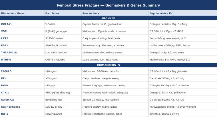
A femoral stress fracture does not arrive without warning — but the warnings are often written in biology, not in pain. Most people who develop one were already running low on something: a mineral, a hormone, a structural protein, or a signaling molecule that keeps bone remodeling in balance. The frustrating part is that standard care rarely looks for any of it. You rest, you take calcium, you return gradually to activity, and sometimes it happens again.
If you have already experienced a femoral stress fracture — or if your situation makes the risk real (high training volume, restrictive eating, hormonal disruption, or a history of multiple stress fractures) — generic advice is unlikely to be sufficient. The bone healed before and it will heal again, but without understanding why it fractured in the first place, the cycle tends to repeat. Two runners doing identical weekly mileage under identical conditions can have completely different fracture risk, because their bones respond differently to load at the cellular and molecular level. Ignoring that difference means fixing the symptom without addressing the mechanism.
What this article offers is a more specific lens. Bone is dynamic tissue, constantly remodeling in response to load, nutrition, hormones, and genetics. When the remodeling balance tips too far toward breakdown — or when the building phase is chronically underpowered — the femur accumulates microdamage faster than it can repair it. Each marker discussed here reveals one distinct part of that equation, and more importantly, each one points toward a specific, targeted intervention.
The article is organized in two main directions. The first, and most immediately actionable, examines seven measurable biomarkers — bone formation and resorption markers, vitamin D status, hormonal signals, and growth factors — that can be tracked through standard blood tests and that respond to diet, exercise, and targeted supplementation. The second looks at six genetic variants that shape your underlying bone biology, including collagen structure, vitamin D receptor efficiency, and estrogen signaling, along with practical plans for each risk variant. Neither direction provides a diagnosis, but together they give you a map that most clinicians never draw.
7 Biomarkers That Reveal Your Femoral Fracture Risk
Most people who develop a femoral stress fracture have at least one measurable biological imbalance that contributed to it. The seven biomarkers below are not exhaustive, but they represent the most clinically useful starting points: consistently supported by human evidence, measurable through standard or specialty labs, and linked to modifiable interventions. The goal is not to chase perfect numbers but to identify the specific lever that, when adjusted, changes the outcome.
Biomarker 1: 25-Hydroxyvitamin D (25-OH D)
Why it matters
Vitamin D is a steroid hormone precursor with receptors in the cells that build and break down bone. When circulating 25-OH D levels fall below adequate ranges, intestinal calcium absorption drops sharply — from roughly 30–40% efficiency to under 15% — and the parathyroid gland compensates by secreting more PTH, which extracts calcium from bone to maintain blood levels. Multiple prospective studies in military recruits and endurance athletes have found that individuals with low vitamin D experience stress fractures at significantly higher rates than those with replete levels. The femur, bearing continuous high-load repetitive stress in runners, cyclists, and soldiers, is particularly unforgiving when bone mineralization is suboptimal. Vitamin D deficiency is one of the most prevalent, most correctable, and most overlooked contributors to stress fracture susceptibility.
How to measure it
A serum 25-OH D test is ordered by most primary care physicians and sports medicine doctors. Cost ranges from $30 to $80 without insurance, and it is frequently included in preventive wellness panels. The NIH Office of Dietary Supplements Vitamin D Fact Sheet defines deficiency below 20 ng/mL (50 nmol/L) and adequacy at 20 ng/mL and above by conventional standards. Many functional medicine practitioners and bone health researchers target 40–60 ng/mL (100–150 nmol/L) as an optimal working range, which is higher than the minimum threshold. Retesting every 3–6 months during active correction is appropriate. A single baseline test is almost always worthwhile before any bone-related protocol.
If the score is low — the plan without supplements
Direct midday sun exposure to large skin areas (arms, legs, back) for 20–30 minutes produces meaningful endogenous vitamin D synthesis in lighter skin tones, though this effect is substantially reduced in darker skin, at latitudes above 35°, or during winter months. Fatty fish consumed 3–5 times per week (salmon, mackerel, sardines, herring), egg yolks, and beef liver all contribute meaningful dietary vitamin D. Reducing factors that chronically suppress vitamin D — including near-zero outdoor time, high-SPF sunscreen during all outdoor exposure, alcohol, and obesity (which sequesters vitamin D in fat tissue) — preserves what is synthesized.
If the score is low — the plan with supplements
Vitamin D3 (cholecalciferol) is the preferred form over D2. For deficient adults, 4,000–5,000 IU daily taken with a fat-containing meal is a widely used starting dose, with adjustments based on retesting. Critically, D3 should be paired with vitamin K2 (MK-7 form, 100–200 mcg/day) to direct calcium appropriately to bone and away from arterial tissue. Magnesium glycinate or malate at 300–400 mg/day is a required cofactor for the conversion of vitamin D to its active form (calcitriol) and is frequently deficient in people with low vitamin D, creating a bottleneck even when D3 supplementation is otherwise adequate. Retest at 12 weeks and adjust dose toward the 40–60 ng/mL target. Avoid self-dosing above 10,000 IU/day without medical monitoring. No cycling is required at standard therapeutic doses.
Biomarker 2: Parathyroid Hormone (PTH)
Why it matters
PTH is the body's calcium emergency signal. When blood calcium is insufficient — typically because vitamin D is inadequate or dietary calcium is chronically low — PTH rises to extract calcium from bone through osteoclast activation. Chronically elevated PTH (secondary hyperparathyroidism) silently erodes cortical bone thickness over months. In stress fracture patients, elevated PTH often reveals the specific driver of bone loss more precisely than vitamin D alone: it confirms that the body has been actively compensating, at bone's expense. PTH is most informative when measured alongside 25-OH D and serum calcium as a triad, since the combination tells a far more complete story than any single value.
How to measure it
PTH is measured via a fasting blood draw (intact PTH assay). Cost ranges from $30 to $80. The normal reference range is typically 15–65 pg/mL. Many bone specialists prefer levels in the lower-to-middle portion of the reference range (20–40 pg/mL). Elevated PTH alongside normal serum calcium strongly suggests vitamin D deficiency or insufficient dietary calcium. Elevated PTH alongside elevated calcium points toward primary hyperparathyroidism — a distinct diagnosis requiring specialist evaluation and potentially parathyroid surgery.
If the score is elevated — the plan without supplements
Increasing dietary calcium from whole food sources is the most direct non-supplement step. Practical sources include dairy (for those who tolerate it), canned sardines or salmon with bones, fortified plant milks, cooked dark leafy greens (bok choy, kale), tahini, and almonds. Vitamin D correction (as above) enables the calcium consumed to be absorbed. Reducing high-sodium processed foods, which increase urinary calcium losses and thereby trigger PTH release, provides additional leverage. Weight-bearing exercise has a mild PTH-suppressing effect and remains appropriate within pain tolerance during recovery.
If the score is elevated — the plan with supplements
Calcium citrate (preferred over carbonate for individuals over 50, or those with low stomach acid or proton pump inhibitor use) at 500 mg per dose, twice daily with meals, provides reliable absorption. Total supplemental calcium should generally stay within 1,000–1,200 mg/day combined with dietary intake, as high-dose supplemental calcium has raised concerns in some cardiovascular meta-analyses. K2 and magnesium remain essential co-factors for proper calcium routing. If PTH remains elevated after 12 weeks of combined vitamin D and calcium correction, investigation for parathyroid disease is warranted regardless of whether a stress fracture has occurred.
Biomarker 3: P1NP — Procollagen Type I N-terminal Propeptide
Why it matters
P1NP is the gold standard bone formation marker, recommended as the reference marker by the International Osteoporosis Foundation. It reflects how actively osteoblasts are laying down new collagen matrix — essentially, the rate at which new bone is being built. In stress fracture patients, low P1NP signals an underpowered repair process: the bone cannot rebuild as fast as it is being damaged under mechanical load. Common drivers of low P1NP include inadequate dietary protein, low sex hormones (especially estrogen in women), glucocorticoid use, energy deficiency, and poor sleep. Measuring P1NP alongside CTX-1 gives a complete picture of the formation-to-resorption balance — two values that are individually meaningful but far more diagnostic when interpreted together.
How to measure it
P1NP is measured from a fasting morning blood draw. Cost ranges from $50 to $120 and may require ordering through a functional medicine physician or specialty reference lab. Premenopausal reference ranges are typically 15–90 µg/L, with significant age- and hormonal-status variation. A single baseline is informative; serial measurements every 3–6 months during an intervention provide meaningful response tracking. P1NP is more stable across a 24-hour period than CTX-1, making it somewhat easier to interpret from a single measurement.
If the score is low — the plan without supplements
Dietary protein is the primary lever: osteoblasts depend on glycine, proline, and hydroxyproline — the building blocks of collagen. Target at least 1.2–1.6 g of protein per kilogram of body weight per day, with emphasis on complete proteins and glycine-rich sources such as bone broth, skin-on poultry, and other connective tissue-containing preparations. Progressive resistance training and impact exercise directly stimulate osteoblast activity, with meaningful P1NP increases measurable within 8–12 weeks of consistent loading. Adequate deep sleep — the phase during which growth hormone is released and bone synthesis is most active — is one of the most underappreciated P1NP modulators.
If the score is low — the plan with supplements
Hydrolyzed collagen peptides at 10–15 g/day, taken with vitamin C (500 mg), have demonstrated in randomized controlled trials the ability to increase bone formation markers and bone density when combined with resistance training. Vitamin C is a required cofactor for hydroxylating proline residues in collagen synthesis; without it, collagen cross-linking is impaired. Creatine monohydrate at 3–5 g/day (no loading phase required, safe for continuous use) has modest but reproducible evidence for supporting bone formation in older adults and is unlikely to cause harm in younger athletes. Copper at 1–2 mg/day supports lysyl oxidase, the enzyme responsible for collagen cross-linking critical to bone matrix integrity. Retest P1NP at 12–16 weeks to assess response. No specific cycling protocol is required for these supplements at standard doses.
Biomarker 4: CTX-1 — C-terminal Telopeptide of Collagen Type I
Why it matters
CTX-1 is the primary bone resorption marker. It measures fragments of type I collagen released when osteoclasts break down old bone matrix. In a healthy remodeling cycle, CTX-1 and P1NP rise and fall together, maintaining approximate balance. In stress fracture scenarios — particularly in athletes during high-load training phases — CTX-1 often outpaces P1NP, meaning bone is being degraded faster than it is being rebuilt. Beyond training overload, elevated CTX-1 is also driven by energy deficiency (RED-S), low estrogen, elevated cortisol, inflammatory states, and vitamin D deficiency. A further nuance: very low CTX-1, typically caused by bisphosphonate medications, can paradoxically increase atypical femoral fracture risk by suppressing the normal removal of microdamage from aging bone matrix — an important clinical distinction.
How to measure it
CTX-1 is highly sensitive to food intake and circadian variation. It must be drawn fasting in the morning — typically before 10 a.m. — for reliable, comparable results. Cost ranges from $50 to $120. In premenopausal women and men under 50, fasting morning CTX-1 above 500–600 pg/mL, particularly alongside a low P1NP, warrants further investigation. Reference ranges vary by lab and hormonal status, and post-menopausal women have higher physiologic reference ranges.
If the score is elevated — the plan without supplements
Training load reduction is often the most direct intervention when elevated CTX-1 coincides with high training volume — the bone remodeling cycle has been overwhelmed by load-induced resorption. Ensuring total caloric adequacy is critical and frequently missed in lean, highly trained individuals: energy deficiency (RED-S) is one of the strongest drivers of uncoupled bone remodeling and it suppresses the formation side while leaving resorption elevated. Increasing dietary protein and calcium, normalizing sleep duration and quality, and addressing any hormonal deficiencies — particularly low estrogen in women or low testosterone in men — directly reduce CTX-1 over weeks.
If the score is elevated — the plan with supplements
Vitamin D3 with K2 (as outlined above) directly modulates osteoclast activity through the RANK/RANK-L/OPG axis. Omega-3 fatty acids (EPA + DHA, 2–3 g/day from fish oil or algal-based sources) have shown anti-inflammatory and modest anti-osteoclastic effects in human trials. Natural options with some evidence include ipriflavone (200 mg three times daily, studied in osteoporosis trials with modest reduction in bone resorption markers) and hesperidin from citrus bioflavonoids. Strontium ranelate has strong clinical evidence for reducing CTX-1 but is a prescription medication in most countries with cardiovascular monitoring requirements. Retest CTX-1 fasting in the morning consistently, always at 12–16 weeks after initiating protocol changes, to assess whether the resorption rate has moderated.
Biomarker 5: Serum Calcium (Corrected or Ionized)
Why it matters
Calcium constitutes roughly 70% of bone mineral by weight, stored as hydroxyapatite crystals in the collagen matrix. Total serum calcium is tightly maintained by the PTH-vitamin D axis — meaning it often remains "normal" even as bone is being continuously depleted to maintain it. That tight regulation is precisely why tracking PTH alongside serum calcium matters: the PTH result often reveals whether normal calcium is being maintained at the cost of bone. Ionized calcium — the biologically active fraction — provides a more accurate picture in athletes, where albumin levels can shift with training volume, artificially altering total calcium calculations. Chronic calcium loss through the urine (driven by high sodium intake, excessive caffeine, or inadequate dietary buffering from vegetables) is a slow but steady drain that, over months, contributes directly to stress fracture risk.
How to measure it
Total serum calcium is included in every basic metabolic panel — cost is typically $10–30 as part of a routine blood draw. Ionized calcium requires a separate measure ($20–50 additional). A 24-hour urine calcium test, or a simpler spot urine calcium-to-creatinine ratio, adds useful context about excretion. The NIH Office of Dietary Supplements calcium fact sheet provides comprehensive data on recommended intakes, dietary sources, and their evidence base.
If the score is borderline — the plan without supplements
Distribute calcium-rich foods across the day rather than in a single large meal, since absorption efficiency drops significantly at higher single doses. Reduce high-sodium processed foods (which increase urinary calcium losses), limit caffeine to moderate amounts, and ensure adequate vitamin D for absorption efficiency. Increase alkalinizing foods (vegetables, fruits) that reduce the acid-buffering demand on bone minerals. Weight-bearing exercise promotes calcium retention by stimulating the bone mechanostat and reducing resorption under normal hormonal conditions.
If the score is borderline — the plan with supplements
Calcium citrate at 500 mg twice daily with meals is well absorbed without requiring stomach acid, making it the preferred form for most adults and universally appropriate for those over 50. Do not exceed 1,500 mg/day total (food plus supplements combined) without medical guidance. Always combine supplemental calcium with magnesium (roughly a 2:1 calcium-to-magnesium ratio) and vitamin K2 to support appropriate mineral routing. Avoid taking calcium supplements simultaneously with iron-rich foods or iron supplements, as they compete for absorption through the same transporters.
Biomarker 6: Sex Hormones — Estradiol and Testosterone
Why it matters
Estrogen and testosterone are the dominant hormonal regulators of adult bone density. Estrogen inhibits osteoclast activity and promotes osteoblast survival; when estrogen levels fall — through menopause, athletic amenorrhea, severe caloric restriction, or hypothalamic suppression — bone resorption accelerates markedly. Low estradiol is among the strongest predictors of femoral stress fracture risk in women, particularly at the femoral neck where the cortical structure is most vulnerable to tensile forces. In men, testosterone converts peripherally to estradiol, which performs the same bone-protective role — low testosterone in male endurance athletes is a well-documented and frequently overlooked stress fracture risk factor. The female athlete triad (now expanded to relative energy deficiency in sport, RED-S) is essentially a hormonal-bone damage pipeline, with menstrual disruption as the visible warning sign and stress fracture as the downstream consequence.
How to measure it
A hormone panel including estradiol, total testosterone, free testosterone, SHBG, LH, and FSH provides the complete picture. Cost ranges from $80 to $200 depending on the panel breadth. In women, testing during the early follicular phase (days 2–5 of the menstrual cycle) produces the most comparable and meaningful estradiol values across time. In men, morning fasting total and free testosterone is standard. Peter Attia consistently recommends including SHBG alongside testosterone to understand the biologically available (free) fraction, as high SHBG can render normal total testosterone functionally low.
If the score is low — the plan without supplements
For women with functional hypothalamic amenorrhea, restoring adequate caloric intake is the highest-leverage intervention — more powerful than any supplement and often more effective than any hormonal treatment. Menstrual function and hormonal output typically recover within weeks of restoring sufficient energy availability, if no underlying pathology exists. Temporarily reducing extreme training volume while correcting the energy deficit is almost always necessary. For men, addressing chronic psychological stress (cortisol directly suppresses testosterone synthesis at the gonadal level), prioritizing sleep quality and quantity (testosterone secretion is strongly nocturnal), and optimizing body composition all support hormonal recovery.
If the score is low — the plan with supplements or equipment
For women, hormonal contraception can partially replace estrogen's bone-protective effects but does not resolve the underlying energy deficit driving amenorrhea, and does not substitute for nutritional correction. Bioidentical estrogen and progesterone therapy is a legitimate medical option evaluated case by case by a specialist. For natural support: ashwagandha (KSM-66 extract, 300–600 mg/day) has demonstrated modest but statistically significant testosterone support in human randomized trials, particularly in men experiencing stress-related suppression. Zinc at 25–45 mg/day with food supports testosterone synthesis; cycle at 8 weeks on, 4 weeks off to prevent copper displacement. Vitamin D3 at replete levels directly supports testosterone production in men who are deficient in it. Any decision regarding hormone replacement therapy — in either sex — requires specialist medical evaluation, appropriate monitoring, and is outside the scope of self-directed supplementation.
Biomarker 7: IGF-1 — Insulin-like Growth Factor 1
Why it matters
IGF-1 is released primarily by the liver in response to growth hormone and is the principal anabolic signal for bone formation. It drives osteoblast proliferation, stimulates collagen synthesis, and coordinates bone matrix production. Low IGF-1 — observed in severe caloric restriction, inadequate protein intake, chronic poor sleep, and GH axis dysfunction — is associated with reduced bone formation markers and increased fracture risk. Thomas Dayspring and Peter Attia both identify IGF-1 as one of the most underused metabolic markers in clinical and preventive medicine, noting that its connection to musculoskeletal integrity is well-established yet rarely discussed in standard care. In athletes, IGF-1 in the lower quartile of the age-adjusted reference range is a meaningful signal even when technically within the normal range — it indicates a submaximal anabolic stimulus that limits bone repair capacity.
How to measure it
Serum IGF-1 is a fasting blood test costing $50–150. Reference ranges are strongly age-dependent — peaking in adolescence, declining through adulthood — making age-adjusted interpretation essential. Functional medicine practitioners typically target the upper-to-middle portion of the age-adjusted reference range rather than accepting "not technically low" as adequate. Repeat testing every 6–12 months during dietary, exercise, or sleep interventions provides meaningful response data.
If the score is low — the plan without supplements
Adequate dietary protein — particularly from complete animal sources — is the most consistent nutritional predictor of IGF-1 outside of clinical GH deficiency. Target at minimum 1.2–1.6 g/kg/day, and ensure overall caloric adequacy; energy deficiency suppresses IGF-1 severely and rapidly. Heavy compound resistance training (squats, deadlifts, loaded carries, weighted step-ups) robustly stimulates IGF-1 independently of diet. Deep, slow-wave sleep is critical: IGF-1 is secreted in pulses during sleep, and unaddressed sleep apnea, poor sleep hygiene, or chronic sleep restriction meaningfully impair this pulse amplitude. Correcting sleep is often the most overlooked IGF-1 intervention.
If the score is low — the plan with supplements or equipment
Zinc and magnesium adequacy — both involved in GH pulse amplitude and pituitary signaling — should be confirmed and corrected before adding other interventions. Sauna use (traditional Finnish or infrared sauna, 3–5 sessions per week, 15–20 minutes at 80–100°C) has emerging human evidence for stimulating growth hormone release and consequently IGF-1, with the effect amplified when combined with resistance training. The mechanism is heat-stress-mediated GH secretion. Deer velvet antler and bovine colostrum have been marketed for IGF-1 support with weak human evidence and are not first-line recommendations. For confirmed GH deficiency with documented low IGF-1, recombinant human growth hormone is a medical option requiring endocrinologist oversight and is not a self-directed intervention.
With these seven biomarkers tracked and addressed systematically, the upstream biology of femoral stress fracture risk becomes substantially clearer. Understanding the genetic layer adds another dimension — particularly for those who correct every measurable imbalance and still struggle.
Genetics Behind Femoral Stress Fractures
Biomarkers reveal what is happening now; genetics explain why some imbalances are harder to correct than others. For individuals with recurrent stress fractures, or those who struggle to improve biomarker profiles despite consistent nutritional and lifestyle effort, genetic variants offer missing context. The six genes below carry the strongest evidence linking them to bone density, structural quality, collagen architecture, and fracture susceptibility. Consumer panels (23andMe, AncestryDNA) report some of these variants; clinical genomics panels from specialty laboratories provide more comprehensive coverage.
Gene 1: COL1A1 — Collagen Type I Alpha 1
Collagen type I forms the organic scaffold of bone. It is the structural framework into which hydroxyapatite crystals deposit, and its cross-link integrity determines bone toughness and resistance to tensile fracture. The COL1A1 Sp1 binding site polymorphism (rs1800012, the "s" allele) alters the ratio of alpha-1 to alpha-2 collagen chains, producing a structurally weaker bone matrix with reduced tensile strength. Individuals carrying the "ss" genotype show significantly higher rates of stress fractures in multiple military cohort studies, and the effect is most pronounced at sites of cortical bone under tensile load — exactly the femoral shaft pattern.
If the gene is a risk variant — the plan without supplements
Optimize collagen synthesis through dietary means: prioritize glycine-rich food sources (bone broth, skin-on poultry, collagen-containing cuts), vitamin C-rich foods with every protein-rich meal (bell peppers, citrus, kiwi), and consistent progressive impact loading to stimulate collagen remodeling in bone tissue. Load the femur progressively rather than in sudden volume spikes; a collagen-vulnerable bone has less tolerance for rapid load escalation than average. Reduce known collagen cross-link disruptors — high blood glucose (through advanced glycation end products, AGEs) and smoking being the most clinically relevant.
If the gene is a risk variant — the plan with supplements
Hydrolyzed collagen peptides at 10–15 g/day, taken 30–60 minutes before exercise with vitamin C (500 mg), have demonstrated in randomized studies the ability to increase bone formation markers when combined with resistance training. Vitamin C is non-negotiable for hydroxylating proline residues; the collagen-to-vitamin C pairing is mechanistically essential, not optional. Copper (1–2 mg/day) and manganese (2–5 mg/day from food or a balanced mineral supplement) support collagen cross-linking enzymes (lysyl oxidase, specifically). Silicon — found in horsetail extract or orthosilicic acid formulations — has emerging evidence for bone collagen synthesis support. Supplementation can be continuous during active training and recovery; no specific cycling is required at standard doses.
Gene 2: VDR — Vitamin D Receptor
Vitamin D only exerts its bone effects if the receptor is functional. Several VDR polymorphisms — particularly FokI (rs2228570), BsmI, ApaI, and TaqI — affect receptor binding affinity and transcriptional efficiency in osteoblasts. The FokI ff genotype produces a receptor protein that is two amino acids longer, with reduced transcriptional activity. Researchers in functional genomics, including perspectives highlighted by Gary Brecka, consistently identify VDR variants as a reason why some individuals fail to improve bone density or resolve secondary hyperparathyroidism despite what appears to be adequate vitamin D supplementation — because the signal simply does not translate as efficiently into the cell.
If the gene is a risk variant — the plan without supplements
Maximize endogenous vitamin D synthesis through regular midday sun exposure on large skin surfaces. Dietary cofactors matter more than usual: magnesium (from dark leafy greens, pumpkin seeds, black beans, dark chocolate) is required for VDR function, and deficiency creates a bottleneck that even good receptor variants cannot bypass. Regular weight-bearing exercise upregulates VDR expression in bone tissue, partially compensating for baseline receptor inefficiency through volume-driven transcriptional activity.
If the gene is a risk variant — the plan with supplements
Individuals with VDR risk variants may need to maintain higher circulating 25-OH D levels — often 55–70 ng/mL rather than the standard 40–50 ng/mL target — to achieve equivalent cellular effect at the receptor level. This may require D3 doses of 5,000–8,000 IU/day, with careful monitoring. Magnesium glycinate at 350–450 mg/day is essential and should be titrated first before adjusting D3 dose. Vitamin K2 (MK-7, 200 mcg/day) completes the D3/K2/Mg triangle. Retest 25-OH D and PTH every 12 weeks during dose adjustment. Long-term monitoring at least twice annually is appropriate once a stable level is achieved.
Gene 3: LRP5 — Low-Density Lipoprotein Receptor-Related Protein 5
LRP5 is the co-receptor of the Wnt signaling pathway — arguably the most important intracellular pathway controlling osteoblast differentiation, survival, and bone mass. Gain-of-function mutations in LRP5 cause extremely high bone density; loss-of-function mutations cause severe osteoporosis. The common polymorphism rs3736228 (the A1330V variant) is associated with modestly reduced bone density and increased fracture risk, as documented across multiple large genome-wide association studies. In practical terms, LRP5 risk variants mean your bone-building signaling is operating with a blunted throttle — adequate loading stimulus is needed, but the bone response per unit of stimulus is somewhat reduced.
If the gene is a risk variant — the plan without supplements
The Wnt/LRP5 pathway is directly activated by mechanical loading. This makes weight-bearing impact exercise — running, jumping, weighted carries, plyometrics — the most direct compensatory intervention available. Even consistent brisk walking meaningfully stimulates Wnt signaling in femoral cortical bone. Extended sedentary periods actively suppress it. If LRP5 is a risk variant, consistent daily loading stimulus (not just three gym sessions per week) matters more than average.
If the gene is a risk variant — the plan with supplements
Sclerostin is the endogenous protein that inhibits LRP5/Wnt signaling; reducing sclerostin activity compensates for LRP5 variants. Mechanical loading is the most powerful sclerostin suppressant known. Boron at 3–6 mg/day modulates mineral and hormonal metabolism in ways that support Wnt pathway activity. Resveratrol (150–500 mg/day from high-polyphenol food sources or supplementation) has demonstrated Wnt pathway activation in human bone cell studies and modest clinical data. Vitamin D itself upregulates LRP5 expression, providing an additional reason to maintain replete vitamin D status in LRP5 variant carriers.
Gene 4: ESR1 — Estrogen Receptor Alpha
Estrogen's bone-protective effects depend entirely on a functional receptor. The ESR1 gene encodes estrogen receptor alpha, and its polymorphisms — particularly XbaI (rs9340799) and PvuII (rs2234693) — affect receptor sensitivity and downstream gene expression in bone tissue. Even with normal circulating estrogen levels, a less sensitive or less efficiently signaling receptor means reduced bone protection. This is a clinically important distinction: a woman with normal estradiol on blood testing but an ESR1 risk variant may have functionally insufficient bone estrogen signaling, which explains why estrogen-level tests alone sometimes fail to predict stress fracture risk accurately.
If the gene is a risk variant — the plan without supplements
Dietary phytoestrogens — isoflavones from fermented soy (tempeh, miso, natto are the most bioavailable forms), and lignans from ground flaxseed — bind to estrogen receptors and can partially compensate for reduced receptor sensitivity by maintaining signaling despite a less efficient receptor. Regular weight-bearing exercise upregulates ER-alpha expression in bone tissue. Maintaining adequate body fat levels — not excessively low — is important in women with ESR1 variants, because adipose tissue is a significant peripheral estrogen producer.
If the gene is a risk variant — the plan with supplements
Soy isoflavone supplements at 40–80 mg/day have been studied in postmenopausal women and show modest but reproducible bone density benefits. DIM (diindolylmethane, 100–200 mg/day) supports estrogen metabolism toward more favorable receptor-active metabolites rather than less active breakdown products. Boron at 3–6 mg/day modestly raises serum estradiol and improves its receptor binding affinity through mechanisms involving sex hormone-binding globulin. For premenopausal women, phytoestrogenic supplements should be cycled — 8–10 weeks on, 2–4 weeks off — and avoided in individuals with hormone-sensitive conditions. These are supportive interventions, not replacements for addressing any underlying energy deficiency driving hormonal suppression.
Gene 5: TNFRSF11B — Osteoprotegerin (OPG)
Osteoprotegerin (OPG) is the decoy receptor that binds to RANK-L and prevents it from activating osteoclasts. The RANK/RANK-L/OPG axis is the central regulatory system of bone remodeling balance: RANK-L drives resorption, OPG inhibits it. Variants in TNFRSF11B that reduce OPG production or function tip this balance toward excessive resorption. These variants are associated with higher fasting CTX-1 levels, accelerated bone loss under high-load training conditions, and increased fracture risk in multiple cohort studies. Elevated inflammatory cytokines (TNF-α, IL-6, IL-1β) — produced in response to overtraining, poor diet, stress, and sleep deprivation — further upregulate RANK-L expression, compounding the effect of an OPG-reducing genotype.
If the gene is a risk variant — the plan without supplements
Anti-inflammatory dietary patterns directly reduce the inflammatory cytokines that upregulate RANK-L expression. A Mediterranean-style diet rich in polyphenols, omega-3 fatty acids from oily fish, and vegetables provides a meaningful and measurable anti-inflammatory effect in human trials. Minimizing chronic psychological stress reduces cortisol-driven RANK-L upregulation. Adequate protein and consistent resistance training support OPG expression by maintaining anabolic hormonal tone.
If the gene is a risk variant — the plan with supplements
Omega-3 fatty acids (EPA + DHA, 2–3 g/day from fish oil or algal-based sources) reduce inflammatory RANK-L stimulation — this is the best-supported intervention for modulating the OPG/RANK-L axis through dietary supplementation. Vitamin K2 (MK-7, 100–200 mcg/day) has direct human evidence for increasing OPG expression in osteoblasts. Curcumin in a highly bioavailable formulation (BCM-95 or liposomal curcumin, 500–1,000 mg/day) inhibits RANK-L signaling in human cell models and has anti-inflammatory human trial data relevant to the OPG/RANK-L axis. Cycle curcumin at 8–12 weeks on, 2–4 weeks off to avoid habituation of the inflammatory suppression pathway. These supplements are synergistic and can be used together safely.
Gene 6: MTHFR — Methylenetetrahydrofolate Reductase
The MTHFR connection to bone is indirect but clinically significant. MTHFR variants — particularly C677T (rs1801133) and A1298C (rs1801131) — impair folate metabolism and methylation, leading to elevated plasma homocysteine. Homocysteine directly interferes with collagen cross-linking: it inhibits lysyl oxidase, the enzyme that forms the structural bonds between collagen fibers in bone matrix. The result is a weaker, less mechanically robust bone scaffold despite normal mineral density. Elevated homocysteine is an independent risk factor for fractures in prospective studies, and its collagen-disrupting mechanism specifically affects the quality of bone matrix in ways that standard DEXA scanning does not detect — meaning bone density may appear normal while structural integrity is compromised.
If the gene is a risk variant — the plan without supplements
Dietary folate from whole food sources — dark leafy greens, liver, lentils, chickpeas — provides the naturally occurring (already reduced) form of folate that bypasses the impaired MTHFR enzyme. Vitamin B12-rich foods (meat, fish, eggs, dairy) are essential metabolic partners in the methylation cycle. Reducing alcohol intake (which impairs folate absorption and depletes B12) and moderating very high methionine intake from excessive red meat without balancing plant-based glycine sources reduces the homocysteine substrate load.
If the gene is a risk variant — the plan with supplements
Methylfolate (5-MTHF, 400–1,000 mcg/day) is the active bypassed form that MTHFR variants cannot efficiently produce from regular folic acid — and notably, supplemental folic acid can accumulate in MTHFR carriers rather than converting, potentially worsening outcomes. Methylcobalamin (active B12, 1,000 mcg/day) is the preferred form over cyanocobalamin. Pyridoxal-5-phosphate (P5P, the active form of B6, 25–50 mg/day) completes the homocysteine remethylation and transsulfuration cycle. Betaine/TMG at 1–2 g/day provides an alternative methylation pathway via the BHMT enzyme as a backup route. Recheck fasting homocysteine levels at 8–12 weeks. Target homocysteine below 8–10 µmol/L for bone protection. No cycling is needed for B vitamins at therapeutic doses; long-term supplementation is appropriate for confirmed variants with elevated homocysteine.
What "Outlive" Gets Right About Bone Health — 10 Things Worth Knowing
Peter Attia's Outlive: The Science and Art of Longevity (2023, written with Bill Gifford) is among the most useful recent books for anyone thinking seriously about musculoskeletal health, bone density, and the biology of aging. It challenges several assumptions that pervade conventional medicine and offers a framework — centered on proactive, quantitative biology — that applies directly to the stress fracture problem. Below are the ten most impactful ideas from the book relevant to femoral stress fractures.
1. Bone density is a longevity metric, not just a fracture metric
Attia frames bone density as central to the "marginal decade" concept: the idea that the quality of your final years depends heavily on the physical reserves you build in your 40s and 50s. For bone, this means the density you carry at 40 determines how much loss you can afford by 80 while remaining above the fragility fracture threshold. A stress fracture at 30 is a warning sign that the reserve-building phase may already be underpowered.
2. DEXA scanning should start far earlier than conventional guidelines suggest
Attia advocates for baseline DEXA scanning in the late 30s or early 40s rather than waiting until postmenopausal standard screening. For athletes with a history of stress fractures, a DEXA well before clinical guidelines recommend is likely to reveal suboptimal trabecular density at the femoral neck before the situation becomes irreversible.
3. Exercise is the single most important longevity intervention — but the type matters
Attia distinguishes between Zone 2 cardio (which improves metabolic health and bone vascularization) and Zone 5 / high-intensity interval work (which stimulates bone adaptation through peak load), and resistance training (which drives osteoblast activation most directly). All three contribute differently to bone health, and an optimal program uses all three. Stress fracture patients often do enormous volumes of one (endurance running) with very little of the others.
4. Protein intake is dramatically underestimated in conventional guidelines
Attia advocates for at least 1.6–2.2 g/kg of protein per day for active individuals, far above the standard RDA of 0.8 g/kg. The RDA, he notes, was designed to prevent deficiency disease, not to support optimal musculoskeletal function, bone collagen synthesis, or IGF-1 maintenance. For bone health specifically, adequate protein is a rate-limiting factor that most people with stress fractures are not meeting.
5. IGF-1 is among the most important measurable biomarkers for musculoskeletal aging
Attia tracks IGF-1 as a core panel marker, not a specialty test. He notes that the age-related decline in IGF-1 directly mirrors the decline in bone formation capacity, muscle anabolism, and repair rate. A low-normal IGF-1 in a 35-year-old athlete is not reassuring — it is a signal worth addressing aggressively.
6. Sleep is anabolic — bone formation peaks overnight
The book dedicates substantial attention to sleep as an anabolic state. Growth hormone is released in pulses during slow-wave sleep; this GH pulse drives the overnight elevation of IGF-1 and P1NP (bone formation markers). Chronic sleep restriction — even subclinical (under 7 hours) — meaningfully impairs this pulse, reducing the overnight bone synthesis rate that the body depends on to repair training microdamage.
7. The relationship between body composition and bone is bidirectional
Attia discusses how extremely low body fat — particularly in women — suppresses estrogen production through reduced peripheral aromatization and hypothalamic signaling. For female athletes especially, the drive to minimize body weight for performance creates a direct hormonal bone cost. Attia frames this not as a nutrition failure but as a systems-level tradeoff that requires deliberate management.
8. Medication side effects on bone are significantly underappreciated
Outlive highlights how multiple common medications impair bone quality: proton pump inhibitors reduce mineral absorption; glucocorticoids suppress osteoblast activity and promote osteoclast survival; SSRIs have emerging evidence for bone density effects; statins impair mitochondrial function in osteoblasts at high doses. For stress fracture patients on any of these medications, a discussion with their prescribing physician about bone impact is warranted.
9. Eccentric loading and impact training are specifically required for cortical bone
Attia distinguishes between types of mechanical stimulus. Cortical bone — which forms the femoral shaft and is the tissue most directly involved in stress fractures — responds best to impact loading and eccentric muscle contractions. Swimming and cycling, while cardiovascularly valuable, provide minimal cortical bone stimulus. Athletes who transition to low-impact training to "protect" a healing stress fracture must understand that this also slows the bone adaptive response.
10. Proactive quantification changes outcomes — reactive medicine does not
The overarching framework of Outlive is that Medicine 3.0 acts on root causes and biological deviations before symptoms demand attention. For bone health, this means tracking the markers in this article before a second fracture forces the conversation, not after. The diagnostic window is years wide; most people do not step through it.
Complementary Approaches Worth Considering
The strategies below have meaningful human evidence for specific aspects of femoral stress fracture management — either accelerating healing, improving bone-protective exercise outcomes, or managing the pain and psychological burden of an extended recovery. They are not replacements for the biomarker and genetic strategies above.
Low-Level Laser Therapy (LLLT) / Photobiomodulation
What it is and why it may be relevant
Photobiomodulation (PBM) uses specific wavelengths of light — typically 630–1,000 nm from laser or LED sources — to stimulate cellular energy production through mitochondrial cytochrome c oxidase absorption. In bone tissue, this translates to enhanced osteoblast activity, improved local circulation, and accelerated inflammatory resolution. For femoral stress fractures, where the primary challenge is the rate of bone remodeling during healing, LLLT is one of the few non-pharmacological interventions with direct mechanistic relevance and emerging clinical evidence.
Supporting evidence and specific protocol
Multiple randomized controlled trials and systematic reviews have examined LLLT for bone healing acceleration and fracture repair, primarily in oral-maxillofacial and orthopedic applications. A systematic review published in the Journal of Photochemistry and Photobiology B: Biology found consistent evidence for enhanced bone regeneration markers following LLLT, with specific benefits at wavelengths of 780–860 nm (near-infrared range). Device parameters in studies typically involve 1–4 J/cm² per session, 2–3 sessions per week during the active healing phase, delivered over the fracture site through a clinical or consumer-grade near-infrared device. Evidence for femoral stress fractures specifically is limited compared to tibial or metatarsal sites, so extrapolation is reasonable but should be noted as such.
How to apply it cautiously
Consumer near-infrared panels (660 nm + 850 nm) are available in the $200–800 range and can be positioned over the lateral thigh for self-directed use. Direct sessions of 10–15 minutes over the fracture site, 3–5 times per week during recovery, represent a reasonable low-risk protocol. LLLT is generally safe; avoid use directly over active hematomas in early acute fracture stages, and consult with a sports medicine physician before initiating if internal fixation hardware is present. The evidence is strong enough to consider, limited enough that it should not replace standard care.
Tai Chi
What it is and why it may be relevant
Tai chi is a low-impact, load-bearing movement practice combining slow, controlled weight-shifting sequences with balance and proprioceptive training. It is relevant to femoral stress fractures on two levels: first, as a weight-bearing modality that provides gentle mechanical loading during the rehabilitation phase when full return to running is not yet appropriate; and second, as a bone density intervention with a meaningful evidence base across populations where other forms of impact training are tolerated poorly.
Supporting evidence and specific protocol
Multiple randomized controlled trials have demonstrated that regular Tai chi practice reduces bone loss and moderately improves bone mineral density in older adults — particularly at the femoral neck. A meta-analysis of nine RCTs found that Tai chi practitioners showed significantly less bone density decline at the hip and spine compared to controls. The Yang style (simplified 24-form) practiced for 45–60 minutes per session, 3–5 times per week, is the most commonly studied protocol. An important secondary benefit for femoral stress fracture patients is fall risk reduction through proprioceptive training, which matters particularly during the return-to-loading phase when bone strength has not fully recovered.
How to apply it cautiously
Tai chi is a low-risk, low-barrier modality appropriate at almost any stage of femoral stress fracture recovery once full weight-bearing is cleared. Beginning with instructor-led sessions (live or video-based) 3 times per week and progressing to daily practice over 4–8 weeks is a practical approach. Avoid forms that include deep lunges or high kicks during early recovery. Tai chi works best as a bridge between early rehabilitation and the gradual reintroduction of more demanding loading — not as a standalone bone-building strategy for high-volume athletes at baseline.
Mindfulness Meditation / MBSR
What it is and why it may be relevant
Mindfulness-Based Stress Reduction (MBSR) is an 8-week structured program combining body scan practice, sitting meditation, and mindful movement. Its relevance for femoral stress fracture recovery operates through two mechanisms: direct pain reduction and cortisol management, both of which have downstream consequences for bone health. Cortisol is a significant bone resorption driver — it suppresses osteoblast activity and promotes RANK-L expression. Chronic psychological stress in athletes and active individuals is a frequently missed contributor to the hormonal milieu that permits stress fractures.
Supporting evidence and specific protocol
MBSR has a Cochrane-reviewed evidence base for chronic musculoskeletal pain reduction, with effect sizes comparable to physical therapy for pain-related outcomes. For bone-specific outcomes, the evidence is more indirect but mechanistically coherent: studies in athletes have found that mindfulness training reduces perceived training stress, improves sleep quality, and measurably reduces cortisol AUC (area under the curve) — all of which reduce the catabolic burden on bone tissue. An 8-week standard MBSR program (2.5 hours/week plus daily 45-minute home practice) is the most studied format.
How to apply it cautiously
The barrier to MBSR is mostly time and commitment rather than risk. Structured programs are available in-person and digitally (through platforms such as Insight Timer, the UCLA Mindful app, or live-trained MBSR teachers). For stress fracture patients facing a frustrating 8–16 week recovery period, MBSR offers both practical pain management and a way to address the cortisol-driven hormonal disruption that may have contributed to the original fracture. Ten to twenty minutes of daily body scan or breath-focused practice can be initiated immediately during recovery, with the full 8-week program being the evidence-based target.
Conclusion
A femoral stress fracture is rarely a simple overuse injury — it is the visible outcome of a biological environment where bone breakdown exceeded bone repair for long enough to produce failure. The seven biomarkers and six genetic variants discussed here represent the most actionable and well-evidenced components of that environment. Some are correctable with straightforward dietary or lifestyle changes. Others require targeted supplementation, hormonal evaluation, or a more fundamental rethinking of training load and energy availability.
The most useful next step is not to pursue all of this simultaneously, but to identify which specific lever matters most for your situation. A basic panel — vitamin D, PTH, P1NP, CTX-1, calcium, a hormone panel, and IGF-1 — ordered through a primary care or sports medicine physician will point toward the highest-priority target. If multiple biomarkers are dysregulated, genetic testing may clarify why correction has been more difficult than expected. Track what changes, retest at 12–16 weeks, and build the data picture before adding the next intervention.
The underlying biology of bone is not mysterious. It is measurable, modifiable, and — for most people who look carefully at it — improvable. That work is worth doing whether you have had one fracture or are trying to ensure you never have your first.
Musculoskeletal Women's Health Men's Health Endocrine & Metabolic
Musculoskeletal: Bone Conditions Sports Injuries