This article contains AI generated content.
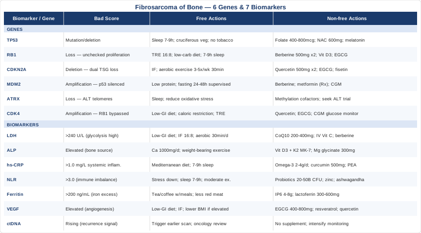
· UpdatedFibrosarcoma of Bone Genes and Biomarkers: 6 Genes and 7 Biomarkers to Track
Introduction
Fibrosarcoma of bone is one of the rarer primary malignant bone tumors, making up fewer than 5% of all bone sarcomas. Because it shares imaging characteristics with other bone lesions and carries no single defining molecular fingerprint visible on standard pathology, it often travels through a long diagnostic workup before a definitive answer arrives. That delay, combined with the limited volume of condition-specific research compared to more common cancers, leaves many patients and caregivers with questions that routine oncology consultations rarely have time to answer in depth.
Standard treatment guidelines — surgery, chemotherapy protocols borrowed from osteosarcoma regimens, and sometimes radiation — are built around population averages. They are the right starting point, but they say little about how your specific tumor behaves, how aggressive it is at the molecular level, or which metabolic and immune signals are driving its progression. Generic advice to "eat well and reduce stress" is not wrong, but it does not give you traction on a condition this specific.
What does give traction is understanding the biology underneath. A handful of measurable blood and tissue markers can tell you and your oncology team whether inflammation is high, whether tumor burden is changing, whether your immune system is holding its ground, and whether certain molecular pathways are especially active. Layered on top of that, knowing which genes are recurrently disrupted in fibrosarcoma of bone gives you a framework for understanding why some tumors behave the way they do — and where emerging targeted research is pointing.
This article is not a substitute for oncology care, and it makes no promises about outcomes. What it does offer is a more granular, science-grounded look at two complementary lenses: the seven most actionable biomarkers to track throughout diagnosis and treatment, and the six key genetic alterations that shape this cancer's behavior. Beyond that, you will find a section on metabolic cancer biology that is reshaping how some researchers think about all solid tumors, practical complementary approaches with real clinical evidence, and an honest summary of what the evidence can and cannot tell you right now.
Summary
This article covers 7 clinically relevant biomarkers — including LDH, ALP, CRP, NLR, ferritin, VEGF, and ctDNA — explaining what each one reveals about fibrosarcoma of bone, how to measure it, what a bad result means, and what can realistically be done about it. It also covers 6 recurrently disrupted genes (TP53, RB1, CDKN2A, MDM2, ATRX, and CDK4), how each shapes tumor behavior, and what lifestyle, supplement, and monitoring strategies have supporting evidence. The bonus genetics section includes specific protocols with cycling guidance and side effects. Beyond standard monitoring, the article summarizes Thomas Seyfried's metabolic cancer framework — a body of work that challenges the purely genetic view of cancer and opens practical nutritional and metabolic levers. Five complementary modalities with genuine clinical evidence round out the picture. If you are navigating a fibrosarcoma of bone diagnosis and wondering what you can actually measure and act on, this is where to start.
7 Biomarkers Worth Tracking in Fibrosarcoma of Bone
Most oncology appointments focus on imaging — MRI, CT, bone scan — to assess disease status. Blood and tissue biomarkers rarely get the same dedicated attention in rare bone sarcomas, partly because condition-specific cutoffs are not well established. But several systemic markers have strong prognostic data in soft tissue and bone sarcomas broadly, and tracking them longitudinally gives you signal that imaging alone cannot provide. Below are the seven most actionable ones, ordered from most accessible to most specialized.
Biomarker 1: Lactate Dehydrogenase (LDH)
Why it matters: LDH is an enzyme involved in anaerobic glycolysis. Tumors with high metabolic activity — particularly those with significant necrosis or rapid proliferation — release large amounts of it. Elevated serum LDH at diagnosis is consistently associated with worse prognosis across multiple sarcoma types, and it has been validated as a prognostic marker in the European Musculoskeletal Oncology Society (EMSOS) framework. In fibrosarcoma of bone specifically, a pre-treatment LDH above the upper limit of normal (roughly 240 U/L in most labs) has been associated with higher metastatic risk and shorter disease-free survival. Research indexed on PubMed supports LDH as one of the most reproducible prognostic markers in bone sarcoma: see relevant literature here.
How to measure it: LDH is included in most comprehensive metabolic panels (CMP). Cost ranges from $15 to $40 for a standalone order, or effectively free when bundled into a broader panel. It is a routine venous blood draw and results are typically available within 24 hours. Ask for LDH isoforms if your oncologist suspects a tissue-specific source, though the total serum value is sufficient for monitoring purposes.
If the score is bad — the plan without supplements: Elevated LDH primarily reflects high tumor glycolytic activity. The most evidence-backed non-supplement intervention is reducing circulating glucose load through dietary modification: a low-glycemic or modified ketogenic diet limits the substrate driving aerobic glycolysis (the Warburg effect). Intermittent fasting (16:8 or 18:6 time-restricted eating) has preclinical support for reducing tumor glucose uptake. Moderate aerobic exercise — 30 minutes most days, at a pace you can sustain through treatment — improves mitochondrial efficiency and lowers resting glycolysis-driven LDH. This is not a cure strategy; it is a metabolic support strategy to be discussed with your oncology team before implementation.
If the score is bad — the plan with supplements or equipment: Coenzyme Q10 (ubiquinol form, 200–400 mg/day) supports mitochondrial oxidative phosphorylation and has shown anti-tumor properties in some in vitro cancer models, though human RCT data in sarcoma is absent. Use CoQ10 cyclically: 8 weeks on, 2 weeks off. Side effects are generally mild (GI upset at high doses). High-dose Vitamin C (intravenous, 25–75 g per infusion, administered by a clinical provider 1–2×/week) has shown tumor-selective pro-oxidant effects in multiple small clinical trials; discuss with your oncologist before pursuing, as it can interact with certain chemotherapeutics. Berberine (500 mg 2×/day with meals) has preclinical evidence for suppressing the Warburg effect via AMPK activation; cycle 8 weeks on, 2 weeks off; monitor liver enzymes if used long-term.
Biomarker 2: Alkaline Phosphatase (ALP)
Why it matters: ALP reflects bone turnover and liver function. In bone sarcomas, elevated serum ALP indicates active osteoblastic activity or significant bone destruction. Multiple studies across osteosarcoma and bone sarcoma broadly show that pre-treatment ALP elevation correlates with advanced disease, worse response to chemotherapy, and reduced overall survival. In fibrosarcoma of bone — which arises from fibroblastic cells within the medullary canal — elevated ALP signals that the periosteal and endosteal environments are under significant disruption. Tracking ALP through treatment provides indirect evidence of local disease control. See: related studies on PubMed.
How to measure it: ALP is part of the standard comprehensive metabolic panel. Cost: $10–30 bundled, $20–45 standalone. If total ALP is elevated, isoenzyme fractionation (bone-specific ALP vs. liver fraction) costs an additional $30–80 and is clinically useful for determining source. Bone-specific ALP (bALP) has greater sensitivity for skeletal disease activity.
If the score is bad — the plan without supplements: If elevated ALP is bone-sourced, the primary goal is reducing skeletal remodeling burden. Weight-bearing exercise appropriate to your fracture risk (cleared by your orthopedic oncologist) supports bone health without excessive remodeling stimulation. Adequate dietary calcium (1000–1200 mg/day from food: dairy, fortified plant milks, leafy greens) and protein (1.2–1.6 g/kg body weight) support bone matrix quality. Eliminate or minimize alcohol (ALP-elevating) and avoid high-dose vitamin A supplements (retinol form > 10,000 IU/day), which increase bone resorption.
If the score is bad — the plan with supplements or equipment: Vitamin D3 (2000–5000 IU/day, adjusted to target serum 25-OH-D of 50–80 ng/mL) directly modulates osteoblast and osteoclast balance. Pair with Vitamin K2 (MK-7 form, 100–200 mcg/day) which directs calcium into bone matrix and away from soft tissue. Both are generally safe long-term with annual monitoring of serum vitamin D, calcium, and parathyroid hormone. Magnesium glycinate (300–400 mg/night) supports both bone mineralization and sleep quality — cycle continuously, no specific break required. Note: discuss bisphosphonate therapy with your oncologist if ALP remains persistently elevated, as these are prescription agents with stronger bone-specific evidence.
Biomarker 3: High-Sensitivity C-Reactive Protein (hs-CRP)
Why it matters: The tumor microenvironment in fibrosarcoma of bone is not biologically isolated — it communicates constantly with systemic immunity and the inflammatory cascade. CRP is the liver's output signal for systemic inflammation, and high-sensitivity CRP (hs-CRP) captures low-grade elevations that standard CRP misses. Elevated hs-CRP in sarcoma patients correlates with higher TNF-alpha and IL-6 levels, both of which promote tumor growth and immune evasion. Studies across soft tissue and bone sarcomas demonstrate that elevated systemic inflammation markers at diagnosis predict inferior outcomes: see relevant studies.
How to measure it: hs-CRP is a simple fasted venous blood draw. Cost: $15–30. Make sure to request the high-sensitivity version, as standard CRP lacks resolution below 5–10 mg/L, which is where prognostically meaningful elevations sit. Target range for a cancer patient: below 1.0 mg/L. Levels between 1–3 mg/L indicate moderate systemic inflammation; above 3 mg/L indicates high-grade systemic inflammation requiring active management.
If the score is bad — the plan without supplements: The Mediterranean dietary pattern has the strongest anti-inflammatory evidence in human studies: emphasize olive oil, oily fish (sardines, mackerel, salmon), colorful vegetables, legumes, and nuts; minimize ultra-processed foods, refined grains, and seed oils high in omega-6. Sleep optimization is critical — hs-CRP rises measurably with less than 6 hours of sleep per night. Target 7–9 hours in a cool, dark room. Low-to-moderate intensity exercise (walking, cycling, swimming) reduces systemic CRP better than high-intensity in cancer patients; 30–45 minutes most days.
If the score is bad — the plan with supplements or equipment: Omega-3 fatty acids (EPA+DHA combined, 2–4 g/day with meals) reduce IL-6 and CRP with strong RCT evidence; use continuously but monitor bleeding time if on anticoagulants. Curcumin with piperine or in phospholipid-complexed form (500–1000 mg/day of the complexed form) reduces NF-kB-mediated inflammation; cycle 3 months on, 1 month off; GI side effects are possible at high doses. Vitamin D (as above). Palmitoylethanolamide (PEA, 600–1200 mg/day) is an endocannabinoid-like molecule with anti-inflammatory effects and very low side-effect profile; use continuously and reassess every 3 months.
Biomarker 4: Neutrophil-to-Lymphocyte Ratio (NLR)
Why it matters: The NLR — calculated from a standard complete blood count (CBC) with differential as neutrophil count divided by lymphocyte count — is one of the most studied and consistently predictive immune biomarkers across solid tumors. A ratio above 3.0–4.0 indicates a systemic immune state dominated by pro-inflammatory, innate immune cells (neutrophils) at the expense of adaptive immune surveillance (lymphocytes). In sarcomas, high NLR at diagnosis predicts lower response rates to chemotherapy and shorter overall survival. It is also a dynamic marker: if NLR rises during treatment, it often reflects immune stress from the tumor or treatment toxicity. See NLR and sarcoma research here.
How to measure it: Derived from a CBC with differential, which costs $20–50 and is ordered routinely during cancer treatment. Calculate by dividing the absolute neutrophil count by the absolute lymphocyte count. Optimal range: below 2.5. Values of 3–5 suggest moderate immune imbalance; above 5 indicates significant inflammatory burden and suppressed adaptive immunity.
If the score is bad — the plan without supplements: High NLR often reflects both tumor-driven inflammation and lifestyle-related immune dysregulation. Chronic psychological stress is one of the most potent drivers of sustained neutrophilia and lymphopenia — structured stress reduction (mindfulness, yoga, social support) has demonstrated direct effects on immune cell ratios in cancer patients. Prioritize 7–9 hours of sleep, as sleep deprivation directly elevates neutrophil activity. Moderate exercise (not intense) supports lymphocyte surveillance; high-intensity workouts in the context of active cancer treatment can transiently spike NLR.
If the score is bad — the plan with supplements or equipment: Probiotics (multi-strain, 20–50 billion CFU/day) modulate gut-associated lymphoid tissue (GALT) and have shown modest but consistent effects on lymphocyte counts in several cancer cohorts; use continuously, well-tolerated. Zinc (15–30 mg/day as zinc glycinate or zinc picolinate) is essential for T-lymphocyte maturation and is commonly depleted during chemotherapy; do not exceed 40 mg/day long-term; use at 8-week intervals with 2-week breaks if concern about copper balance. Selenium (100–200 mcg/day as selenomethionine) supports natural killer cell activity; do not exceed 400 mcg/day; use cyclically. Ashwagandha (KSM-66 extract, 300–600 mg/day) is an adaptogen with clinical evidence for reducing cortisol-driven neutrophilia; cycle 2–3 months on, 1 month off; contraindicated in some autoimmune contexts — verify with your oncologist.
Biomarker 5: Serum Ferritin
Why it matters: Ferritin is typically framed as an iron storage marker, but in malignancy it carries a dual meaning. Tumor cells upregulate ferritin synthesis to secure iron for rapid proliferation (iron is essential for ribonucleotide reductase, a key enzyme in DNA synthesis). Elevated serum ferritin in sarcoma patients therefore often reflects both the iron-hoarding behavior of tumor cells and a systemic acute-phase inflammatory response. Studies in multiple solid tumors link ferritin above 200–300 ng/mL with poorer outcomes, independent of standard inflammatory markers. Iron also participates in ferroptosis — a form of regulated cell death that some emerging anti-cancer strategies attempt to exploit. Explore ferritin and cancer research here.
How to measure it: A standalone serum ferritin test costs $20–40. Always pair it with transferrin saturation and serum iron to distinguish true iron overload from inflammatory pseudoelevation. Target ferritin: 50–150 ng/mL for cancer patients (avoid both deficiency and excess). Retesting every 8–12 weeks during active treatment is reasonable.
If the score is bad — the plan without supplements: If ferritin is elevated above 200 ng/mL and the elevation is iron-driven rather than purely inflammatory, reduce dietary iron bioavailability: drink tea or coffee with meals (tannins bind iron), eat iron-containing foods alongside calcium-rich foods, and reduce red and processed meat to 2–3 times per week maximum. Do not take iron supplements unless you are genuinely anemic and your oncologist specifically recommends it.
If the score is bad — the plan with supplements or equipment: IP6 (inositol hexaphosphate, 4–8 g/day on an empty stomach) acts as a natural iron chelator and has a modest body of clinical evidence suggesting both iron-binding and anti-tumor effects in certain cancers; cycle 3 months on, 1 month off; avoid taking alongside mineral supplements as it binds broadly to divalent metals. Lactoferrin (apo-lactoferrin form, 300–600 mg/day) sequesters free iron in a bioavailable-limiting way and has some immunomodulatory evidence in cancer; well-tolerated; use continuously. Note: pharmaceutical iron chelation (deferoxamine) is a medical intervention requiring oncologist oversight; do not pursue independently.
Biomarker 6: Vascular Endothelial Growth Factor (VEGF)
Why it matters: Solid tumors beyond a few millimeters require angiogenesis — new blood vessel formation — to supply oxygen and nutrients. VEGF is the primary driver of this process, and fibrosarcoma of bone is no exception. Elevated serum or plasma VEGF correlates with higher microvascular density in the tumor, greater metastatic potential, and resistance to certain chemotherapeutic agents. In several sarcoma studies, high pre-treatment VEGF is associated with early pulmonary metastasis, which is the dominant pattern of distant spread in fibrosarcoma of bone. Anti-VEGF strategies (bevacizumab, pazopanib, regorafenib) are being studied across sarcoma subtypes precisely because of this relationship: read related literature here.
How to measure it: Serum or plasma VEGF is measured by ELISA and costs $200–500 at specialized labs. It is not routinely ordered in standard oncology panels, but it is available through most academic medical centers and reference labs. Fasting before collection reduces variability. Platelet contamination during blood processing can artificially elevate VEGF (platelets store large quantities); request platelet-poor plasma if comparing values across time.
If the score is bad — the plan without supplements: VEGF is suppressed in hypoxic states, but intermittent moderate hypoxia (altitude training, breath-hold work) does the opposite. Instead, focus on reducing the upstream drivers of VEGF signaling: high insulin and IGF-1 (driven by high glycemic load diets) are potent VEGF inducers. A low-glycemic diet and intermittent fasting (16:8 at minimum) reduce VEGF through insulin sensitization. Reducing BMI if elevated also lowers circulating VEGF via reduction of adipose-derived growth factors.
If the score is bad — the plan with supplements or equipment: Green tea extract (EGCG, 400–800 mg/day, standardized to 50%+ EGCG) has multiple mechanisms of VEGF suppression and has shown anti-angiogenic effects in several pre-clinical sarcoma models; cycle 8 weeks on, 2 weeks off; can elevate liver enzymes at high doses — monitor. Resveratrol (trans-resveratrol, 250–500 mg/day) inhibits HIF-1alpha, a key upstream VEGF inducer; use cyclically; take with a fat-containing meal for absorption. Quercetin (500–1000 mg/day) synergizes with EGCG on VEGF pathways; well-tolerated; cycle with the EGCG schedule.
Biomarker 7: Circulating Tumor DNA (ctDNA)
Why it matters: Liquid biopsy — the detection of tumor-derived DNA fragments in peripheral blood — represents one of the most significant advances in oncology monitoring of the past decade. For fibrosarcoma of bone, ctDNA offers two things that imaging cannot: an earlier signal of recurrence (ctDNA rises before lesions are visible on CT or MRI) and molecular characterization of tumor evolution, allowing identification of resistance mutations during treatment. While ctDNA assays are not yet standard of care for fibrosarcoma specifically (the rarity of the tumor limits large cohort validation studies), the technology is rapidly advancing and is available through commercial platforms. See ctDNA and sarcoma studies here.
How to measure it: Commercial liquid biopsy platforms (Guardant360, Foundation Medicine FoundationOne Liquid CDx, Tempus xF) offer ctDNA analysis from a standard blood draw. Cost: $500–2000 per test; insurance coverage is improving but variable. For monitoring purposes rather than initial diagnosis, the test is typically repeated every 3–6 months or at clinical decision points. A positive result (detectable ctDNA) requires clinical interpretation — not every fragment detected signals active progression.
If ctDNA is rising — the approach: A rising ctDNA signal between scheduled imaging should trigger an earlier scan rather than a change in treatment based on liquid biopsy alone. It is primarily an alerting tool, not a standalone treatment guide. There are no supplements that directly lower ctDNA — the ctDNA signal reflects tumor DNA shedding, which diminishes when the tumor burden decreases. The clinical response is to intensify monitoring, discuss whether re-biopsy for resistance mutation profiling is warranted, and review current treatment efficacy with your oncology team.
With these seven markers tracked consistently through treatment and surveillance, you gain a multi-dimensional view of tumor biology that imaging alone cannot provide. The next layer of understanding comes from what the tumor's own genes are doing.
The Genetic Landscape of Fibrosarcoma of Bone
Fibrosarcoma of bone does not have a single defining chromosomal translocation the way synovial sarcoma (SS18-SSX) or Ewing sarcoma (EWSR1-FLI1) do. It is instead characterized by a complex karyotype — multiple chromosomal gains, losses, and point mutations that accumulate across several key cancer-related genes. Understanding which of these pathways are altered in your tumor helps explain its behavior and opens the door to targeted therapeutic considerations. The following six genes are the most recurrently altered in fibrosarcoma of bone based on available genomic studies.
Gene 1: TP53 — The Guardian of the Genome
What it does: TP53 encodes p53, the most important tumor suppressor protein in human biology. In its normal state, p53 halts the cell cycle when DNA damage is detected, initiates DNA repair mechanisms, and triggers apoptosis if damage is irreparable. In fibrosarcoma of bone, TP53 mutations or deletions are found in a significant minority of cases and correlate with high-grade histology, genomic instability, and resistance to conventional chemotherapy. When p53 function is lost, cells accumulate damage without checkpoint control. See TP53 and sarcoma literature.
If the gene is bad — the plan without supplements: Without functional p53, the most important intervention is minimizing sources of ongoing DNA damage. Eliminate tobacco exposure entirely. Minimize ionizing radiation beyond medically necessary exposure. Optimize sleep (7–9 hours) as deep sleep is the primary window for DNA repair via NER (nucleotide excision repair) pathways. A diet rich in cruciferous vegetables (broccoli, Brussels sprouts, cabbage) provides sulforaphane, which activates Nrf2-mediated antioxidant pathways that partially compensate for reduced p53-directed repair. Avoid excessive alcohol, which generates acetaldehyde — a direct DNA damaging agent.
If the gene is bad — the plan with supplements or equipment: Folic acid or 5-MTHF (400–800 mcg/day depending on MTHFR status) supports DNA methylation and nucleotide biosynthesis, reducing replication errors; use continuously. Resveratrol (250–500 mg/day) has been shown in cell studies to partially upregulate p53-independent apoptotic pathways via SIRT1; cycle 8 weeks on, 2 weeks off; take with food containing fat. N-acetylcysteine (NAC, 600–1200 mg/day) supports glutathione synthesis and reduces oxidative DNA damage; generally well-tolerated; use continuously; avoid in active tumor treatment phases without oncologist input as it may also protect some tumor cells from oxidative chemotherapy. Melatonin (10–20 mg at night) is a potent endogenous antioxidant and has independent anti-tumor data in several cancer models; well-tolerated at these doses; start low (3 mg) and titrate.
Gene 2: RB1 — The Cell Cycle Gatekeeper
What it does: The retinoblastoma gene RB1 produces pRb, a protein that prevents cells from entering S-phase (DNA synthesis) by sequestering the E2F family of transcription factors. Loss of RB1 function removes this brake, allowing cells to proliferate unchecked even when conditions are not favorable. RB1 loss is found in a subset of fibrosarcomas and is associated with high-grade tumors and poor prognosis. Importantly, it intersects with the CDK4/CDK6 pathway — when RB1 is intact, CDK4 inhibitors work by preventing RB1 phosphorylation; when RB1 is lost, CDK4 inhibitors lose their primary mechanism. See RB1 research in bone sarcoma.
If the gene is bad — the plan without supplements: Caloric restriction and time-restricted eating (16:8 minimum) reduce CDK activity through several nutrient-sensing pathways (mTOR, AMPK) that partially compensate for RB1 deficiency. Reducing dietary refined carbohydrates reduces insulin-driven PI3K/Akt signaling, which otherwise drives cell cycle progression through RB1-independent pathways. Adequate sleep (the recommendation is the same as above: 7–9 hours) is the most underappreciated lever for reducing uncontrolled cell cycle activity during the day.
If the gene is bad — the plan with supplements or equipment: Berberine (500 mg twice daily with meals) activates AMPK and inhibits mTOR, creating a metabolic environment less supportive of unchecked cell cycle entry; cycle 8 weeks on, 2 weeks off; can lower blood glucose significantly — monitor if diabetic. Vitamin D3 (as above, targeting 50–80 ng/mL serum) has direct effects on cell cycle arrest via the vitamin D receptor, acting in part through pathways independent of RB1. EGCG (as above) reduces E2F-mediated transcription through multiple upstream targets; cycle with the RB1/EGCG schedule.
Gene 3: CDKN2A — The p16 Brake
What it does: CDKN2A encodes two tumor suppressors from the same locus: p16INK4a (which inhibits CDK4 and CDK6, keeping RB1 hypophosphorylated and active) and p14ARF (which stabilizes p53 by inhibiting MDM2). Deletion or silencing of CDKN2A therefore simultaneously removes two independent tumor suppression mechanisms. It is among the most frequently deleted loci in fibrosarcoma of bone and strongly correlates with aggressive behavior. See CDKN2A research.
If the gene is bad — the plan without supplements: The same metabolic interventions that address RB1 loss apply here, because p16 normally feeds into the CDK4/RB1 axis. Intermittent fasting is particularly relevant: it reduces IGF-1 and insulin — both of which drive CDK activity through upstream signals. Physical activity (aerobic exercise 3–5×/week, 30–45 minutes) independently upregulates p21, another CDK inhibitor that can partially compensate for p16 loss.
If the gene is bad — the plan with supplements or equipment: Quercetin (500 mg twice daily) and EGCG act as natural CDK4/6 inhibitors and have in vitro evidence in CDKN2A-deleted cancer cells; cycle together with the broader EGCG schedule above. Fisetin (500–1000 mg/day with a fatty meal) is a senolytic flavonoid with CDK inhibitory and pro-apoptotic properties, with emerging human evidence mostly from aging research; cycle 2–3 days per month as a pulse dosing strategy rather than continuous use; well-tolerated.
Gene 4: MDM2 — The p53 Suppressor
What it does: MDM2 is amplified in a meaningful subset of bone sarcomas and acts by tagging p53 for proteasomal degradation. Even when TP53 itself is structurally normal, MDM2 amplification effectively silences it. This is important: MDM2-amplified tumors with wild-type p53 are theoretically responsive to MDM2 inhibitors (a class of drugs in active clinical development). Distinguishing MDM2 amplification from TP53 mutation is therefore a critical step in molecular characterization of the tumor. See MDM2 sarcoma literature.
If the gene is bad — the plan without supplements: The primary goal is reducing upstream stimulation of MDM2 expression. IGF-1 signaling strongly upregulates MDM2 transcription — reducing IGF-1 through dietary caloric restriction (reducing total protein to 0.8–1.0 g/kg on non-exercise days), limiting dairy and highly processed foods, and prioritizing plant-based protein sources is the most accessible dietary lever. Fasting of 24–48 hours (medical supervision recommended, especially during treatment) has been shown to reduce IGF-1 by 30–50% in clinical studies.
If the gene is bad — the plan with supplements or equipment: Berberine (500 mg twice daily; cycle as above) reduces IGF-1/PI3K signaling and has been shown to reduce MDM2 expression in some cancer cell lines. Metformin (500–1000 mg/day) — a prescription medication, not an over-the-counter supplement — reduces both IGF-1 and MDM2 through AMPK activation and is being studied in multiple sarcoma trials; discuss with your oncologist. For lifestyle-based equipment: continuous glucose monitors (CGM, $70–150/month) provide real-time feedback on glycemic patterns and enable tighter metabolic control, which is one of the most practical ways to suppress IGF-1 signaling chronically.
Gene 5: ATRX — Chromatin Remodeling and Telomere Maintenance
What it does: ATRX is a chromatin remodeling protein that plays a critical role in maintaining telomere integrity and regulating gene expression through histone H3.3 incorporation. In fibrosarcoma and other sarcomas, ATRX loss or mutation is associated with the Alternative Lengthening of Telomeres (ALT) phenotype — a telomerase-independent mechanism that allows cancer cells to extend their telomeres and thus achieve replicative immortality. ALT-positive tumors have distinct biological behavior and may respond differently to certain therapeutic approaches. See ATRX and ALT in sarcoma.
If the gene is bad — the plan without supplements: ATRX loss cannot be compensated through lifestyle alone in any direct molecular sense. The practical implication is that the tumor is using non-canonical telomere maintenance, which means telomerase inhibitors would likely be ineffective as standalone strategies. From a supportive biology standpoint, maintaining genomic stability through the strategies already outlined (sleep, oxidative stress reduction, DNA repair support) remains relevant. Discuss ATRX status with your oncologist — it may influence clinical trial eligibility.
If the gene is bad — the plan with supplements or equipment: There is no well-validated supplement protocol for ALT compensation. Epigenetic support through methylation cofactors (folate, vitamin B12, betaine) may support broader chromatin health but has no specific ATRX evidence. The most important intervention here is clinical: ask whether your tumor has been tested for ALT (FISH-based detection of APBs, ultrabright telomere spots) and whether any clinical trials targeting ALT-positive tumors are open. This is an area of active research and clinical opportunity.
Gene 6: CDK4 — Cell Cycle Amplification
What it does: CDK4 (cyclin-dependent kinase 4) is frequently amplified in high-grade bone sarcomas and acts by phosphorylating and inactivating RB1, releasing cells into S-phase. When CDK4 is amplified, its activity becomes unchecked even when p16 (CDKN2A) is functional — effectively overwhelming the CDK4/6 inhibitory mechanism. CDK4 amplification often co-occurs with MDM2 amplification on chromosome 12q, and this co-amplification is a recognized molecular signature in a subset of high-grade fibrosarcomas and dedifferentiated liposarcomas. See CDK4 research in sarcoma.
If the gene is bad — the plan without supplements: The same caloric restriction and low-glycemic dietary strategies that reduce CDK activity at the signaling level apply here. Of note: CDK4 amplification is the most pharmacologically tractable of the six genes discussed — FDA-approved CDK4/6 inhibitors (palbociclib, ribociclib, abemaciclib) are in active clinical trials for sarcoma. Ask your oncologist about CDK4 testing by FISH or next-generation sequencing and whether CDK4/6 inhibitor trial enrollment is appropriate.
If the gene is bad — the plan with supplements or equipment: Natural flavonoids with CDK4 inhibitory properties include quercetin, fisetin, and EGCG (all detailed above). Intermittent fasting remains the most impactful non-pharmaceutical CDK4 reducer through AMPK-mediated mechanisms. As with MDM2, use a CGM to monitor and minimize postprandial glucose spikes, which are primary drivers of the insulin/CDK4 signaling cascade.
Connecting these genetic and biomarker findings to a broader framework for understanding cancer biology leads naturally to one of the most interesting — and practically useful — bodies of work in oncology thinking: the metabolic theory of cancer.
What Thomas Seyfried's Research on Cancer Metabolism Can Change About Your Approach
Thomas Seyfried, a professor of biology at Boston College, has spent decades building and refining a thesis that most cancer researchers in the genetic mainstream have resisted but that is increasingly difficult to dismiss: that cancer is fundamentally a metabolic disease, arising from mitochondrial dysfunction, and that the genetic mutations typically described as "driving" cancer are largely downstream consequences rather than root causes. His book Cancer as a Metabolic Disease: On the Origin, Management, and Prevention of Cancer (Wiley, 2012) synthesizes decades of research and challenges the field's almost exclusive focus on genetics.
Here are the ten most practically impactful ideas from his framework and the broader work it has inspired:
1. Cancer Cells Rely on Fermentation, Not Oxidative Phosphorylation
The Warburg effect — the observation by Otto Warburg in the 1920s that cancer cells ferment glucose to lactate even in the presence of oxygen — is not merely a quirk of tumor metabolism. Seyfried argues it is the foundational metabolic lesion of cancer. Most cancer cells have dysfunctional mitochondria and cannot efficiently run oxidative phosphorylation; they compensate by upregulating glycolysis and glutamine fermentation. This is why elevated LDH (a glycolysis-associated enzyme) is such a consistent prognostic marker.
2. Glucose and Glutamine Are the Two Fuels Cancer Depends On
By restricting both primary fuels, you can theoretically starve the metabolic engine driving most tumors. Seyfried's lab and others have demonstrated significant tumor suppression in animal models using ketogenic diets (which restrict glucose-derived energy) combined with DON (a glutamine antagonist) or other glutamine-targeting strategies. Human clinical trials using ketogenic diets in glioblastoma have shown partial feasibility; data in sarcoma is limited but biologically plausible.
3. Caloric Restriction and Ketosis Reduce IGF-1 and Insulin — Both Tumor Growth Signals
Fasting and caloric restriction reduce serum insulin and IGF-1 by 30–60% within days. Both hormones activate mTOR and PI3K/Akt pathways that drive tumor proliferation. This is not speculation — it is why both intermittent fasting and modified ketogenic diets are showing up in cancer clinical trial combinations. The practical implication: managing your glycemic environment is a direct metabolic oncology intervention.
4. The Press-Pulse Therapeutic Strategy
Seyfried and colleagues have proposed a framework they call Press-Pulse: a chronic background "press" of metabolic restriction (ketogenic diet, caloric restriction) combined with periodic acute "pulse" interventions (chemotherapy, hyperbaric oxygen, specific metabolic drugs). The press weakens the metabolic foundation of tumor cells; the pulse delivers maximum cytotoxicity to cells already metabolically compromised. This is an active area of clinical investigation.
5. Hyperbaric Oxygen Therapy (HBOT) Exploits the Metabolic Vulnerability
Normal cells thrive under hyperoxic conditions; cancer cells with dysfunctional mitochondria and dependence on anaerobic glycolysis are selectively stressed. Seyfried's group has published preclinical evidence showing HBOT reduces tumor growth and metastasis in combination with metabolic restriction. Several cancer centers now offer HBOT as an adjunct to standard treatment. Cost ranges from $150–400 per session; a typical protocol is 20–40 sessions. Discuss with your oncologist before pursuing, as certain tumor types may have complex interactions with HBOT.
6. Mitochondrial Transplantation and Mitophagy Are Emerging Therapeutic Targets
Seyfried's framework predicts that restoring mitochondrial function could reverse or halt tumor progression. Supporting mitophagy (the cellular process of clearing damaged mitochondria) through fasting, exercise, and certain phytonutrients is therefore not merely supportive care — it is mechanistically targeted. Fasting triggers mitophagy within 12–18 hours through AMPK and PINK1/Parkin pathways.
7. Most Tumor Cells Cannot Survive Without Glucose or Glutamine — Normal Cells Can
This is the therapeutic selectivity argument: healthy cells can run on ketone bodies (from fat) even when glucose is restricted, but most cancer cells cannot efficiently use ketones for ATP production. This creates a metabolic window of selectivity that does not exist with most cytotoxic drugs. The practical implication: ketogenic diet as an adjunct to treatment may widen the therapeutic index by selectively weakening tumor cells while sparing normal tissue.
8. The Ketogenic Diet for Cancer Is Not the Same as Ketogenic Diet for Weight Loss
Therapeutic ketogenic diets in cancer research use protein moderation (to reduce glutamine precursors and limit gluconeogenesis), not just carbohydrate restriction. Fat sources matter — medium-chain triglycerides (MCT oil) are ketogenic without requiring high dietary fat intake and are being studied specifically in cancer-associated ketosis protocols. Do not design a cancer-adapted ketogenic diet from standard fitness sources — work with a metabolic oncology dietitian.
9. The Metabolic Framework Does Not Replace Standard Treatment — It Complements It
Seyfried and his colleagues are explicit: the metabolic approach is an adjunct to surgery, chemotherapy, and radiation, not a replacement. The strongest published evidence so far is in glioblastoma and mouse tumor models. In fibrosarcoma of bone, there are no condition-specific randomized trials of metabolic therapy — but the underlying biology is not tumor-type specific. Applying these principles under oncology supervision is scientifically reasonable; abandoning standard treatment in favor of them is not.
10. Psychological and Social Stress Drive Cortisol, Which Drives Metabolic Dysfunction
Elevated cortisol chronically elevates blood glucose (through hepatic gluconeogenesis), suppresses lymphocyte activity, and promotes inflammatory signaling. In Seyfried's metabolic framework, chronic stress is not merely a quality-of-life problem — it is a metabolic tumor-support mechanism. Structured stress reduction (MBSR, sleep, social connection, nature exposure) therefore belongs inside the metabolic oncology framework, not as a soft add-on but as a metabolic management strategy.
Beyond these metabolic strategies, a growing evidence base supports several complementary non-pharmacological approaches that address pain, anxiety, and immune function in cancer patients.
Complementary Approaches with Meaningful Clinical Evidence
For a rare tumor like fibrosarcoma of bone, condition-specific RCTs for complementary therapies do not exist. The following five modalities are selected from broader cancer populations where rigorous human evidence supports measurable benefits in domains highly relevant to fibrosarcoma patients: pain management, anxiety reduction, immune support, and quality of life.
Mindfulness-Based Stress Reduction (MBSR)
MBSR is an 8-week structured program developed by Jon Kabat-Zinn that combines body scanning, sitting meditation, and gentle movement. In cancer populations, its mechanisms are relevant to fibrosarcoma management at multiple levels: it reduces cortisol, lowers CRP and IL-6, improves NK cell activity, and reduces the perception of pain and treatment-related anxiety. This is not soft or tangential — reducing cortisol directly improves the immune surveillance environment and the glycemic profile, both of which matter in cancer.
A landmark RCT published in Psychoneuroendocrinology (Carlson et al., 2007) followed breast and prostate cancer patients through MBSR and found significant reductions in cortisol and IL-6 alongside improved sleep quality. A subsequent Cochrane-level meta-analysis of MBSR in oncology found consistent improvements in depression, anxiety, fatigue, and quality of life across heterogeneous cancer types: See the supporting literature here.
For fibrosarcoma patients, MBSR is most practically applied during and after chemotherapy, when anxiety, fatigue, and sleep disruption are highest. Programs are available in-person (8 weekly sessions of 2.5 hours plus a full-day retreat) and online (comparable efficacy in several recent trials). The Palouse MBSR program is a validated, low-cost online version. Side effects are negligible; the main barrier is time investment. Begin week 1 of any treatment cycle if possible.
Music Therapy
Music therapy uses live or recorded music interventions delivered by a trained music therapist to address anxiety, pain, and emotional well-being. Its mechanism in cancer care involves modulation of the autonomic nervous system, reduction of salivary cortisol, and distraction/reframing of pain perception. In oncology wards specifically, music therapy has been studied in procedural pain, chemotherapy nausea, and generalized treatment anxiety.
A Cochrane review (Music interventions for improving psychological and physical outcomes in people with cancer, Bradt et al., 2021) analyzed 29 trials involving over 1,400 cancer patients and found moderate evidence for reductions in anxiety and pain, with small effects on mood and quality of life: See the Cochrane evidence base here.
For fibrosarcoma patients undergoing surgical or chemotherapy procedures, music therapy is most accessible as receptive listening sessions (patient-selected or therapist-curated playlists) during infusions or pre-procedure preparation. Hospital-based music therapy is increasingly available at cancer centers; if not, structured self-directed sessions (30–60 minutes of binaural or slow-tempo instrumental music at 60 BPM or below) during stressful treatment periods offer a practical replication. No adverse effects are documented.
Massage Therapy
Massage therapy — specifically light-touch Swedish or oncology massage adapted for cancer patients — has one of the strongest evidence bases among complementary modalities for reducing pain and fatigue in cancer patients. In bone and soft tissue sarcoma patients, musculoskeletal pain from the tumor site, post-surgical scarring, and chemotherapy-induced peripheral neuropathy are all relevant targets.
An RCT conducted at Memorial Sloan Kettering Cancer Center (Cassileth and Vickers, 2004; Cancer) found immediate significant reductions in pain, fatigue, anxiety, nausea, and depression following oncology massage in over 1,000 cancer inpatients: See supporting evidence here.
For fibrosarcoma patients, oncology massage should be performed only by a therapist specifically trained in oncology massage (not standard deep tissue) to avoid applying pressure directly to tumor sites, lymph nodes at risk, or areas with bone compromise. Sessions of 30–60 minutes weekly are a reasonable protocol during treatment; bi-weekly during surveillance phases. Avoid massage over surgical sites until full healing is confirmed by your surgeon (typically 8–12 weeks post-operation). The main caution in bone sarcoma: if cortical bone integrity is compromised, vigorous soft tissue work near the affected area requires oncologist clearance.
Guided Imagery
Guided imagery involves the use of structured mental visualization — typically facilitated by an audio recording or practitioner — to create a focused relaxation response and reduce pain perception. Its relevance in cancer care relates to both psychoneuroimmunological mechanisms (modulation of cortisol, autonomic nervous system balance) and direct pain gate modulation. For fibrosarcoma patients dealing with post-operative or disease-related bone pain, guided imagery offers a non-pharmacological pain adjunct with essentially zero side effects.
Clinical evidence for guided imagery in cancer pain management includes several RCTs demonstrating significant reductions in pain severity scores. A study by Syrjala et al. in Pain (1995, and replicated in subsequent work) found that cancer patients using guided imagery plus relaxation training during bone marrow transplant reported significantly lower pain and nausea scores than controls: See the relevant literature here.
For practical application: audio-guided sessions of 15–20 minutes twice daily work best when practiced consistently rather than intermittently. Apps such as Insight Timer include dedicated cancer-focused guided imagery tracks. The Simonton imagery protocol — developed specifically for cancer patients and involving visualization of immune cells targeting the tumor — has decades of use in oncology supportive care. Practice during high-anxiety periods: before imaging results, during infusion, and post-surgical recovery. No special training required; immediate accessibility is a key advantage.
Breathing-Based Therapies
Controlled breathing techniques — including slow diaphragmatic breathing, box breathing, and physiological sighing — activate the parasympathetic nervous system through vagal afferents, reducing heart rate variability imbalance, cortisol, and sympathetic arousal. In cancer patients, dysregulated autonomic tone driven by treatment stress, anxiety, and pain is a common complication. Chronic sympathetic dominance suppresses NK cell activity and elevates pro-inflammatory cytokines — a direct interface with the tumor-immune relationship.
Clinical evidence supports breathing-based therapy for cancer-related anxiety and fatigue. A 2019 RCT published in Psycho-Oncology found that 4 weeks of structured diaphragmatic breathing significantly reduced cortisol, anxiety, and fatigue in cancer patients receiving chemotherapy: See supporting research here. The Andrew Huberman lab has also published on the physiological sigh (double inhale through the nose followed by a long exhale) as the fastest known real-time intervention for reducing subjective stress.
For fibrosarcoma patients, a practical protocol is: 5–10 minutes of slow coherent breathing (5-second inhale, 5-second exhale) twice daily, plus physiological sighs on demand during acute anxiety moments (e.g., before medical procedures, awaiting results). This requires no equipment, no cost, no supervision, and can be started immediately. For deeper structured work, 8-week programs in pranayama yoga breathing or clinical respiratory biofeedback (typically $50–120 per session at trained providers) provide a more immersive version of the same physiological mechanism.
Conclusion
Fibrosarcoma of bone is a biologically complex tumor, and the information available to any individual patient has historically been limited to what imaging and histopathology can show. The seven biomarkers discussed here — LDH, ALP, hs-CRP, NLR, ferritin, VEGF, and ctDNA — give you a richer, longitudinal signal of what the disease and your body are doing between scans. The six genes — TP53, RB1, CDKN2A, MDM2, ATRX, and CDK4 — explain the molecular logic behind tumor behavior and point toward both pharmacological and lifestyle-level interventions. The metabolic framework from Seyfried's work adds another dimension entirely: it suggests that the metabolic environment you create through diet, fasting, sleep, and stress management is not passive context but an active biological variable in cancer outcomes.
None of this replaces surgical oncology, chemotherapy, or radiation where they are indicated. What it does is give you a set of measurable, actionable levers to work with alongside standard treatment. The next smart step is to bring your biomarker list to your oncology team and ask which ones can be incorporated into routine monitoring. Get your tumor's molecular profile if it has not already been sequenced. Work with an integrative oncology dietitian on dietary metabolic optimization. And track what changes when you do — because in a rare cancer with limited population-level data, your own longitudinal record is among the most valuable evidence you have.
Cancer & Oncology Endocrine & Metabolic
Musculoskeletal: Bone Conditions
Autoimmune: Inflammatory Conditions
Cancer & Oncology: Bone Cancer