This article contains AI generated content.
· UpdatedFibrous Dysplasia Genes And Biomarkers: 4 Genes And 6 Biomarkers To Track
Introduction
Living with fibrous dysplasia means navigating a condition that most doctors encounter only a handful of times in their careers. Whether you were diagnosed recently or have been managing this for years, you probably know what it feels like to be handed a treatment plan built primarily around fracture prevention and pain management — with very little said about what is actually happening at a biological level, or how to track it meaningfully over time.
Fibrous dysplasia is driven by a somatic mutation in the GNAS gene that leads to abnormal bone replacement by fibrous tissue. But the downstream consequences of that mutation — on bone turnover, mineral regulation, and hormonal signaling — vary significantly from person to person, and even from bone to bone within the same person. Two patients carrying the same diagnosis can have profoundly different trajectories. Generic reassurance like "monitor it with imaging every year" rarely captures why some lesions remain stable for decades while others progress, why bone pain flares unpredictably, or what metabolic patterns predict a worse outcome.
That's where biomarkers and genetics add genuine precision. They don't replace imaging or specialist oversight, but they create a dynamic layer of information that routine monitoring misses. Tracking specific markers of bone turnover, phosphate regulation, and inflammation gives both you and your physician a real-time view of what the skeleton is doing right now — not just what a scan documented six months ago.
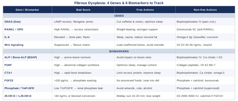
This article takes two main approaches. The first covers 6 practical biomarkers you can follow with blood and urine tests — what each one reveals, how to obtain it, and what may help if a value is off. The second examines 4 key genes and molecular pathways underlying fibrous dysplasia, translating current research into actionable steps. Both frameworks, used together, offer a more complete and useful picture of a condition that continues to surprise even its most experienced researchers.
6 Biomarkers to Track in Fibrous Dysplasia
Bone is not static tissue. It is continuously remodeling — building and breaking down in a cycle that, in fibrous dysplasia, is fundamentally disrupted. The six biomarkers below reflect different dimensions of that disruption: from the pace of bone turnover to the hormonal signals that go wrong when fibrous lesions begin producing factors affecting the entire body. Tracking these consistently, and watching trends over time rather than interpreting isolated values, is where the real clinical value lies.
Biomarker 1: Alkaline Phosphatase and Bone-Specific ALP
Why it matters: Alkaline phosphatase is one of the most routinely measured markers of bone activity, and in fibrous dysplasia it frequently runs high. Total ALP reflects osteoblast activity — when bone-forming cells are overactive, as they chronically are within FD lesions, ALP rises. The bone-specific fraction (BSAP) is more precise because it isolates the skeletal contribution from the liver, making it a cleaner signal of disease activity in the skeleton.
In clinical practice, elevated ALP correlates with the extent of skeletal involvement and with metabolic activity of lesions. For patients managing polyostotic disease — where multiple bones carry fibrous tissue — BSAP functions as a useful summary signal of overall skeletal burden. It is one of the most widely used markers to assess response to bisphosphonate therapy and to detect periods of accelerated disease activity between imaging studies.
How to measure it: Total ALP is included in the standard comprehensive metabolic panel (CMP), making it accessible and low-cost ($10–$30 as part of a panel). Bone-specific ALP is ordered separately through specialty labs and typically costs $50–$150. No special preparation is required beyond standard blood draw protocols. Measure at the same time of day across visits, since ALP shows mild diurnal variation.
If the score is high: the plan without supplements
Elevated ALP in an FD patient without liver disease points to elevated bone turnover from active lesions. The most impactful non-supplement intervention is bisphosphonate therapy — specifically intravenous pamidronate or zoledronic acid — administered under specialist supervision. These agents reduce both resorption and the compensatory spike in formation markers, bringing ALP down in a meaningful proportion of patients.
Beyond medication: avoiding high-impact mechanical loading of affected bones is essential, as fractures spike remodeling acutely and will drive ALP higher. Ensuring adequate weight-bearing exercise on unaffected limbs and bones, through swimming, gentle cycling, or upper-body resistance work, maintains bone quality in normal segments without traumatizing lesion sites.
If the score is high: the plan with supplements or equipment
Calcium and vitamin D are foundational before and during bisphosphonate therapy — without them, bisphosphonates work poorly, and their absence allows secondary hyperparathyroidism to amplify resorption. Calcium citrate (500–1000 mg/day in divided doses, with meals) paired with vitamin D3 (2000–4000 IU/day, adjusted to blood levels) is a standard starting point. Magnesium (200–400 mg/day, glycinate or malate form) is an often-overlooked cofactor that supports mineral metabolism broadly.
Red light therapy (photobiomodulation) at 630–850 nm wavelengths has shown early evidence for promoting osteoblast activity and reducing local inflammatory signaling in bone healing contexts. Evidence specific to FD lesions does not yet exist, but using a calibrated device 3–5 times per week on accessible affected areas represents a low-risk addition to a broader bone support protocol.
Biomarker 2: P1NP — The Bone Formation Marker
Why it matters: P1NP (procollagen type 1 N-terminal propeptide) is released into the bloodstream as osteoblasts lay down new collagen — the structural backbone of bone matrix. It is one of the most sensitive and specific markers of bone formation currently available, preferred by the International Osteoporosis Foundation and metabolic bone specialists for monitoring treatment response.
In fibrous dysplasia, where fibrous tissue is being produced instead of organized lamellar bone, P1NP reflects the abnormal collagen synthesis occurring within lesions as well as whatever normal bone formation is occurring elsewhere in the skeleton. Tracking P1NP over sequential tests — not just reading a single value — is where the information becomes clinically useful: a declining P1NP trend after initiating treatment signals a favorable response, while a rising trend during apparent stability should prompt reassessment.
How to measure it: P1NP is a fasting blood test, ideally drawn in the morning. Specialty labs and many hospital systems offer it for $50–$150. Reference ranges vary by lab, age, and sex; in adult FD patients, values above the upper limit of normal are common during active disease phases. Some direct-to-consumer longevity panels now include it.
If the score is high: the plan without supplements
Elevated P1NP alongside elevated ALP suggests active lesion metabolism. Bisphosphonates remain the most evidence-backed pharmacological intervention. Beyond medication, sleep quality has a meaningful impact on bone formation that is rarely discussed in FD management: growth hormone pulses during slow-wave sleep are a primary driver of osteoblast activity, and disrupted sleep — whether from pain, stress, or untreated sleep apnea — blunts this anabolic signal and chronically elevates cortisol, which promotes resorption. Prioritizing 7–9 hours of quality sleep, managing light exposure in the evening, and addressing pain as a barrier to sleep are practical steps.
If the score is high: the plan with supplements or equipment
Collagen peptides (10–15 g/day) have modest evidence for supporting bone formation quality by providing glycine and proline substrates, with some data suggesting benefit in reducing bone turnover velocity in populations with elevated markers. Vitamin K2 in the MK-7 form (100–200 mcg/day) supports carboxylation of osteocalcin, improving how newly formed bone matrix mineralizes — an important downstream step that bisphosphonates do not directly address. Both are low-risk additions to a supplement protocol. Cycling K2 is generally not necessary at these doses.
Biomarker 3: CTX-I — Bone Resorption in Real Time
Why it matters: CTX-I (beta-CTX, or C-telopeptide of type I collagen) is released into blood when osteoclasts break down old bone matrix. It is the counterpart to P1NP: together, these two markers give a picture of both sides of the remodeling equation — how fast bone is being formed and how fast it is being destroyed. In fibrous dysplasia, the mutant Gsα protein promotes osteoclast recruitment through RANKL signaling, meaning resorption is often disproportionately elevated. High CTX-I in an FD patient signals accelerated breakdown of both lesional and adjacent normal bone.
Monitoring the P1NP/CTX-I ratio over time is particularly informative. In a healthy remodeling state, these markers move in rough proportion; in active FD, CTX-I often rises faster than P1NP, reflecting the osteoclast dominance that characterizes the disease.
How to measure it: CTX-I is highly sensitive to food intake — it must be measured fasting, in the morning, ideally before 10 a.m. Even a light meal can suppress values by 20–30%, making non-fasting results uninterpretable. Blood draw cost: $50–$150 through specialty labs. Some direct-to-consumer platforms include it in bone health panels.
If the score is high: the plan without supplements
Bisphosphonates act primarily by inhibiting osteoclast function and are the most targeted available intervention for elevated CTX-I. Beyond prescription management: reducing excessive protein intake above 2.2 g/kg/day (which can increase acid load and secondary resorption), correcting phosphate and vitamin D status, and ensuring adequate sleep are all meaningful levers. Moderate weight-bearing exercise on unaffected limbs temporarily increases CTX-I acutely but improves bone quality over months by stimulating formation; the net effect over a 3-to-6-month period is beneficial.
If the score is high: the plan with supplements or equipment
Calcium timing matters: taking calcium citrate with meals blunts the nocturnal resorption spike that occurs as the body draws from bone to maintain serum calcium during overnight fasting. Magnesium deficiency is associated with elevated PTH-driven resorption — many FD patients with phosphate dysregulation also have secondary mineral imbalances — making magnesium repletion a logical first step before attributing elevated CTX-I solely to FD lesion activity.
Omega-3 fatty acids (EPA+DHA, 2–3 g/day from fish oil or algae-derived sources) reduce NF-κB-driven osteoclast activation. This is not a substitute for bisphosphonate therapy in significant disease, but it provides a meaningful supportive effect with a favorable safety profile. High-dose fish oil may slightly increase bleeding time; note this before any planned surgical procedures.
Biomarker 4: FGF23 — The FD-Specific Hormone
Why it matters: FGF23 (fibroblast growth factor 23) is a bone-derived hormone that regulates phosphate balance by acting on the kidneys. In fibrous dysplasia, lesional cells produce excess FGF23 — and the more extensive the skeletal disease burden, the higher the circulating levels tend to run. Elevated FGF23 causes the kidneys to waste phosphate in urine and simultaneously suppresses the renal enzyme that activates vitamin D, producing a combination that significantly worsens the underlying bone disease.
This phenomenon was documented in a landmark paper by Riminucci and colleagues published in the Journal of Clinical Investigation (2003), which established the mechanistic link between fibrous dysplasia lesions and systemic phosphate dysregulation (Riminucci et al., J Clin Invest 2003). Patients with high FGF23 and polyostotic disease frequently report amplified bone pain and faster skeletal deterioration — making this one of the most actionable and FD-specific biomarkers to monitor.
How to measure it: Intact FGF23 (C-terminal) is a specialty blood test requiring specific sample handling and processing. Cost ranges from $100–$300 depending on the lab. It requires a physician order and is not part of standard panels. In an FD patient with polyostotic disease, intact FGF23 above 100 pg/mL in the context of low or low-normal serum phosphate is clinically meaningful.
If the score is high: the plan without supplements
No over-the-counter intervention directly blocks FGF23 production. What can be addressed without prescription: eliminating dietary phosphate offenders that worsen the systemic picture — heavily processed foods with phosphate additives, dark cola beverages (phosphoric acid), and high-phosphate convenience foods. Treating iron deficiency, which independently elevates FGF23 through a well-characterized mechanism, is a simple and often overlooked lever in patients who have not been screened for it.
If FGF23 is high but serum phosphate remains within range, increasing monitoring frequency is the appropriate immediate step. If high FGF23 is paired with confirmed low phosphate, specialist consultation is urgent — untreated phosphate wasting accelerates fracture risk significantly.
If the score is high: the plan with supplements or equipment
Phosphate supplementation, when serum phosphate is low, must be physician-supervised in FD. Standard protocols use potassium phosphate (250–500 mg elemental phosphate, 3–4 times per day), always paired with calcitriol (active vitamin D) to support intestinal calcium absorption and prevent secondary hyperparathyroidism. Without calcitriol, isolated phosphate supplementation can worsen calcium balance. Burosumab, an anti-FGF23 monoclonal antibody approved for X-linked hypophosphatemia, is under investigation in FD-associated hypophosphatemia and early compassionate-use data are promising — this is currently a specialist-managed option in clinical trial contexts.
Iron repletion in deficient patients (ferrous bisglycinate orally, or intravenous iron in severe cases) is worth addressing since iron deficiency independently drives FGF23 elevation — normalizing iron can meaningfully reduce FGF23 in some patients before any other intervention.
Biomarker 5: Serum Phosphate and TmP/GFR
Why it matters: Given that FGF23 excess in FD causes renal phosphate wasting, serum phosphate is a critical downstream indicator. But serum phosphate alone is frequently misleading — it shifts substantially with meals, exercise, and time of day. A more refined and diagnostically powerful measure is TmP/GFR (tubular maximum phosphate reabsorption relative to GFR), calculated from simultaneous fasting blood and urine phosphate and creatinine. This ratio reveals how efficiently the kidneys are actually retaining phosphate.
Low TmP/GFR is the hallmark of FGF23-mediated phosphate wasting and can detect early renal phosphate leak before frank hypophosphatemia develops in serum — making it a more sensitive early-warning tool than serum phosphate alone, particularly in patients with extensive but not yet severe polyostotic disease.
How to measure it: Serum phosphate is part of the comprehensive metabolic panel ($10–$30). TmP/GFR calculation requires a fasting morning blood draw plus a timed two-hour urine collection; lab cost approximately $50–$100. The calculation itself is: TmP/GFR = serum phosphate − [(urinary phosphate × serum creatinine) / urinary creatinine]. Some nephrology and metabolic bone labs report this directly.
If the score is low: the plan without supplements
Eliminate phosphate-depleting habits: excess alcohol consumption, chronic use of aluminum- or magnesium-containing antacids (which bind dietary phosphate in the gut), and ultra-processed dietary patterns low in whole-food phosphate sources. Dark cola soft drinks are a specific and often underappreciated driver — the phosphoric acid load contributes to urinary phosphate losses in patients already wasting phosphate renally.
Increasing whole-food protein sources — eggs, meat, fish, legumes — provides organic phosphate in better-regulated forms than the inorganic phosphate additives prevalent in processed foods. This dietary shift is unlikely to normalize TmP/GFR in significant FD, but it reduces unnecessary nutritional aggravators.
If the score is low: the plan with supplements or equipment
Phosphate supplementation is always physician-supervised in FD due to the risk of secondary hypocalcemia and worsening hyperparathyroidism if administered without concurrent calcitriol. The standard paired protocol (phosphate + calcitriol) normalizes serum phosphate and supports bone mineralization in phosphate-wasting FD patients, though it does not address the root cause of FGF23 excess.
Burosumab (anti-FGF23 monoclonal antibody) targets the root hormonal driver rather than supplementing the depleted mineral downstream, and is now the most mechanistically rational treatment for this specific abnormality — though access outside clinical trials remains limited in FD specifically.
Biomarker 6: 25-Hydroxyvitamin D and the Active Form
Why it matters: Vitamin D deficiency in fibrous dysplasia creates problems through two distinct pathways. First, like all metabolic bone diseases, FD worsens when vitamin D is depleted — inadequate 25-OH-D means reduced calcium absorption, secondary hyperparathyroidism, and accelerated resorption. Second, in patients with high FGF23, the renal enzyme that converts 25-OH-D to its active form (1,25-dihydroxyvitamin D, or calcitriol) is specifically suppressed — meaning a patient can have normal 25-OH-D on routine testing while being functionally vitamin D insufficient at the tissue level.
For this reason, measuring both 25-OH-D and 1,25-dihydroxyvitamin D in FD patients with suspected phosphate wasting is important, since the two can diverge significantly and lead to very different management decisions. This nuance is consistently highlighted by researchers in the NIH metabolic bone disease unit and in precision medicine frameworks for bone health.
How to measure it: 25-OH-D is widely available ($30–$80). 1,25-dihydroxyvitamin D is a specialty test ($75–$150). For FD management, target 25-OH-D between 40–60 ng/mL (100–150 nmol/L). Testing every 6 months during active management is reasonable; more frequently when adjusting supplementation.
If the score is low: the plan without supplements
Deliberate midday sun exposure (10–20 minutes on arms and legs, between 10 a.m. and 2 p.m., without sunscreen) is the most natural source of vitamin D substrate. However, in FD patients with high FGF23, sun-derived 25-OH-D still cannot be effectively converted to calcitriol at the kidney — making sun exposure necessary but insufficient as the only intervention. Weight loss in overweight individuals improves vitamin D bioavailability, since the vitamin is stored in adipose tissue and sequestered away from circulation.
If the score is low: the plan with supplements or equipment
Vitamin D3 (cholecalciferol) at 2000–5000 IU/day is a reasonable starting dose, adjusted to achieve 40–60 ng/mL 25-OH-D. Crucially: in FD patients with elevated FGF23 and confirmed low 1,25-dihydroxyvitamin D despite normal 25-OH-D, supplementing D3 alone is insufficient — calcitriol (prescription active vitamin D) is required because the conversion step is blocked at the kidney. This distinction is clinically significant and routinely missed in primary care settings.
Magnesium (200–400 mg/day, glycinate or malate) is required as a cofactor for vitamin D activation. Studies have shown that vitamin D supplementation is substantially less effective in magnesium-deficient individuals. Testing serum or red blood cell magnesium alongside vitamin D is worthwhile, particularly in FD patients with phosphate dysregulation who may have multiple mineral imbalances.
The Genetic and Molecular Architecture of Fibrous Dysplasia
Understanding the molecular drivers of fibrous dysplasia doesn't require genetic testing in most cases — the primary mutation is already known. What varies between patients is how downstream pathways respond to that mutation, and how aggressively each is dysregulated. These four genetic and molecular factors explain much of the variability in disease severity, progression rate, and treatment response. They also reveal where intervention has the most biological leverage.
GNAS and the Gsα Mutation: The Root Cause
What it is: Fibrous dysplasia is caused by an activating somatic mutation in GNAS, the gene encoding the alpha subunit of the stimulatory G protein (Gsα). The mutation — almost universally at codon R201 (arginine substituted by histidine or cysteine) — prevents the GTPase function of Gsα from turning itself off. This leaves adenylyl cyclase chronically activated, cAMP perpetually elevated in affected osteoprogenitor cells, and normal differentiation permanently arrested in a fibrogenic state.
The mutation is somatic (post-zygotic), occurring in a single progenitor cell after fertilization. Early embryonic timing leads to widespread skeletal involvement and McCune-Albright syndrome; later timing produces monostotic disease in a single bone segment. This explains why FD does not follow Mendelian inheritance patterns.
What it affects: Every major downstream consequence of FD — elevated IL-6, RANKL-driven osteoclastogenesis, suppressed Wnt signaling, excess FGF23, and failure of lamellar bone formation — traces back to this single point. The mutation cannot currently be corrected at the somatic level with available clinical tools, making downstream pathway management the practical target.
If the gene is bad: the plan without supplements
Since the GNAS mutation is fixed in affected cells, management targets its consequences. Bisphosphonates (pamidronate IV or zoledronic acid IV) directly counteract the excessive osteoclast activity downstream of mutant Gsα. Protocols typically involve infusions every 3–6 months, with interval titrated to bone turnover marker response. Imaging (X-ray for cortical integrity, MRI when soft tissue involvement is suspected) combined with biomarker tracking provides the most complete current monitoring framework.
Reducing factors that amplify cAMP signaling more broadly across the body is a supportive but meaningful step: chronic excess caffeine (which inhibits phosphodiesterase, extending cAMP signals), chronic psychological stress (which stimulates Gs-coupled adrenergic receptors), and sleep deprivation (which broadly dysregulates hormonal signaling) all add systemic load on a pathway already constitutively overactive in FD lesions.
If the gene is bad: the plan with supplements or equipment
No supplement reverses or meaningfully compensates for GNAS mutation activity in FD lesions. However, addressing modifiable amplifiers of cAMP-mediated inflammation is rational: omega-3 fatty acids reduce downstream inflammatory amplification, and curcumin at pharmacological doses (1–3 g/day of phospholipid-complexed form for bioavailability) has shown cAMP-modulating effects in cell studies — though human data in GNAS-mutant disease is essentially absent. This is a speculative addition, not a primary intervention.
RANKL and OPG: The Osteoclast Control Switch
What it is: Mutant Gsα in FD lesional cells promotes RANKL (receptor activator of NF-κB ligand) expression while simultaneously suppressing OPG (osteoprotegerin), RANKL's natural inhibitor. The resulting imbalance creates a strongly pro-resorptive environment — osteoclasts are recruited excessively, bone matrix is broken down faster than it can be replaced, and lesional tissue expands. Elevated CTX-I on blood testing is the downstream readout of this pathway being overactive.
The degree of RANKL/OPG imbalance likely contributes to how aggressively a given lesion behaves — and estrogen status in women modulates this pathway significantly, which may partly explain why some women with FD report accelerated progression around perimenopause.
If the score is bad: the plan without supplements
Weight-bearing physical activity and adequate estrogen (in women) are among the most potent natural OPG-supportive signals. Estrogen deficiency — from menopause, hypothalamic amenorrhea due to low body weight, or other causes — sharply worsens the RANKL/OPG ratio. Addressing estrogen status in women with FD is clinically important and often underemphasized in bone disease management outside of menopause specialists.
Reducing chronic dietary patterns that drive NF-κB activity — ultra-processed foods, excess refined sugars, high omega-6 load from industrial seed oils — decreases a key upstream driver of RANKL expression.
If the score is bad: the plan with supplements or equipment
Omega-3 fatty acids (EPA+DHA, 2–3 g/day) suppress NF-κB activity and have shown modest OPG-supportive effects in small human trials. Vitamin K2 (MK-7 form, 100–200 mcg/day) has been associated with improved OPG expression in osteoblasts in human observational studies. Both are low-risk additions to a bone health protocol in FD.
Denosumab — a monoclonal antibody directly blocking RANKL — is used off-label in FD and is the most targeted pharmacological tool for this specific pathway. It is administered subcutaneously every 6 months under specialist supervision. A notable consideration: stopping denosumab abruptly in bone disease can trigger a rebound increase in resorption markers and fracture risk; any initiation or cessation requires careful specialist management.
IL-6: The Inflammation Amplifier Behind Bone Pain
What it is: Elevated cAMP in FD lesional cells drives substantial IL-6 production. IL-6 is a cytokine that promotes osteoclast differentiation, sustains systemic inflammation, and sensitizes pain pathways — it is one of the key mediators of the "flare" episodes of acute bone pain that many FD patients experience, particularly in craniofacial lesions. Beyond acute flares, persistently elevated IL-6 contributes to the fatigue, diffuse aching, and cognitive fog that some patients describe well beyond what can be explained by local lesion pain alone.
Measuring high-sensitivity CRP (hsCRP) as a proxy for systemic IL-6 activity is an inexpensive and practical approach — direct IL-6 measurement is available but more expensive and less standardized across labs. hsCRP below 1 mg/L is the target range for low systemic inflammatory burden.
If the score is bad: the plan without supplements
Anti-inflammatory lifestyle modifications are the most accessible levers: improving sleep quality (IL-6 surges with even partial sleep deprivation), reducing visceral adiposity (adipose tissue is a sustained IL-6 source independent of FD), and managing chronic psychological stress (which activates sympathetic-IL-6 signaling through norepinephrine). Regular heat stress through sauna use (3–4 sessions per week, 15–20 minutes at 80–100°C) has been associated with reduced systemic inflammatory markers including IL-6 in Finnish epidemiological studies — a low-cost, accessible tool with an improving evidence base.
If the score is bad: the plan with supplements or equipment
Boswellic acids (Boswellia serrata extract, 300–500 mg standardized to 60% AKBA, twice daily) specifically inhibit 5-lipoxygenase, reducing leukotriene-mediated inflammation alongside IL-6. Clinical trial evidence for inflammation reduction exists in arthritis populations; FD-specific data is absent but the mechanism is biologically plausible. Cycle 8–12 weeks on, 4 weeks off. Omega-3 fatty acids (3 g/day) and curcumin-piperine combination (1 g curcumin / 5–10 mg piperine, twice daily with food) provide modest but consistent IL-6 suppression in multiple human randomized trials. High-dose fish oil may mildly increase bleeding time; consider this before any surgical intervention.
Wnt Signaling: The Bone Formation Deficit
What it is: Normal organized bone formation depends critically on intact Wnt/β-catenin signaling in osteoblasts. In fibrous dysplasia, mutant Gsα suppresses this pathway — contributing directly to the failure of lamellar bone formation and the persistence of the abnormal fibrous matrix. Instead of mature osteoblasts producing organized collagen and mineralized tissue, FD progenitor cells arrest in an immature, fibrogenic differentiation state. This is why bisphosphonates — which primarily target osteoclast resorption — do not fully normalize bone architecture in FD: they reduce breakdown but cannot rescue the formation deficit.
Emerging research into sclerostin inhibitors (romosozumab, which is approved for osteoporosis and indirectly activates Wnt signaling by blocking its endogenous suppressor SOST) may eventually be explored in FD. This is not a current approved application but represents a biologically rational avenue that researchers are increasingly interested in.
If the score is bad: the plan without supplements
Mechanical loading is the most potent non-pharmacological activator of Wnt signaling in bone. Impact and resistance exercise stimulate osteocytes to suppress sclerostin, releasing the brake on Wnt and activating osteoblast differentiation. Even in FD patients where direct loading of lesional bone is contraindicated, loading unaffected bone — upper-body resistance work if lower limbs carry lesions, or lower-limb walking if lesions are in the arms or ribs — stimulates paracrine Wnt signals and improves the overall skeletal environment.
Avoiding glucocorticoids (oral or inhaled steroids) wherever clinically possible is important — glucocorticoids powerfully suppress Wnt signaling and dramatically worsen bone quality.
If the score is bad: the plan with supplements or equipment
Vitamin D3 at adequate levels (40–60 ng/mL 25-OH-D) supports Wnt co-receptor expression at the osteoblast surface. Inositol (myo-inositol, 2–4 g/day) intersects with downstream insulin-Wnt signaling pathways and has been studied in bone contexts, though evidence in FD is preliminary. These are supportive measures, not primary interventions.
Bone Longevity Through Biomarkers: Lessons From Peter Attia's Outlive
Peter Attia's 2023 book Outlive: The Science and Art of Longevity does not address fibrous dysplasia directly, but it offers one of the most rigorous and practical frameworks currently available for thinking about bone metabolism, biomarker-driven monitoring, and why standard bone assessment is insufficient for complex patients. The following ten ideas, drawn from Attia's framework and adapted to FD, are among the most clinically actionable for someone managing this condition.
1. Static DEXA Misses Dynamic Bone Metabolism
A bone density scan tells you where mineral density sits at a point in time. It tells you nothing about whether bone is being lost rapidly or slowly, or whether a lesion is metabolically active versus quiescent. Bone turnover markers — P1NP, CTX-I, ALP — provide the dynamic layer that DEXA cannot. In FD, where structural bone quality is abnormal independent of density, these dynamic markers are arguably more important than the scan itself.
2. Phosphate Is a Forgotten Vital Sign
Most physicians check calcium routinely while neglecting phosphate in the absence of renal disease or known FD. In FD, phosphate and the TmP/GFR ratio are arguably more important than calcium because FGF23 specifically targets phosphate reabsorption. Attia's longevity framework consistently flags metrics that standard care ignores — phosphate belongs on that list for every FD patient regardless of disease severity.
3. Insulin Resistance Worsens Bone Turnover
Attia documents extensively how insulin resistance drives systemic NF-κB-mediated inflammation and worsens bone resorption markers. In FD patients who are sedentary due to pain, insulin resistance may compound the baseline IL-6 and RANKL excess from FD lesions. Optimizing metabolic health through dietary carbohydrate quality and resistance training — not just skeletal management — directly reduces the inflammatory burden on bone.
4. Zone 2 Cardio Protects Bone Metabolism Without Fracture Risk
Sustained low-intensity aerobic exercise (Zone 2, roughly conversational pace) improves mitochondrial function, lowers systemic IL-6, and improves metabolic markers without the impact risk of higher-intensity activities. For FD patients who cannot tolerate walking or jogging on affected limbs, upper-body ergometers, swimming, or seated cycling provide equivalent metabolic benefit.
5. Protein Intake Is Protective, Not Harmful, for Bone
The older clinical intuition that dietary protein "acidifies" the body and leaches bone calcium has been substantially refuted. Adequate protein intake (1.6–2.2 g/kg body weight per day) supports both muscle mass and bone collagen formation. For FD patients who may be deconditioned due to pain-limited mobility, protein adequacy is a foundational priority that often receives insufficient attention.
6. Sleep Is When Bone Rebuilds
Growth hormone release during slow-wave sleep is a primary driver of osteoblast activity and bone formation. Disrupted sleep — from pain, stress, or sleep apnea — blunts this anabolic window and chronically elevates cortisol, which promotes resorption. Tracking sleep architecture with a wearable device (WHOOP, Oura, Garmin) can reveal whether poor sleep quality is a hidden driver of elevated bone turnover markers.
7. Vitamin D Optimization Requires the Full Picture
Attia emphasizes that simply raising 25-OH-D without addressing magnesium cofactor status, K2 for matrix mineralization, and the downstream activation step is incomplete supplementation. In FD with high FGF23, this is especially true: checking 1,25-dihydroxyvitamin D alongside 25-OH-D reveals whether the conversion pathway is functional or blocked.
8. Creatine Supports Bone Through Muscle Mass
Creatine monohydrate (3–5 g/day, no cycling required) is primarily associated with muscle performance, but skeletal muscle mass is one of the strongest independent predictors of long-term bone health outcomes. For FD patients unable to perform heavy resistance training, creatine can partially offset sarcopenia-related bone mass loss by preserving what lean mass is attainable within physical constraints.
9. HbA1c Belongs in the Bone Health Panel
Glycated hemoglobin reflects average blood glucose over approximately three months. Elevated HbA1c is an independent predictor of bone fragility, likely through collagen glycation and oxidative stress impairing matrix quality. In FD patients with chronic pain limiting activity, glycemic management is often deprioritized — tracking HbA1c adds an important metabolic dimension to the monitoring picture.
10. Trend Data Beats Single Data Points
A single P1NP value of 65 ng/mL tells you almost nothing in isolation. P1NP trending from 90 to 65 ng/mL over six months while on bisphosphonates tells you the treatment is working. Building a longitudinal biomarker dashboard — even just four markers measured every six months — transforms monitoring from reactive to genuinely proactive management.
Complementary Approaches Worth Considering
The three modalities below have meaningful human evidence for contexts directly relevant to fibrous dysplasia — bone quality, fracture healing, and chronic skeletal pain management. None replace conventional management; all can complement it with a favorable risk profile.
Low-Level Laser Therapy and Photobiomodulation
Low-level laser therapy (LLLT) — also called photobiomodulation — uses specific red and near-infrared wavelengths (630–850 nm) to stimulate cellular energy production through mitochondrial cytochrome c oxidase. In bone biology, LLLT has been studied for its ability to promote osteoblast proliferation, reduce oxidative stress in bone tissue, and accelerate fracture repair — mechanisms directly relevant to fibrous dysplasia, where bone quality is chronically impaired and healing is often delayed.
A systematic review published in Photomedicine and Laser Surgery (Sella et al., 2016) found that LLLT significantly accelerated bone repair across multiple animal model studies, with emerging human data showing benefit in alveolar bone regeneration, post-surgical healing, and distraction osteogenesis. Evidence in FD specifically does not yet exist in published form. The mechanistic rationale, however, is sound: reducing oxidative stress in lesional bone cells and supporting whatever residual normal osteoblast activity is present within affected regions.
For practical application in FD: a commercially available dual-wavelength red light panel (660 nm and 850 nm) used for 10–15 minutes per session, 4–5 times per week over accessible affected bone areas, is a low-cost and low-risk supportive protocol. Maintain device distance of 6–12 inches from the skin. Avoid applying near active tumors or eyes. This should be viewed as an adjunct to conventional management, not a replacement.
Mindfulness-Based Stress Reduction for Chronic Skeletal Pain
Pain in fibrous dysplasia operates on multiple levels simultaneously: nociceptive pain from bone deformation and microfractures, inflammatory pain from IL-6 and prostaglandins in active lesions, and — over time — central sensitization, where the nervous system itself becomes amplified in its pain signaling independent of peripheral tissue damage. Standard analgesic management often addresses the first two but rarely the third. Mindfulness-Based Stress Reduction (MBSR) is one of the few non-pharmacological approaches with robust randomized trial evidence specifically for central sensitization and chronic pain amplification.
A landmark randomized trial by Cherkin et al. published in JAMA Internal Medicine (2016) found that an 8-week MBSR program significantly reduced chronic low back pain and functional limitations compared to usual care, with effects maintained at 26 weeks — effects that exceeded those of usual medical care (Cherkin et al., JAMA Internal Med 2016). While not conducted in FD, the underlying mechanism — reducing cortical pain amplification, improving emotional modulation of pain, and lowering the affective and cognitive components of chronic pain — applies broadly to skeletal pain of any origin where central sensitization has taken root.
For FD patients: a structured 8-week MBSR program is the evidence-based entry point. Programs are available online through institutional providers (UMASS Center for Mindfulness, UCLA Mindful Awareness Research Center) and via apps with guided MBSR tracks. Starting with 10 minutes of daily body scan practice and building to 30–45 minutes over 4 weeks is realistic for patients limited by pain-related fatigue. Effects accumulate; measurable benefits typically begin between weeks 4 and 8.
Gentle Yoga for Mechanical Loading and Functional Maintenance
Yoga — specifically restorative and gentle styles adapted to bone fragility — offers FD patients something rare: a structured movement practice that can be individually modified to skeletal limitations while still providing the mechanical stimulation that bone tissue needs to activate Wnt signaling and maintain non-lesional bone quality. Upper-body weight bearing in supported positions (tabletop, modified downward dog, plank variations with weight on hands) loads the arms and spine without applying force to affected femoral or tibial lesion sites.
A randomized controlled trial by Lu et al. (2016) published in Topics in Geriatric Rehabilitation found that a structured yoga program improved balance, functional mobility, and bone turnover markers in older adults with bone density concerns. Direct FD-specific evidence is absent, but the mechanistic and functional rationale transfers: preserving muscle-bone interaction, improving proprioception to reduce fall and fracture risk, and maintaining joint mobility around structurally weakened skeletal segments.
Practically: 2–3 sessions per week of restorative or osteoporosis-adapted yoga with a teacher experienced in bone fragility conditions offers the best risk-benefit balance. Chair yoga or mat-based yoga with props (blocks, bolsters) minimizes impact risk significantly. Avoid inversions, unsupported deep forward bends, and any axial loading of known lesion sites. Programs specifically developed for bone fragility — such as Margaret Martin's "Yoga for Better Bones" framework — provide a well-adapted protocol with clearly defined contraindications.
Conclusion
Fibrous dysplasia is a condition where the primary molecular cause is well understood, but its downstream consequences vary enough between patients that management must be individualized to be effective. The six biomarkers covered here — alkaline phosphatase, P1NP, CTX-I, FGF23, serum phosphate with TmP/GFR, and vitamin D — provide a dynamic and practical window into how the disease is behaving right now, and how well any given intervention is actually working. The four genetic and molecular pathways — GNAS, RANKL/OPG, IL-6, and Wnt signaling — explain the mechanisms behind those numbers and point toward where targeted action has the most leverage.
Better information does not replace specialist care, but it enables more productive clinical conversations, helps detect concerning trends before they become fractures or functional crises, and gives you meaningful choices to make between appointments. The most concrete next step is to work with a metabolic bone specialist or endocrinologist familiar with FD to establish a baseline panel of the biomarkers above — then commit to testing them consistently every 6 months to build the trend data that turns individual values into a genuinely informative picture.
Musculoskeletal Endocrine & Metabolic
Musculoskeletal: Bone Conditions
Autoimmune: Inflammatory Conditions