This article contains AI generated content.
Fibular Collateral Ligament Enthesopathy: 6 Genes And 6 Biomarkers To Track
Lateral knee pain that keeps circling back to the same spot — right where the fibular collateral ligament anchors into bone — tends to outlast the generic advice handed out for it. Rest, ice, a few weeks of physical therapy, maybe a brace. For a lot of people that works. For a meaningful subset, it doesn't, and the pain returns as soon as load returns, sometimes worse than before.
Part of the reason is that "enthesopathy" is not one condition. It can be a purely mechanical overuse injury in an otherwise healthy ligament attachment, or it can be the first visible sign of a systemic inflammatory process that happens to prefer entheses — the exact sites where ligaments and tendons fuse into bone. Two people with an identical MRI report can be dealing with entirely different biology, and generic rehab protocols are written for the average case, not for either of them specifically.
This article looks at what is actually known about the genetic and biochemical variables that shape how well a fibular collateral ligament enthesis tolerates load, repairs itself, and resists inflammatory flare-ups. It draws on the kind of thinking popularized by researchers like Ali Torkamani, who has spent years studying how genetic variants translate into real physiological risk, and by health educators like Gary Brecka, who push people to test rather than guess. None of this replaces an orthopedic or rheumatologic evaluation. It does give you a more precise set of questions to bring to one.
The honest, grounded version of hope here is simple: better information changes decisions. Knowing whether your collagen genes run toward stiffer or more compliant tissue, or whether an inflammatory genetic marker is in play, changes how aggressively you load, what you supplement, and when you escalate care. This piece covers the genetic side in depth, then a shorter biomarker-tracking section, a research-backed protocol from tendon physiology research, and a look at complementary approaches with real evidence behind them.
Summary
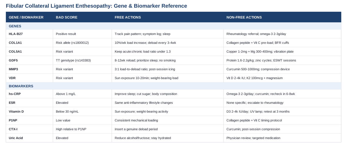
Fibular collateral ligament enthesopathy sits at an unusual intersection: part sports-medicine overuse injury, part potential window into systemic inflammatory disease. Six genes come up repeatedly in the research — from collagen-structure genes like COL1A1 and COL5A1 to the immune marker HLA-B27, which reframes a "stubborn ligament" as something worth a rheumatology conversation, not just more physical therapy. Alongside the genetics, six blood-based biomarkers — from hs-CRP to vitamin D to bone-collagen turnover markers repurposed for soft tissue — offer a cheaper, faster way to track what's actually happening in the tissue right now, without needing a genetic test at all.
The sections below break down what each gene and each biomarker actually indicates, what a reasonable low-cost first step looks like, and what a more aggressive supplement-or-equipment-based plan looks like if the simpler approach isn't enough. There's also a research-backed loading and nutrition protocol drawn from tendon physiology work that most people never hear about from a standard physical therapist, plus a look at which complementary therapies have genuine evidence behind them for lateral knee and enthesis-related pain — and which don't.
What Recent Genetic Research Suggests About Ligament Enthesis Health
Ligament and tendon genetics is a real, if young, field. Most of the strongest evidence comes from sports-genetics cohorts studying Achilles tendinopathy and ACL rupture rather than the fibular collateral ligament specifically, simply because those are the injuries large cohorts have been built around. The underlying biology — collagen fibril assembly, matrix remodeling, and inflammatory regulation at entheses — applies across ligament and tendon attachment sites, so the findings are reasonably transferable, with the caveat that direct fibular-collateral-ligament data is still sparse. One gene on this list, HLA-B27, is different in kind: it isn't about collagen structure at all, and its presence changes the entire framing of "why does this enthesis keep flaring."
HLA-B27: The Immune Marker That Reframes Everything
HLA-B27 is not a collagen gene. It codes for a cell-surface protein involved in presenting peptides to the immune system, and certain subtypes are strongly associated with the spondyloarthritis family of conditions — a group of diseases where enthesitis (inflammation at ligament and tendon attachment sites) is a defining feature rather than a side effect of overuse. In one detailed case series, enthesopathy was a prominent feature in roughly 75% of HLA-B27-associated spondyloarthritis cases presenting in childhood, and the pattern of attachment-site inflammation described there overlaps meaningfully with what shows up on imaging in adult lateral knee enthesopathy (HLA-B27-associated spondyloarthritis and enthesopathy, clinical and radiographic observations). More recent immunology work has clarified the likely mechanism: self-peptides displayed by HLA-B27 can resemble microbial peptides closely enough to trigger autoreactive T-cell activity at entheses (HLA-B27 and spondyloarthritis: at the crossroads of innate and adaptive immunity).
If the Gene Is Positive: The Plan Without Supplements
A positive result doesn't mean disease is present — plenty of carriers never develop spondyloarthritis — but it does mean a lateral knee enthesopathy that isn't resolving with standard rehab deserves a rheumatology referral rather than another round of physical therapy alone. Practically: track whether pain is worse with rest and improves with movement (an inflammatory pattern), watch for other enthesitis sites (Achilles insertion, plantar fascia, elbow), keep a symptom log covering morning stiffness duration, and bring it to the referral conversation. Sleep consistency and reducing ultra-processed food intake are reasonable, low-cost steps that support general inflammatory regulation while that workup happens.If the Gene Is Positive: The Plan With Supplements or Equipment
Omega-3 fatty acids (EPA/DHA, roughly 2–3g combined daily) have the best-supported anti-inflammatory supplement profile and are reasonable to run continuously, reassessing via hs-CRP every 3 months; watch for easy bruising or GI upset, and flag it to a physician if on blood thinners. Vitamin D optimization (see the biomarker section below) is worth pairing with this, since low vitamin D and inflammatory joint disease frequently travel together. Beyond that, this is a case where equipment matters less than escalation of care: a formal rheumatology workup, possibly including inflammatory markers and imaging, is the actual next step, not a longer supplement stack.COL1A1: Collagen Type I Structure and Stiffness
COL1A1 encodes the alpha-1 chain of type I collagen, the dominant structural protein in ligament tissue. A functional variant near the gene's Sp1 transcription factor binding site (rs1800012) alters how much of one collagen chain type gets produced relative to another, which affects tissue stiffness. A meta-analysis across nineteen case-control studies found this variant associated with altered risk of sports-related tendon and ligament injury, with one allele consistently trending protective (Association of COL1A1 polymorphism rs1800012 with tendon and ligament injuries).If the Gene Is Bad: The Plan Without Supplements
The practical implication of a less favorable variant is that the ligament may tolerate abrupt load spikes less well. The fix is unglamorous: structured load progression. Follow roughly a 10% weekly increase rule for any running, cutting, or loaded knee-flexion activity, and insist on a deload week every third or fourth week rather than pushing continuously. Isometric holds at the knee (wall sits, banded lateral holds) 3–4 times weekly build tolerance without the shear stress of pivoting sports.If the Score Is Bad: The Plan With Supplements or Equipment
A collagen-peptide-plus-vitamin-C loading protocol, taken roughly 45–60 minutes before targeted rehab loading sessions (detailed further in the podcast-derived protocol below), 3–4 times weekly in 8–12 week blocks, is the most evidence-aligned supplement approach here. Blood flow restriction (BFR) cuffs allow lower-load conditioning that still stimulates adaptation, useful during early rehab when full mechanical load isn't tolerated yet. Side effects are minimal at these doses; vitamin C above roughly 2g can cause GI upset, so stay near 1g per collagen dose.COL5A1: Fibril Assembly and Tissue Compliance
COL5A1 regulates how type I and type V collagen fibrils assemble together, effectively setting fibril diameter and, downstream, how compliant or stiff the tissue behaves under load. A meta-analysis of Caucasian cohorts found specific COL5A1 variants consistently associated with altered tendon-ligament injury risk, in both protective and risk-increasing directions depending on the specific polymorphism (COL5A1 gene polymorphisms and risk of tendon-ligament injuries: a meta-analysis).If the Gene Is Bad: The Plan Without Supplements
Because COL5A1 variants shift the injury-risk curve rather than guaranteeing injury, the highest-leverage move is monitoring training load ratios rather than avoiding load. Track acute:chronic workload (this week's training volume against the trailing 4-week average) and keep it under roughly 1.3 to avoid the spike-related injury zone. Add consistent mobility work for the hip and ankle, since compensatory stress at the knee often originates from stiffness elsewhere in the chain.If the Score Is Bad: The Plan With Supplements or Equipment
Copper and magnesium are cofactors for lysyl oxidase, the enzyme that crosslinks collagen fibrils — a deficiency in either can compound a less favorable COL5A1 profile. A reasonable approach is 1–2mg copper and 300–400mg magnesium daily, reassessed every 2–3 months, staying well under the 10mg/day copper upper limit. Vibration-plate or eccentric-loading equipment (a slant board for knee-flexion eccentric work) can be cycled in 6-week blocks; evidence for the vibration piece specifically is preliminary, so treat it as adjunct, not core.GDF5: Growth Signaling for Joint and Ligament Tissue
GDF5 (growth differentiation factor 5) regulates joint and connective tissue development and repair signaling. The rs143383 variant sits in the gene's 5' regulatory region and measurably lowers GDF5 expression in TT-genotype carriers, who show roughly double the risk of Achilles tendon pathology compared to C-allele carriers in case-control data (GDF5 rs143383 polymorphism and musculoskeletal degenerative disease susceptibility: a meta-analysis). Interestingly, this same variant hasn't shown a clear link to ACL rupture specifically, suggesting its influence is more about chronic overuse repair capacity than acute traumatic failure.If the Gene Is Bad: The Plan Without Supplements
Lower baseline repair signaling means rehab timelines should be planned generously rather than compressed. Give any lateral knee enthesopathy flare a minimum 8–12 week structured reload before judging whether an intervention worked. Prioritize sleep — deep sleep is when growth-hormone-driven tissue repair peaks — and eliminate smoking and heavy alcohol use, both of which independently blunt connective tissue repair signaling.If the Score Is Bad: The Plan With Supplements or Equipment
Adequate protein intake (1.6–2.2g/kg bodyweight daily) supplies the raw material that lagging growth signaling still needs to act on, and zinc (15–30mg daily, cycled 8 weeks on/4 off to avoid copper depletion) supports several of the same repair pathways. Extracorporeal shockwave therapy (ESWT), delivered in clinic, has decent evidence in tendinopathy for stimulating local repair response and is reasonable to consider after conservative measures stall — typically 3–6 weekly sessions. Expect transient soreness and bruising after each session; it's not a first-line step, but a legitimate escalation.MMP3: Matrix Remodeling and Degradation Balance
MMP3 encodes an enzyme that breaks down extracellular matrix components as part of normal tissue remodeling. Certain MMP3 variants shift this balance toward excess degradation, and research has found these variants associated with Achilles tendinopathy risk, with an interaction effect alongside COL5A1 genotype that either compounds or offsets the risk depending on which alleles are present together (Variants within the MMP3 gene are associated with Achilles tendinopathy: possible interaction with COL5A1).If the Gene Is Bad: The Plan Without Supplements
If matrix degradation tends to run ahead of matrix synthesis, the priority is avoiding degradation-dominant training patterns — heavy eccentric spikes without adequate recovery windows. Structure loading in a roughly 3:1 load-to-deload week ratio rather than continuous progressive overload, and use post-session icing or compression to blunt the acute inflammatory degradation phase after harder sessions.If the Score Is Bad: The Plan With Supplements or Equipment
Curcumin (500–1000mg daily with piperine for absorption, cycled 8 weeks on/2 off) has modest evidence for modulating inflammatory matrix breakdown; green tea catechin extracts have shown MMP-modulating activity in laboratory studies, though direct human ligament evidence is limited, so treat this as a lower-confidence add-on rather than a core piece. Pneumatic compression devices used post-training can mechanically support the resorption phase. Avoid stacking high-dose green tea extract with other liver-processed supplements, since case reports of hepatotoxicity exist at high cumulative doses.VDR: Vitamin D Receptor and Inflammatory Regulation
The vitamin D receptor gene doesn't just matter for bone; VDR signaling regulates inflammatory cytokine activity and matrix metalloproteinase expression in connective tissue generally. Research examining sports-related soft tissue injuries found VDR gene polymorphisms associated with altered inflammatory cytokine profiles (IL-1, IL-6, IL-10) and fracture and soft-tissue healing trajectories (VDR gene polymorphisms, inflammatory cytokines, and healing in sports-related injuries).If the Gene Is Bad: The Plan Without Supplements
Regular, moderate sun exposure (roughly 10–20 minutes midday, several times weekly, adjusted for skin tone and latitude) supports endogenous vitamin D production even when receptor sensitivity is reduced. Weight-bearing activity itself upregulates local VDR expression in connective tissue, so consistent loading is doing double duty here.If the Score Is Bad: The Plan With Supplements or Equipment
Because receptor sensitivity may be reduced, some people with unfavorable VDR variants need higher circulating 25-OH-D to get the same cellular effect — 2,000–4,000 IU daily is a reasonable starting dose, retested via blood work at 3 months and adjusted to a target serum level (detailed in the biomarker section). Pair with vitamin K2 (100mcg daily) and magnesium, since both are involved downstream of vitamin D metabolism. Watch for hypercalcemia with sustained high-dose supplementation (above 10,000 IU/day) — a periodic calcium check is a reasonable safeguard, not a reason to avoid supplementing at moderate doses.Genetics sets the ceiling on how a given ligament attachment responds to load and repairs itself, but it doesn't tell you what's happening in the tissue this month. That's where tracking a smaller set of blood-based biomarkers earns its place — faster to act on, cheaper to repeat, and useful even without ever running a genetic panel.
Biomarkers Worth Tracking for Ligament and Enthesis Health
None of these markers were designed specifically for ligament enthesopathy — most come from broader inflammatory or bone-metabolism research, borrowed here because type I collagen dominates both bone and ligament tissue and the inflammatory pathways overlap substantially. This is the kind of pragmatic, evidence-adjacent tracking approach favored by clinicians like Peter Attia and lipidologists like Thomas Dayspring and Allan Sniderman in the cardiometabolic space, applied here to a musculoskeletal question instead.
High-Sensitivity CRP (hs-CRP)
Elevated hs-CRP reflects systemic inflammation and has been specifically associated with tendon pathology — one study found hs-CRP above 1 mg/L improved diagnostic accuracy for tendon tearing when combined with other variables (High-sensitivity CRP and tearing of the long head of the biceps tendon). How to measure it: a standard blood draw, widely available, typically $15–$40 out of pocket or bundled into a metabolic panel. If the score is bad, the plan without supplements: address sleep debt, reduce ultra-processed food and added sugar, and manage body composition, all of which independently move hs-CRP. If the score is bad, the plan with supplements or equipment: omega-3s (2–3g EPA/DHA daily, continuous with quarterly recheck) and curcumin as described above; retest in 6–8 weeks rather than sooner, since hs-CRP moves slowly.Erythrocyte Sedimentation Rate (ESR)
ESR is a cruder, older inflammation marker than hs-CRP but remains clinically useful, particularly as a second data point when hs-CRP is borderline or when an inflammatory arthropathy is being ruled in or out. How to measure it: simple blood draw, usually $10–$25, often ordered alongside hs-CRP. If the score is bad, the plan without supplements: the same anti-inflammatory lifestyle levers as hs-CRP; persistent elevation despite lifestyle changes is a stronger signal to pursue the HLA-B27 and rheumatology pathway described earlier rather than to keep adjusting diet. If the score is bad, the plan with supplements or equipment: there isn't a specific supplement that reliably lowers ESR independent of treating the underlying driver — this marker's real value is in escalation decisions, not self-directed treatment.25-Hydroxyvitamin D
Vitamin D status affects tendon and ligament inflammation directly; research on rotator cuff injury has shown vitamin D's immunomodulatory role in tendon healing, decreasing matrix metalloproteinase activity that would otherwise degrade repair tissue (Vitamin D and the immunomodulation of rotator cuff injury). How to measure it: blood draw, $30–$60 uninsured, included in most comprehensive panels. If the score is bad, the plan without supplements: regular sun exposure and weight-bearing activity, as above. If the score is bad, the plan with supplements or equipment: 2,000–4,000 IU D3 daily, targeting a serum level of roughly 40–60 ng/mL, retested every 3 months until stable, then every 6–12 months; a UV lamp is a reasonable equipment substitute in low-sunlight climates, used per manufacturer exposure guidelines to avoid skin burns.P1NP (Procollagen Type I N-Terminal Propeptide)
P1NP reflects active type I collagen synthesis. It's a bone-formation marker by design, but since ligament is also predominantly type I collagen, it's used off-label by some sports-medicine researchers as a rough proxy for whole-body collagen-building activity, including the spike triggered by the loading-and-nutrition protocol described in the next section (Use of CTX-I and PINP as bone turnover markers: National Bone Health Alliance recommendations). How to measure it: specialty lab blood draw, $50–$120, not always covered by insurance for non-osteoporosis indications. If the score is bad (low), the plan without supplements: consistent mechanical loading is the primary synthesis stimulus — undertraining the ligament suppresses this marker as much as overtraining damages tissue. If the score is bad, the plan with supplements or equipment: the vitamin-C-plus-collagen-peptide timing protocol below is specifically designed to raise this marker; retest after 8–12 weeks of consistent protocol use.CTX-I (C-Terminal Telopeptide of Type I Collagen)
CTX-I is the resorption counterpart to P1NP — it rises when collagen is being broken down. Tracked alongside P1NP, it gives a rough sense of whether tissue turnover is net-building or net-breaking-down at a given point in a rehab cycle. How to measure it: specialty lab, similar cost range to P1NP, ideally drawn fasting and at a consistent time of day given known daytime variation. If the score is bad (persistently high relative to P1NP), the plan without supplements: insert a genuine deload period — this pattern often reflects accumulated under-recovered training rather than a single bad session. If the score is bad, the plan with supplements or equipment: the same anti-degradation levers described under MMP3 (curcumin, post-session compression) apply here; reassess the P1NP:CTX-I ratio rather than either marker in isolation.Uric Acid
Uric acid isn't a ligament-specific marker, but it earns a place on this list because crystal arthropathy (gout, pseudogout) can produce peri-articular pain and swelling near the fibular head that mimics a mechanical enthesopathy, and elevated uric acid is an easy, cheap way to rule that differential in or out. How to measure it: standard blood draw, $10–$20, included in most comprehensive metabolic panels. If the score is bad, the plan without supplements: reduce alcohol (particularly beer) and high-fructose beverage intake, and maintain hydration. If the score is bad, the plan with supplements or equipment: this is a case where a physician conversation about targeted medication (if genuinely elevated and symptomatic) is more appropriate than a supplement stack — self-treating suspected gout with anti-inflammatory supplements alone risks missing a straightforward diagnosis.Tracking a handful of these numbers over a few months tells you far more about where an enthesopathy actually stands than a single office visit can. But numbers alone don't tell you how to load the tissue day to day — for that, the more interesting recent work comes from tendon and ligament physiology research rather than clinical labs.
The Collagen-Loading Protocol That Changes How You Think About Rehab Timing
A recurring theme in tendon and ligament physiology research, popularized in long-form conversations like the Huberman Lab podcast episode featuring UC Davis researcher Dr. Keith Baar, is that connective tissue responds to very specific timing cues that most standard physical therapy protocols never mention. The core science behind this comes from a controlled human trial, and it's worth understanding in detail because it directly informs how to apply several of the supplement plans above.
1. Tendons and Ligaments Adapt on a Much Slower Clock Than Muscle
Muscle protein turns over in days; collagen-based tissue turns over in months. This is the single biggest reason generic "give it six weeks" rehab timelines under-deliver for ligament issues specifically — the tissue frequently hasn't finished remodeling yet when a course of PT ends.2. There's a Real, Measurable Window to Spike Collagen Synthesis
A randomized, double-blind crossover trial found that consuming vitamin-C-enriched gelatin about an hour before a short bout of intermittent activity measurably increased collagen synthesis markers compared to placebo (Vitamin C-enriched gelatin supplementation before intermittent activity augments collagen synthesis). The timing, not just the ingredient, was the active variable.3. The Loading Stimulus Doesn't Need to Be Large
In that trial, just six minutes of jump-rope-style intermittent activity was enough to trigger the synthesis response when paired with the supplement timing — a useful reminder that the mechanical signal required is a trigger, not a full workout.4. Dose-Response Is Real for Collagen Peptides
More recent work found the collagen synthesis response to acute resistance exercise was significantly greater with 30g of hydrolyzed collagen compared to 15g or 0g in resistance-trained young men (Collagen synthesis response to resistance exercise: dose comparison of hydrolyzed collagen), suggesting the 15g dose commonly recommended may be a floor rather than an optimum for some people.5. Chronic Anti-Inflammatory Use Can Blunt the Adaptation Signal
The inflammatory cascade after loading is part of the signal that drives remodeling. Leaning on NSAIDs continuously around every training session, rather than for acute flare management, risks dulling the exact process the loading protocol is trying to stimulate.6. Complete Rest Is Frequently the Wrong Default
Baar's research consistently pushes back against total immobilization for enthesis-related pain, favoring controlled, progressive loading. Entheses respond to mechanical signal; removing it entirely doesn't protect the tissue so much as let it deteriorate quietly.7. Heat and Local Blood Flow Before Loading May Matter
Baar's working hypothesis, based on tissue-level physiology rather than a specific large human trial, is that warming the tissue before loading (a short warm-up, heat pack, or light aerobic activity) improves collagen's mechanical compliance and reduces injury risk during the loading window — a plausible mechanism worth applying practically, even without a definitive RCT behind the specific claim.8. Sleep Quality Is a Structural Variable, Not Just a Recovery Nicety
Growth hormone release during deep sleep supports the same collagen synthesis pathways this protocol is trying to activate through nutrition and timing. Undermining sleep quietly undercuts the rest of the plan.9. Consistency Beats Intensity
Because collagen turnover is slow, a modest protocol run consistently for 8–12 weeks outperforms an intense but inconsistent one. This is the practical reason the plans throughout this article are framed in cycles and blocks rather than single interventions.10. Genetics Set the Ceiling, Timing and Nutrition Determine How Close You Get to It
None of this protocol overrides an unfavorable COL1A1, COL5A1, or GDF5 variant — but it does determine whether that genetic ceiling is actually reached. This is the throughline connecting the genetics section, the biomarker section, and this protocol: genes set potential, tracked biomarkers show current status, and timed loading-and-nutrition protocols close the gap between them.Nutrition and loading timing address the biological side of recovery. A few complementary, non-invasive approaches also have real evidence behind them for lateral knee pain and enthesis-adjacent conditions, and are worth knowing which ones actually hold up.
Complementary Approaches With Real Evidence Behind Them
Low-Level Laser Therapy (Photobiomodulation)
Low-level laser therapy (LLLT), also called photobiomodulation, uses specific wavelengths of red or near-infrared light believed to influence mitochondrial activity and local inflammatory signaling in tendon and ligament tissue. For a fibular collateral ligament enthesopathy, the appeal is that it's non-invasive, can be applied directly over the fibular head insertion, and doesn't compete with a concurrent loading program the way some other passive modalities might.A systematic review and meta-analysis of randomized controlled trials found that low-level red and near-infrared photobiomodulation improved pain and function outcomes in tendinopathy, though the same review was candid that high-quality evidence supporting its routine use is still limited (Low-level red and near-infrared photobiomodulation for pain and function in tendinopathy: systematic review and meta-analysis).
Realistically, LLLT is best positioned as an adjunct rather than a standalone treatment — used 2–3 times weekly for 4–6 weeks alongside, not instead of, a structured loading program, and ideally administered by a clinician familiar with correct dosimetry, since underdosing is a common reason home devices underperform in practice.
Massage Therapy (Deep Transverse Friction)
Deep transverse friction massage, developed by Dr. James Cyriax, applies focused, perpendicular pressure across the fiber direction of a tendon or ligament, intended to prevent disorganized fibrous adhesions and encourage more normally aligned scar tissue during healing. It's one of the few manual therapy techniques studied specifically at lateral knee tendinitis, which is anatomically adjacent to where fibular collateral ligament enthesopathy presents.A Cochrane systematic review looking specifically at lateral elbow and lateral knee tendinitis found the existing trial evidence too limited to confirm a clear benefit over other physical therapy interventions (Deep transverse friction massage for treating lateral elbow or lateral knee tendinitis: Cochrane review) — worth stating plainly rather than overselling.
Given that honest evidence gap, deep transverse friction is reasonable as a short trial (2–3 sessions weekly for 3–4 weeks) alongside active rehab, not as a substitute for it, and should be stopped if it increases rather than reduces local irritation.
Biofeedback-Assisted Gait Retraining
Real-time biofeedback devices measure joint loading or movement patterns during walking or running and give the user immediate feedback to consciously adjust their gait. Since fibular collateral ligament stress is heavily influenced by frontal-plane knee mechanics (varus loading, in particular), correcting the movement pattern that's loading the ligament excessively can matter as much as anything applied directly to the tissue.Research using real-time estimation of tibiofemoral joint contact forces has demonstrated that biofeedback can meaningfully and immediately alter knee loading patterns during gait (Biofeedback for gait retraining based on real-time estimation of tibiofemoral joint contact forces), and broader systematic review evidence supports gait retraining generally for knee-loading conditions.
A practical version of this doesn't require a research lab: many physical therapy clinics now offer wearable sensor-based gait feedback, typically run over 6–8 sessions across several weeks, with the goal of a lasting movement-pattern change rather than an ongoing dependency on the device.
Tai Chi for Knee Proprioception
Tai chi's slow, controlled, weight-shifting movements place sustained demand on knee joint position sense — the body's ability to sense and correct knee alignment without visual feedback. Since ligaments (including the fibular collateral ligament) function partly as proprioceptive sensors, not just mechanical restraints, improving this system can reduce the repetitive micro-loading that drives an enthesopathy to flare.A pilot randomized controlled trial in knee osteoarthritis found that a 12-week, twice-weekly tai chi program measurably improved knee proprioception compared to an attention-control stretching and education group (The influence of tai chi exercise on proprioception in patients with knee osteoarthritis: pilot randomized controlled trial), with a related trial replicating improved joint position sense at 24 weeks of practice.
For a lateral knee enthesopathy, a similar twice-weekly, 45–60 minute practice sustained over 12 or more weeks is a reasonable target — this is a slow-adaptation intervention, not one to judge after a handful of sessions.
Conclusion
A fibular collateral ligament enthesopathy that isn't responding to standard care is usually telling you something specific, not just being stubborn. Collagen-structure genes like COL1A1, COL5A1, and GDF5 shape how the tissue tolerates and repairs from load. MMP3 and VDR variants shift the balance between building and breaking down matrix. And HLA-B27 changes the entire diagnostic picture, from a mechanical overuse problem to a potential systemic inflammatory one worth a rheumatology conversation. Tracked alongside that, hs-CRP, vitamin D, and collagen turnover markers give a faster, cheaper read on what's happening in the tissue right now, without waiting on genetic testing.
None of this replaces a clinical evaluation, and none of it promises a fixed timeline — connective tissue changes slowly, and the honest expectation is measured in months, not weeks. The realistic next step is straightforward: get a baseline set of the biomarkers above drawn, discuss whether the pattern of symptoms and their response to rest versus activity warrants an HLA-B27 check, and build a loading plan with a clinician who's willing to work in structured blocks rather than open-ended rest. That combination of information and patience is what actually moves a stubborn enthesopathy forward.
Musculoskeletal: Bone Conditions Joint Conditions