This article contains AI generated content.
· UpdatedFibular Head Bursitis – 5 Genes And 6 Biomarkers To Track
Introduction
If you have been dealing with pain, swelling, or tenderness on the outer side of your knee — right over that bony prominence where the fibula meets the knee — you already know how surprisingly disruptive this condition can be. Fibular head bursitis is the inflammation of the small fluid-filled sac that cushions the area between the fibular head, the biceps femoris tendon, and the surrounding tissue. It is not the most commonly discussed joint condition, which means most people with it end up receiving the same recycled advice: rest, ice, anti-inflammatories, and wait.
That approach works for some people, some of the time. But for others, the inflammation persists, returns after periods of improvement, or shows up alongside other joint problems. When that happens, the standard explanation — you irritated the bursa — does not tell you why your body is struggling to resolve it, or why someone else doing the same activity bounces back in days while you are still dealing with it weeks later.
This article takes a different approach. It looks at the biological inputs that shape how your body creates and resolves inflammation: the measurable markers in your blood that signal what is actually happening in your tissue, and the genetic variants that can quietly tip your immune response toward chronic inflammation. Neither of these is a cure, but both give you something actionable — specific numbers to track, specific levers to pull.
Two practical angles are covered here. The first focuses on six key biomarkers you can measure today to understand the inflammatory drivers behind your condition. The second examines five genes that research suggests may influence tissue healing, collagen stability, and inflammatory regulation — each with a concrete plan. Further on, you will also find a summary of one of the most relevant podcasts on joint inflammation and recovery, along with complementary physical approaches that have human clinical support.
Summary
This article covers six blood biomarkers — hsCRP, IL-6, uric acid, 25-OH vitamin D, the AA:EPA ratio, and homocysteine — chosen for their direct relevance to how fibular head bursitis develops and persists. For each, you will find how to measure it, what a problematic result looks like, and a concrete improvement plan both with and without supplementation. On the genetics side, five key gene variants are explored — IL6, TNF, COL1A1, MTHFR, and VDR — with protocols tailored to each variant. Further sections cover what the Andrew Huberman Lab podcast series with Dr. Andy Galpin reveals about inflammation and tissue recovery, and three complementary modalities with clinical support for musculoskeletal inflammation. The article closes with a practical action plan to start today.
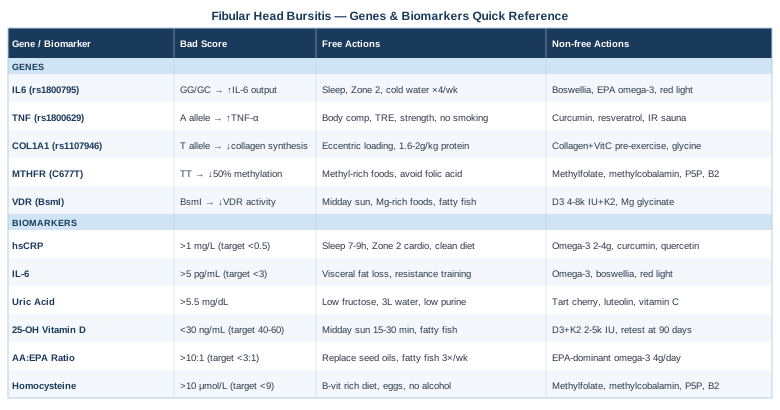
6 Biomarkers That Can Reveal the Root Causes of Your Fibular Head Bursitis
Biomarkers do not tell you that you have fibular head bursitis — your pain already does that. What they can reveal is why your inflammatory response is not resolving efficiently, whether a systemic inflammatory process is feeding local tissue irritation, and which nutritional or metabolic deficiencies may be quietly undermining your recovery. The six below are chosen for practical availability, cost-effectiveness, and direct relevance to bursitis physiology.
Biomarker 1: High-Sensitivity C-Reactive Protein (hsCRP)
Why it matters: hsCRP is produced by the liver in response to cytokine signaling, particularly IL-6. It is one of the most reliable available markers of systemic low-grade inflammation. In people with bursitis that keeps returning or fails to fully resolve, elevated hsCRP often indicates that the body is in a persistently pro-inflammatory state — making even minor mechanical triggers sufficient to re-ignite local inflammation.
Optimal range and what a bad result reveals: Peter Attia and most longevity-oriented clinicians target hsCRP below 0.5 mg/L. Values between 1 and 3 mg/L indicate moderate systemic inflammation; above 3 mg/L indicates a meaningful inflammatory burden outside of acute infection. In the context of chronic bursitis, a result above 1 mg/L is worth addressing actively.
How to measure it: A standard blood panel with the high-sensitivity version (not standard CRP) through any primary care physician or direct-to-consumer lab. Cost: $15–$40 USD depending on the lab. It should be measured in a resting, non-infected state — avoid testing during a cold or acute illness.
If the score is bad — the plan without supplements: The strongest lifestyle levers for lowering hsCRP are sleep quality (aim for 7–9 hours with consistent timing), eliminating ultra-processed seed oils from the diet (replacing with olive oil and animal fats), reducing refined carbohydrate load, time-restricted eating, and zone 2 cardiovascular exercise four to five times per week at 150–180 minutes total. Even modest weight loss in those with visceral adiposity can reduce hsCRP by 30–40% within 12 weeks.
If the score is bad — the plan with supplements or equipment: Omega-3 fatty acids (EPA+DHA, 2–4g per day with the largest meal, continuous use, monitor for mild GI upset or bruising at the high end). Curcumin with piperine (500–1000mg standardized curcumin extract daily with food; cycle 8 weeks on, 2–4 weeks off; rare GI side effects; avoid with blood thinners at high doses). Magnesium glycinate (300–400mg at night, continuous; supports systemic inflammation reduction; very well tolerated). Quercetin (500–1000mg daily with food; cycle similarly to curcumin; generally well tolerated).
The landmark JUPITER trial (Ridker et al., 2008, NEJM) established hsCRP as a primary clinical target for inflammatory risk, separate from lipid levels — a paradigm shift worth understanding if your hsCRP is persistently elevated.
Biomarker 2: Interleukin-6 (IL-6)
Why it matters: IL-6 is a key pro-inflammatory cytokine that directly drives the acute phase response, stimulates CRP production, and plays a central role in synovial and bursal inflammation. When the bursa around the fibular head is chronically irritated, local immune cells release IL-6, which then enters systemic circulation. Elevated serum IL-6 in the absence of acute infection suggests either persistent local inflammation, visceral adiposity (fat tissue is a major IL-6 source), or an overactive immune regulatory pattern.
Optimal range and what a bad result reveals: Normal fasting IL-6 is typically below 2.0–3.0 pg/mL. Values above 5 pg/mL in non-acute states are increasingly associated with musculoskeletal inflammation chronicity and impaired tissue healing.
How to measure it: Serum IL-6 can be ordered through integrative medicine physicians or direct-to-consumer services. It is not always on standard panels and may cost $50–$120 USD. It requires careful sample handling — inform your lab you need the high-sensitivity cytokine assay.
If the score is bad — the plan without supplements: Visceral fat reduction is the single most impactful non-pharmacological intervention for systemic IL-6, as adipose tissue is a direct IL-6 secretor. Resistance training (three sessions per week, compound movements) reduces IL-6 levels independently of weight change. Cold water immersion (cold shower or 10–15 minutes in 15°C water, three to four times per week) has shown consistent evidence for downregulating pro-inflammatory cytokines in recovery contexts.
If the score is bad — the plan with supplements or equipment: EPA-rich omega-3 supplementation (same protocol as for hsCRP) specifically downregulates IL-6 production. Boswellia serrata (AKBA-standardized extract, 100–200mg AKBA content daily with food; 12-week cycle then reassess; very low side effect profile; do not combine with anticoagulants). A red light therapy panel (660nm and 850nm wavelengths, 10–20 minutes over the affected knee area daily; evidence for reducing local IL-6 production in periarticular tissue; no side effects at standard exposure).
Biomarker 3: Uric Acid
Why it matters: Uric acid is often associated only with gout, but elevated serum uric acid — even at sub-gout levels — contributes to crystalline microdeposition in periarticular tissues including bursae. These microcrystals act as persistent irritants, triggering neutrophil-driven inflammation. Fibular head bursitis has been documented in association with crystal-induced inflammatory arthropathies, and checking uric acid in cases of recurrent or disproportionately painful bursitis is clinically meaningful.
Optimal range and what a bad result reveals: While conventional labs flag uric acid only above 7.0 mg/dL (males) or 6.0 mg/dL (females), many functional clinicians consider above 5.5 mg/dL worth addressing in someone with active joint inflammation. Values in the 6–7 mg/dL range with recurrent bursitis warrant further evaluation including synovial fluid analysis if possible.
How to measure it: Part of a standard comprehensive metabolic panel. Cost: included in most routine blood work, or $10–$25 standalone. Fasting is recommended for best accuracy.
If the score is bad — the plan without supplements: Reduce fructose significantly — high-fructose corn syrup and alcohol are the primary dietary drivers of uric acid elevation. Adequate hydration (minimum 2–3 liters of water daily) actively promotes uric acid renal clearance. Limiting organ meats and high-purine shellfish (sardines, anchovies, mussels) in the context of already elevated levels is reasonable. These changes can reduce uric acid by 0.5–1.5 mg/dL within four to six weeks.
If the score is bad — the plan with supplements or equipment: Tart cherry extract (480mg anthocyanin-equivalent daily, continuous use; well-documented for uric acid reduction and anti-inflammatory effects; occasional GI sensitivity). Luteolin-rich extracts (from artichoke leaf or chamomile, 300–500mg daily, 8-week cycles). Vitamin C at modest doses (500–1000mg daily) has shown modest uric acid-lowering effects in short-term trials and is very low risk. Higher dose supplementation requires medical supervision due to potential urate-calcium oxalate interactions.
Biomarker 4: 25-OH Vitamin D
Why it matters: Vitamin D is not just a bone mineral — it functions as an immune modulator, regulating inflammatory gene expression in macrophages and synovial cells. Deficiency is associated with impaired tissue healing, increased inflammatory cytokine production, and musculoskeletal pain syndromes. Research suggests that the majority of patients with persistent musculoskeletal pain, including periarticular conditions, are vitamin D deficient.
Optimal range: Most integrative and longevity-oriented clinicians (Attia, Rhonda Patrick) target 40–60 ng/mL. Values below 30 ng/mL indicate deficiency by functional standards; below 20 ng/mL is classified as severe deficiency by most labs.
How to measure it: A 25-OH vitamin D serum test, available with any blood panel. Cost: $30–$75 standalone. Test twice yearly — once in late winter, once in early autumn — to understand your seasonal pattern.
A 2003 study (Plotnikoff & Quigley, Mayo Clinic Proceedings) found that the vast majority of patients presenting with persistent, nonspecific musculoskeletal pain had severe vitamin D deficiency — a finding with direct relevance to bursitis that does not resolve normally.
If the score is bad — the plan without supplements: Midday sun exposure on large skin surface areas (arms, legs, back) for 15–30 minutes depending on skin tone and latitude. This alone can raise vitamin D by 3–10 ng/mL per week in optimal conditions. Dietary sources (fatty fish, egg yolks, beef liver) contribute modestly but rarely enough to correct deficiency alone.
If the score is bad — the plan with supplements or equipment: Vitamin D3 with K2 (2000–5000 IU D3 with 100–200mcg MK-7 K2 daily; take with the fattiest meal of the day; continuous use is generally safe at these doses with regular monitoring; K2 is essential to prevent arterial calcification at higher D3 doses). Retest after 90 days. Some individuals with the VDR gene variant (discussed in the genetics section) require higher doses — up to 8000 IU — to achieve the same serum level; this should be supervised by a physician.
Biomarker 5: Arachidonic Acid to EPA Ratio (AA:EPA)
Why it matters: The ratio between arachidonic acid (AA), the primary pro-inflammatory omega-6 fatty acid, and eicosapentaenoic acid (EPA), the key anti-inflammatory omega-3, directly determines whether your cellular environment tilts toward inflammation resolution or inflammation amplification. Prostaglandins and leukotrienes derived from AA drive the inflammatory cascade in bursitis. An unfavorable AA:EPA ratio — common in Western dietary patterns — creates a biochemical background that predisposes bursal tissue to prolonged inflammation after any mechanical trigger.
Optimal range: An AA:EPA ratio below 3:1 is considered anti-inflammatory by functional medicine standards. The typical Western diet produces ratios of 20:1 to 25:1. Many inflammatory conditions correlate with ratios above 10:1.
How to measure it: The OmegaCheck or Omega-3 Index test (Omega Quant, Cleveland HeartLab) measures full fatty acid profile including AA:EPA ratio. Cost: $60–$110 direct-to-consumer. A simple Omega-3 Index test (measures EPA+DHA as percentage of red blood cell fatty acids) is a related and cheaper option ($40–$80).
If the score is bad — the plan without supplements: Dietary fat quality is the primary lever. Replacing seed oils (soybean, sunflower, corn, canola) with olive oil, avocado oil, and butter/ghee significantly reduces AA substrate. Eating two to three servings of fatty cold-water fish per week (salmon, mackerel, sardines) raises EPA. Results take 8–12 weeks to appear in red blood cell measurements.
If the score is bad — the plan with supplements or equipment: EPA-dominant omega-3 supplementation (4g total EPA+DHA per day with at least 60% EPA fraction; with meals; continuous; monitor for slight blood-thinning effect at high doses; avoid with prescription anticoagulants without physician guidance). Reducing linoleic acid intake (seed oil elimination is the most powerful dietary intervention here) within 8–12 weeks can measurably shift the AA:EPA ratio even without supplementation.
Biomarker 6: Homocysteine
Why it matters: Homocysteine is a sulfur-containing amino acid produced during methionine metabolism. Elevated homocysteine is associated with chronic low-grade vascular and tissue inflammation, impaired collagen cross-linking, and reduced tissue repair capacity. In the context of musculoskeletal health, high homocysteine compromises the structural integrity of connective tissue — including the periarticular structures surrounding the fibular head — and amplifies inflammatory signaling in soft tissue. It is particularly relevant for people with the MTHFR gene variant (covered in the next section).
Optimal range: Functional medicine practitioners, including those who follow Thomas Dayspring's cardiovascular risk approach, target homocysteine below 8–9 µmol/L. Conventional labs flag only above 15 µmol/L, but evidence suggests risk increases meaningfully above 10 µmol/L for tissue health outcomes.
How to measure it: Serum homocysteine is available on most comprehensive blood panels or as a standalone test. Cost: $25–$60. Fasting is recommended.
If the score is bad — the plan without supplements: Dietary strategies center on adequate protein quality and B-vitamin richness: eggs, leafy greens, legumes, liver, and fish. Reducing alcohol, which inhibits B12 absorption, is important. Ensuring adequate methionine cycling through protein adequacy is also relevant.
If the score is bad — the plan with supplements or equipment: The methylated B-vitamin trio is the most evidence-supported intervention: methylfolate (400–800mcg 5-MTHF daily), methylcobalamin (500–1000mcg B12 daily), and pyridoxal-5-phosphate (P5P) (25–50mg B6 daily). These are all continuous, with very good safety profiles at these doses. Avoid folic acid (non-methylated) if you carry an MTHFR variant, as it can paradoxically worsen methylation. Riboflavin (B2, 100–400mg) is also meaningful for homocysteine in MTHFR carriers. Reassess homocysteine in 8–12 weeks.
Moving from what the body is doing now to what your genes may predispose you toward opens a related but distinct line of inquiry — one that helps explain why two people with similar diets and activity levels can have very different inflammatory tendencies.
What Recent Genetics Research Suggests About Fibular Head Bursitis
Genetics does not determine destiny in inflammatory conditions — but it does set the playing field. Certain gene variants alter how aggressively your immune system responds to tissue injury, how efficiently your body builds and repairs collagen, and how well you process key anti-inflammatory nutrients. Understanding these variants allows for targeted interventions rather than generic lifestyle advice. The following five genes have meaningful human evidence supporting their relevance to musculoskeletal inflammation and tissue repair. Note: early-stage or animal-only evidence is flagged where applicable.
The most practical way to access your personal variants is through a direct-to-consumer genotyping service (23andMe, AncestryDNA) and then run the raw data through a nutrigenomics or health interpretation platform. Clinically validated panels are also available through functional medicine practitioners.
Gene 1: IL6 (rs1800795)
What it does: The IL6 gene encodes interleukin-6, a central inflammatory cytokine. The rs1800795 variant (C/G polymorphism in the IL6 promoter region) influences baseline IL-6 transcription. The GG genotype is associated with higher IL-6 production, while CC is associated with lower. Individuals with the GG or GC genotype may produce stronger and more sustained IL-6 responses to tissue injury, which can translate to more persistent bursal inflammation after mechanical insult.
Evidence status: Multiple human association studies; relevant within the broader context of inflammatory gene panels.
If the gene is bad — the plan without supplements: Prioritize all four inflammatory lifestyle pillars: sleep quality, zone 2 aerobic training (which reduces systemic IL-6 at rest despite transiently raising it during exercise), dietary seed oil elimination, and active stress reduction. Cold water immersion (three to four times per week, 10–15 minutes at 15°C or cooler) has consistent evidence for post-exercise cytokine downregulation and is particularly relevant for IL-6 modulation in carriers of the high-expression variant.
If the score is bad — the plan with supplements or equipment: Boswellia serrata (100–200mg AKBA standardized, with food, 12-week cycle, then reassess; direct evidence for IL-6 pathway inhibition). EPA-rich omega-3 supplementation at the higher end of the range (3–4g EPA+DHA daily). Red or near-infrared light therapy over the affected knee joint (660nm/850nm, 15–20 minutes daily) — evidence for local reduction in pro-inflammatory cytokines in periarticular tissue.
Gene 2: TNF (rs1800629)
What it does: TNF encodes tumor necrosis factor-alpha, a master regulator of the inflammatory cascade. The rs1800629 A allele (A/G variant in the TNF promoter) is associated with elevated TNF-alpha production and has been linked in multiple studies to inflammatory arthropathies, tendinopathies, and bursitis-type conditions. Carriers of the A allele tend to have more robust pro-inflammatory responses to mechanical tissue stress, with longer resolution times.
Evidence status: Well-documented in human association studies for inflammatory joint conditions. Considered by researchers including those studying Ali Torkamani's work on inflammatory genetic profiling.
If the gene is bad — the plan without supplements: The most impactful behavioral intervention for TNF-alpha modulation is body composition management — visceral adipose tissue is a primary TNF-alpha source. A time-restricted eating window (8–10 hours) combined with strength training three times per week has direct evidence for reducing TNF-alpha production. Avoiding smoking and minimizing alcohol exposure is critical, as both significantly amplify TNF-driven inflammatory signaling.
If the gene is bad — the plan with supplements or equipment: Curcumin (standardized BCM-95 or phytosome form, 500–1000mg daily with food; 8 weeks on, 2–4 weeks off; avoid with anticoagulants). Resveratrol (250–500mg daily; cycle 8 weeks on, 4 weeks off; mild GI sensitivity possible; generally well-tolerated). Quercetin (500mg twice daily with food; cycle 8 weeks; synergistic with resveratrol and curcumin in TNF-pathway modulation). Infrared sauna (3–4 sessions per week, 20–30 minutes at 55–70°C; consistent evidence for TNF-alpha reduction with regular use; contraindicated in active acute inflammation).
Gene 3: COL1A1 (rs1107946)
What it does: COL1A1 encodes the alpha-1 chain of type I collagen — the primary structural protein in tendons, ligaments, bursae, and periarticular connective tissue. The rs1107946 variant (T allele in the promoter) is associated with reduced collagen type I synthesis and has been linked to increased susceptibility to soft tissue injury, slower tendon/bursa healing, and higher recurrence rates in bursitis and tendinopathy. This is a particularly relevant gene for fibular head bursitis, given the structural involvement of the biceps femoris tendon and bursa.
Evidence status: Human studies, particularly in sports medicine research on connective tissue injuries.
If the gene is bad — the plan without supplements: Loading protocols that stimulate collagen synthesis are central: progressive eccentric exercise targeting the biceps femoris and knee flexor chain (3 sets of eccentric contractions, three times per week; low load, slow tempo). This mechanical loading is the primary stimulus for collagen gene expression in periarticular tissue. Adequate dietary protein (1.6–2.0g per kg body weight per day) provides the amino acid substrate — glycine and proline in particular.
If the gene is bad — the plan with supplements or equipment: Hydrolyzed collagen peptides (10–15g daily; take 30–60 minutes before exercise with vitamin C 50–150mg to maximize collagen synthesis signaling; continuous; very well-tolerated; minimal side effects). Vitamin C (50–150mg alongside collagen supplementation; do not exceed 1000mg chronically without reason, as very high doses may impair collagen crosslinking). Glycine supplementation (3–5g at night; collagen is 1/3 glycine by weight; supports both tissue repair and sleep quality; safe for long-term use). Silicon from orthosilicic acid formulations — emerging but promising evidence for collagen gene expression stimulation in periarticular tissues.
Gene 4: MTHFR (rs1801133, C677T)
What it does: MTHFR encodes methylenetetrahydrofolate reductase, an enzyme critical for converting folate to its active methylated form. The C677T variant — especially in the TT homozygous form — reduces enzyme efficiency by 50–70%, impairing methylation and leading to elevated homocysteine. As described in the biomarker section, elevated homocysteine compromises connective tissue integrity and amplifies chronic inflammatory signaling in soft tissue. Gary Brecka, who focuses heavily on methylation genetics, considers MTHFR one of the highest-impact genetic variants for systemic inflammation and tissue health.
Evidence status: MTHFR variants are among the most extensively studied genetic polymorphisms in humans; homocysteine-raising effects are well-established; the downstream tissue implications are supported by strong epidemiological evidence.
If the gene is bad — the plan without supplements: Dietary methyl donors become critical: eggs (choline), leafy greens (natural folate), beets (betaine), and organ meats (B12, folate, riboflavin). Avoiding synthetic folic acid — which competes with natural folate at enzyme sites and is poorly converted in TT carriers — is important; read all fortified food labels. Alcohol elimination is particularly impactful in MTHFR carriers, as alcohol directly inhibits methylation pathways.
If the gene is bad — the plan with supplements or equipment: Methylfolate (5-MTHF, 400–1000mcg daily; use the methylated form only — not folic acid; continuous; rare activation-type side effects possible in very sensitive individuals — start low). Methylcobalamin (1000mcg B12 daily; methylated form, sublingual preferred). P5P (pyridoxal-5-phosphate, 25–50mg B6 daily; important cofactor in the methylation-homocysteine pathway). Riboflavin B2 (100–200mg daily; specifically reduces homocysteine in TT MTHFR carriers; well-tolerated). TMG (trimethylglycine/betaine, 1–2g daily with food; alternative methylation donor pathway; very well-tolerated). Reassess serum homocysteine at 8–12 weeks.
Gene 5: VDR (rs1544410, BsmI)
What it does: VDR encodes the vitamin D receptor, which mediates virtually all genomic effects of vitamin D including immune modulation, inflammatory gene suppression, and musculoskeletal maintenance. The BsmI variant (rs1544410) affects VDR activity and has been associated in multiple studies with altered inflammatory responses, increased susceptibility to musculoskeletal conditions, and reduced vitamin D effectiveness per unit of serum concentration. In practical terms: carriers may need substantially higher serum 25-OH vitamin D levels to achieve the same anti-inflammatory effect as non-carriers.
Evidence status: Well-supported in human studies across musculoskeletal and immune contexts; included in most nutrigenomic clinical panels.
If the gene is bad — the plan without supplements: Maximize endogenous vitamin D synthesis through regular midday sun exposure. Additionally, magnesium adequacy becomes even more important for VDR-variant carriers, as magnesium is required for both vitamin D conversion and receptor signaling. Dietary vitamin D sources (fatty fish, egg yolks, beef liver) should be maximized. Reducing gut permeability — through elimination of processed foods and focus on fiber and fermented foods — improves fat-soluble vitamin absorption.
If the gene is bad — the plan with supplements or equipment: Higher-dose vitamin D3 may be required: 4000–8000 IU daily (always with MK-7 K2, 200mcg; with the fattiest meal; monitor 25-OH vitamin D every 3 months until stable; target 50–70 ng/mL rather than the standard 40–60 ng/mL). Magnesium glycinate (400mg at night; required cofactor for vitamin D metabolism; very well-tolerated). Consider a vitamin D lamp (UVB narrowband, 311nm) for high-latitude winter months — a practical alternative to supplementation that activates the D3 synthesis pathway naturally.
Alongside these targeted biological insights, there is a broader framework worth understanding — one that ties together movement, inflammation, and tissue recovery in a practical, research-grounded way.
The Andrew Huberman Podcast That May Change How You Think About Joint Inflammation
The Huberman Lab podcast's multi-part series with Dr. Andy Galpin — exercise scientist at Cal State Fullerton — is one of the most clinically dense and actionable resources on tissue repair, recovery, and inflammatory management in the public domain. The series spans several episodes and draws on dozens of peer-reviewed studies. For someone dealing with a persistent inflammatory joint condition like fibular head bursitis, the concepts below are among the most impactful.
1. Inflammation Is Not the Enemy — But Chronic Inflammation Is
Acute inflammation after tissue injury is necessary and purposeful — it clears debris and signals repair. The problem is when the inflammatory response fails to resolve. Many people with persistent bursitis are not in an acute repair state; they are stuck in low-grade chronic inflammation. The goal is not to eliminate inflammation but to ensure it resolves fully.
2. Sleep Is the Most Potent Recovery Intervention
Both Huberman and Galpin emphasize that the majority of connective tissue repair and anti-inflammatory cytokine release (including IL-10 and TGF-beta) occurs during slow-wave sleep. Consistently sleeping fewer than 7 hours — or having fragmented sleep — measurably increases circulating CRP, IL-6, and impairs tissue healing rates. Addressing sleep quality is not optional for bursitis recovery.
3. The Inflammatory Window After Exercise Matters
Taking NSAIDs or high-dose antioxidants immediately after therapeutic exercise blunts the inflammatory signal required to stimulate collagen synthesis and tissue remodeling. If you are doing loading exercise for connective tissue rehabilitation, avoid anti-inflammatory supplements within two hours post-exercise.
4. Zone 2 Cardio Is Anti-Inflammatory at Rest — Even Though It Raises IL-6 Acutely
Four to five weekly sessions of sustained low-intensity aerobic exercise (where you can hold a conversation but are mildly breathless) reduces resting IL-6, CRP, and TNF-alpha substantially over time, despite transiently raising IL-6 during the session itself. This training effect is one of the most robust anti-inflammatory interventions available without medication.
5. Cold Exposure Timing Determines Whether It Helps or Hurts
Cold water immersion immediately after strength training inhibits hypertrophy and collagen synthesis (by blunting the necessary inflammatory signal). But cold exposure at least six hours after training, or on rest days, consistently reduces inflammatory markers and accelerates systemic recovery without the trade-off. For bursitis specifically: local ice application within 15 minutes of injury or irritation is appropriate; routine cold immersion is better scheduled away from therapeutic exercise sessions.
6. The Protein Timing and Type for Connective Tissue Is Different From Muscle
Muscle protein synthesis peaks within two hours of exercise with any complete protein source. Collagen synthesis, however, is best stimulated by gelatin- or collagen-peptide-based protein taken 45–60 minutes before the loading session, alongside vitamin C. This window-based approach is not intuitive but has emerging clinical support.
7. Respiratory Pattern Directly Modulates Inflammatory State
Huberman discusses how chronic mouth breathing and upper chest breathing patterns maintain a low-grade autonomic activation that keeps pro-inflammatory cytokines elevated. Nasal breathing, particularly extended exhalation-focused breathing (box breathing or 4-7-8 patterns), activates the parasympathetic nervous system and has shown measurable effects on IL-6 and CRP in short-term trials.
8. Eccentric Loading Remodels Bursae and Periarticular Tissue
Galpin's research synthesis points to eccentric (lengthening) muscle contractions as the primary driver of collagen remodeling and tendon/bursa structural adaptation. For fibular head bursitis, this means progressive eccentric work for the posterior chain (particularly the biceps femoris) is more therapeutically effective than passive rest or concentric-only training.
9. Dehydration Impairs Synovial and Bursal Fluid Quality
Even mild dehydration (1–2% body weight in fluid) reduces synovial fluid viscosity and bursal tissue hydration. Galpin recommends a minimum of body weight (lbs) × 0.5 = ounces of water per day, increasing to 0.65–0.70× with regular training. The majority of joint lubrication quality is directly tied to chronic hydration status.
10. The Most Overlooked Recovery Variable Is Hip and Ankle Mobility
Restrictions in hip flexor flexibility and ankle dorsiflexion create compensatory mechanical load patterns that are transferred to the outer knee — the exact location of the fibular head bursa. Galpin and Starrett both emphasize that resolving upstream mobility deficits is more durable than treating the local site of pain alone.
Complementary Approaches With Clinical Evidence
The three modalities below are selected specifically for their human clinical evidence in the context of musculoskeletal and periarticular inflammatory conditions. They are presented as adjuncts to, not replacements for, the biomarker and lifestyle interventions described above.
Low-Level Laser Therapy (LLLT) / Photobiomodulation
What it is and why it may be relevant: Low-level laser therapy (also called photobiomodulation) delivers specific wavelengths of red (630–670nm) and near-infrared (800–850nm) light to tissue, stimulating mitochondrial activity, reducing pro-inflammatory cytokine production locally, and accelerating tissue repair. For fibular head bursitis, LLLT can reduce local IL-6 and prostaglandin production in the bursa itself while promoting blood flow and resolution of the inflammatory cycle without systemic side effects.
Supporting evidence and protocol: A systematic review by Bjordal et al. (Australian Journal of Physiotherapy, 2003) found clinically meaningful pain reduction in chronic joint disorders with location-specific LLLT doses. Clinical protocols typically involve 4–8 joules per point delivered over the inflamed bursa, two to three sessions per week for four to six weeks. Physiotherapy clinics offering LLLT cost approximately $50–$150 per session; at-home near-infrared panels (660nm/850nm) are available from $150–$400 for devices from reputable manufacturers and allow daily self-treatment.
Realistic application for fibular head bursitis: Position the LLLT device or near-infrared panel directly over the fibular head (outer knee, just below the joint line). Treat for 10–15 minutes per session. Start every other day for the first two weeks, then daily maintenance. Discontinue if skin sensitivity develops. LLLT is generally safe in the hands of a physiotherapist; at-home panels should be from a manufacturer providing wavelength certification. Avoid applying over active infection or known malignancy.
Massage Therapy
What it is and why it may be relevant: Therapeutic soft-tissue massage to the structures surrounding the fibular head — particularly the biceps femoris tendon, peroneal nerve path, and lateral knee musculature — can reduce the mechanical tension that perpetuates bursal irritation. Beyond local mechanical effects, massage has documented effects on reducing substance P, cortisol, and pro-inflammatory cytokines with regular sessions, addressing the systemic component of inflammatory chronicity.
Supporting evidence and protocol: A study in the Journal of Alternative and Complementary Medicine (Rapaport et al., 2012) demonstrated significant reductions in circulating IL-5, IL-10, and inflammatory markers following a single 45-minute Swedish massage, with cumulative effects over multiple sessions. For periarticular bursitis, deep tissue techniques targeting the lateral hamstring and iliotibial band attachments around the fibular head — combined with gentle decompression of the peroneal nerve path — are the most specifically relevant. One to two sessions per week for six to eight weeks, then reassessing frequency based on symptom response.
Realistic application for fibular head bursitis: Seek a licensed massage therapist or physiotherapist with experience in lower extremity soft tissue work. Communicate clearly the location and nature of the bursitis — direct pressure over an acutely inflamed bursa is contraindicated; the work should target the surrounding musculature and tendon attachments rather than the bursa directly. Between sessions, gentle self-massage with a foam roller or massage ball to the lateral hamstring and proximal fibula region (not directly over the bony prominence) can extend the benefits. Avoid during acute, highly inflamed phases.
Mindfulness Meditation and MBSR
What it is and why it may be relevant: Mindfulness-Based Stress Reduction (MBSR), developed by Jon Kabat-Zinn at the University of Massachusetts Medical School, is an eight-week structured program combining meditation, body scanning, and gentle movement. For chronic musculoskeletal pain, the relevance is two-fold: MBSR measurably reduces cortisol and inflammatory cytokines (including IL-6 and TNF-alpha), which feed the chronic inflammatory state underlying persistent bursitis; and it reduces the central sensitization component of chronic pain that can make local tissue irritation disproportionately painful.
Supporting evidence and protocol: A landmark randomized controlled trial published in Psychosomatic Medicine (Davidson et al., 2003) demonstrated that an 8-week MBSR program produced significant reductions in pro-inflammatory markers and improved immune function. For musculoskeletal pain specifically, MBSR has shown clinically meaningful pain reduction and improved function in randomized trials. The standard protocol is 2.5 hours per week of guided practice plus a day-long retreat at week six, over eight weeks. Free MBSR programs are available through Jon Kabat-Zinn's recorded courses and the University of Massachusetts online program.
Realistic application for fibular head bursitis: Begin with 10–15 minutes of body-scan meditation daily, specifically spending time with neutral, observational awareness of sensations around the outer knee — not trying to change the pain, simply noticing it without catastrophizing. Build to 20–30 minutes over two to three weeks. At four to six weeks, introduce formal sitting meditation alongside the body scan. The pain neuroscience benefit is most relevant for people who have had bursitis for more than eight weeks and notice that anxiety, catastrophic thinking, or sleep disruption are amplifying the pain experience.
Conclusion
Fibular head bursitis is a localized condition with systemic roots. Understanding which inflammatory biomarkers are elevated, which genetic variants may be lowering your threshold for persistent inflammation, and which lifestyle and supplementation levers are most relevant to your biology is a genuinely different approach than generic rest-and-anti-inflammatories — and the evidence supports its value.
The clearest next step is to start with two or three of the six biomarkers: hsCRP, uric acid, and 25-OH vitamin D are the most accessible and actionable starting points. From there, each result tells you where to apply effort. If your biomarkers are addressed and recovery is still slow, reviewing your genetics adds another layer of precision.
As always, work with a physician who is willing to engage with the specific data you bring — ideally one familiar with functional or integrative approaches. The goal is not to self-medicate but to arrive at conversations with your healthcare provider armed with better questions and more specific information.
Musculoskeletal: Joint Conditions Tendon & Ligament Conditions
Autoimmune: Inflammatory Conditions Connective Tissue Conditions