This article contains AI generated content.
Fibular Head Enthesopathy: 5 Genes and 6 Biomarkers to Track
When Outside-of-the-Knee Pain Doesn't Add Up
Pain at the fibular head, the small bony knob on the outside of your knee where the biceps femoris tendon and the lateral collateral ligament anchor into bone, has a way of outlasting the advice meant to fix it. You rest it, it calms down. You go back to running, cycling, squatting, or simply standing for long shifts, and it flares again. A scan might say enthesopathy, the medical term for a damaged or irritated tendon-to-bone or ligament-to-bone junction, and then the trail often goes cold. Rest, ice, maybe a brace, maybe a cortisone injection. Nobody asks why this particular junction, in this particular body, keeps breaking down.
That gap is real. Generic advice for enthesopathy is built for the average case, and the average case is not you. Two people with the same MRI finding can have completely different underlying drivers: one may have a mechanical overload problem from gait or training, another may be dealing with a smoldering systemic inflammatory process, a metabolic environment that is quietly degrading connective tissue, or an inherited variation in how their tendons and ligaments are built and repaired. Treating all three the same way explains why so many people cycle through physical therapy, injections, and rest without a lasting answer.
This article takes a more specific approach. Instead of another list of stretches, it looks at the biological signals, measurable biomarkers and, to a lesser extent, inherited genetic variants, that plausibly influence how well your fibular head enthesis tolerates load and how well it repairs itself. None of this replaces a clinical diagnosis or an orthopedic or rheumatologic workup, especially if pain is present at multiple entheses or alongside other symptoms. But it can turn a vague, discouraging problem into a short list of concrete things worth checking.
None of what follows is a guaranteed fix, and there is no single test that "solves" enthesopathy. What it offers instead is better information: a clearer picture of which of your internal systems, inflammatory, metabolic, or structural, might be working against you, and specific, evidence-based steps for each one. The biomarker approach below is the most actionable starting point, backed by routine blood work most labs already offer. The genetic angle, covered afterward, adds useful context for anyone who wants to understand inherited risk, and a look at recent tendon-science research and complementary approaches rounds out a fuller picture of what actually moves the needle.
Summary
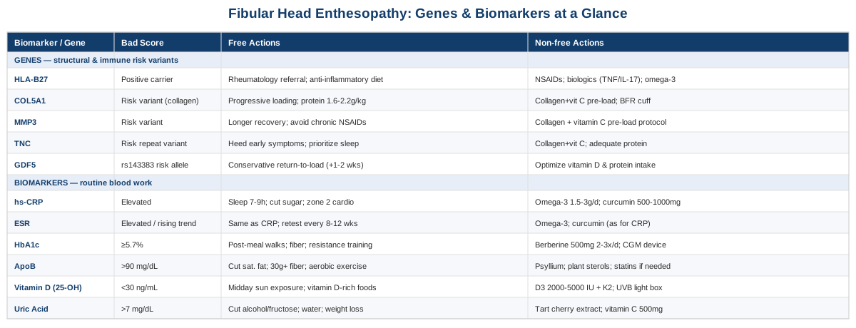
Fibular head enthesopathy rarely has one cause, and the six biomarkers below, from inflammatory markers to a specific cholesterol particle count, can reveal whether your body is fighting silent inflammation, running high blood sugar, or missing raw materials your tendons need to rebuild. Some of these numbers are ones your doctor has probably never mentioned in the context of knee pain, yet they show up repeatedly in tendon and enthesitis research. Beyond the blood work, five inherited gene variants help explain why some people develop stubborn entheseal problems from ordinary loads while others never do, and a look at recent tendon-science research reveals a counterintuitive fact: the loading approach many people are told to avoid is often the one that works best, provided it is dosed correctly for an insertional problem like this one. A short section on complementary approaches with real supporting evidence, and a practical checklist to close, rounds out the picture.
The Biomarkers That Matter Most for a Stubborn Enthesis
Enthesopathy at the fibular head is a mechanical problem on the surface, but the tissue that fails under load is not operating in isolation. Tendon and ligament entheses are metabolically active structures, and their capacity to withstand and recover from load is shaped by systemic inflammation, blood sugar control, lipid particle burden, vitamin D status, and uric acid levels. The six biomarkers below are not exotic. Most are available through a standard blood draw, and several are already on the panel your primary care doctor orders every year, just rarely interpreted through this lens.
1. High-Sensitivity CRP and ESR: Is There a Low-Grade Fire Burning?
High-sensitivity C-reactive protein (hs-CRP) and erythrocyte sedimentation rate (ESR) are the two oldest, cheapest markers of systemic inflammation, and they matter here because enthesitis, inflammation specifically at the tendon or ligament attachment to bone, is a hallmark feature of the seronegative spondyloarthropathies (ankylosing spondylitis, psoriatic arthritis, reactive arthritis, and inflammatory bowel disease-associated arthritis). The fibular head is not the most classic enthesitis site on formal indices, but it is a recognized one, and when enthesitis shows up at multiple sites, including a knee, heel, or elbow at the same time, it is a meaningful clue that the process is systemic rather than purely mechanical. Research on HLA-B27-positive enthesitis-related arthritis has found that these patients tend to run measurably higher inflammatory markers and lower hemoglobin at diagnosis than HLA-B27-negative patients, which is one reason rheumatologists lean on CRP and ESR when enthesitis does not behave like a simple overuse injury (see Prevalence of HLA-B27, clinical characteristics and treatment outcomes in children with enthesitis-related arthritis).
A persistently elevated CRP or ESR in someone with fibular head pain, especially alongside other joint or entheseal symptoms, psoriasis, eye inflammation, or bowel symptoms, is a signal to loop in a rheumatologist rather than only a sports medicine physician or physical therapist. In someone without those systemic features, a mildly elevated CRP more often reflects visceral fat, poor sleep, or metabolic strain than joint-specific disease, but it is still worth normalizing, since systemic inflammation slows tendon repair regardless of its source.
How to measure it
Both are simple blood draws. hs-CRP costs roughly 15 to 35 US dollars out of pocket through direct-to-consumer lab services, and is included in most standard metabolic panels ordered by a physician. ESR runs about 10 to 20 dollars and is often ordered alongside CRP when inflammatory joint disease is suspected. Neither requires fasting.
If the score is high: the plan without supplements or equipment
Prioritize consistent sleep (7 to 9 hours), since even a few nights of restriction measurably raises CRP. Reduce ultra-processed food and added sugar, which drive visceral adiposity, a major contributor to background inflammation. Add two to three weekly sessions of zone 2 cardio (brisk walk, easy cycling) for 30 to 45 minutes, which lowers CRP over 8 to 12 weeks in most people. Retest hs-CRP and ESR after 8 to 12 weeks of these changes rather than after a few days, since inflammatory markers move slowly.
If the score is high: the plan with supplements or equipment
Omega-3 fatty acids (EPA/DHA, 1.5 to 3 grams daily combined, with food) have the most consistent evidence for modestly lowering CRP over 8 to 12 weeks; cycle continuously but reassess yearly, and reduce the dose or stop if you notice easy bruising or you are on blood thinners. Curcumin with piperine (500 to 1,000 mg daily) has more modest, less consistent evidence but a good safety profile at these doses; avoid if you have gallstones or are on anticoagulants. If ESR and CRP remain high after 3 months of lifestyle changes and supplementation, this is the point to see a rheumatologist rather than escalate supplements further.
2. HbA1c: Is Your Blood Sugar Quietly Stiffening Your Tendons?
Hemoglobin A1c reflects average blood sugar over roughly three months, and it matters for enthesopathy because chronic hyperglycemia drives advanced glycation end-products (AGEs) that cross-link collagen, making tendons and ligaments stiffer, more brittle, and less able to remodel under load. This is not a diabetes-only phenomenon. Research shows that people with HbA1c in the prediabetic range, not just diagnosed diabetics, carry meaningfully elevated odds of lower-extremity tendon injury (see Is Elevated HbA1c Level Associated with Achilles Tendon Contracture Development in Diabetic Foot Patients?). For someone with a stubborn fibular head enthesopathy and an HbA1c creeping toward 5.7 percent or higher, this is a legitimate contributing factor worth addressing, not a coincidence.
How to measure it
A standard blood draw, no fasting required, though fasting glucose is often drawn alongside it. Cost is roughly 15 to 35 dollars direct-to-consumer, and it is included in nearly every annual physical panel.
If the score is high: the plan without supplements or equipment
Walk for 10 minutes after meals, which measurably blunts post-meal glucose spikes. Shift carbohydrate intake toward fiber-rich, minimally processed sources and away from liquid sugar and refined starches. Add two weekly sessions of resistance training, which improves insulin sensitivity independent of weight change. Recheck HbA1c after 3 months, since red blood cell turnover means it will not move meaningfully sooner.
If the score is high: the plan with supplements or equipment
Berberine (500 mg, two to three times daily with meals) has trial evidence comparable to metformin for glucose control in some populations; cycle in 8 to 12 week blocks with a break, and stop if you experience GI upset, which is the most common side effect. A continuous glucose monitor (roughly 50 to 100 dollars per month) is the single most useful piece of equipment here, since it shows in real time which specific foods and habits spike your glucose, letting you personalize the diet changes above rather than guess. If HbA1c stays at or above 5.7 percent despite 3 to 6 months of consistent changes, this warrants a conversation with a physician about further metabolic workup.
3. ApoB: The Particle Count That Predicts More Than Cholesterol
Apolipoprotein B (ApoB) counts the number of atherogenic lipoprotein particles (LDL, VLDL, IDL) in your blood, and researchers including Peter Attia, Thomas Dayspring, and Allan Sniderman have argued for years that it is a more accurate risk marker than standard LDL-cholesterol, because a small number of large, cholesterol-rich particles and a large number of small, cholesterol-poor particles can produce the same LDL-C number while carrying very different risk (see Apolipoprotein B Particles and Cardiovascular Disease: A Narrative Review). The connection to enthesopathy is not theoretical: in familial hypercholesterolemia, one of the most visible clinical signs is a thickened, sometimes painful Achilles tendon xanthoma, a direct deposit of cholesterol particles inside tendon tissue, and these xanthomas track with the burden of circulating atherogenic particles (see Achilles tendon xanthomas are associated with the presence and burden of subclinical coronary atherosclerosis in heterozygous familial hypercholesterolemia). Very few people with fibular head enthesopathy have familial hypercholesterolemia, but the underlying mechanism, that lipid particles infiltrate and stiffen tendon tissue, applies on a smaller scale to ordinary elevated ApoB as well.
How to measure it
A blood draw, ideally fasting for the cleanest read, though ApoB is less fasting-sensitive than triglycerides. Direct-to-consumer cost runs 20 to 50 dollars as a standalone marker, or 100 to 200 dollars as part of an advanced lipid panel (which typically also includes Lp(a), a one-time genetic-driven marker worth checking once in your life). This is one of the more "advanced" biomarkers on this list in the sense that many standard physicals still default to LDL-C rather than ApoB, so you may need to specifically request it.
If the score is high: the plan without supplements or equipment
Reduce saturated fat intake and replace refined carbohydrates with fiber (aim for 30+ grams daily), both of which meaningfully lower ApoB-containing particle counts within 6 to 8 weeks. Add regular aerobic exercise, which modestly improves particle clearance. Address abdominal obesity specifically, since visceral fat strongly correlates with elevated ApoB independent of total weight.
If the score is high: the plan with supplements or equipment
Psyllium fiber (5 to 10 grams daily with water) has solid trial evidence for lowering LDL/ApoB particle counts; take consistently, and introduce gradually to avoid bloating. Plant sterols/stanols (2 grams daily with meals) modestly lower ApoB and are well tolerated long-term. If ApoB remains high (commonly cited target is under 90 mg/dL for most adults, lower if additional cardiovascular risk factors are present) despite 3 months of the above, this is a conversation for a physician about statins or other lipid-lowering therapy, which are far more effective than any supplement at this stage and have decades of safety data.
4. 25-Hydroxyvitamin D: The Nutrient Tendons Quietly Depend On
Vitamin D does more in tendon tissue than support bone. It regulates collagen synthesis, modulates local inflammation, and protects tendon cells from oxidative stress through vitamin D receptors expressed directly in tendon tissue. A large retrospective analysis of over 300,000 patients found that vitamin D-deficient individuals had roughly 2.5 times the incidence of distal biceps tendinopathy compared with matched controls (see Association of Vitamin D Deficiency With Distal Biceps Injury: A Retrospective Analysis of 336,320 Patients). While that study looked at a different tendon, the underlying biology, that vitamin D supports the collagen-producing and repair capacity of tendon tissue broadly, plausibly extends to the fibular head enthesis as well, and deficiency is common enough (especially in northern climates and in anyone who trains mostly indoors) that it is worth ruling out.
How to measure it
A standard blood draw measuring 25-hydroxyvitamin D, the storage form. Direct-to-consumer cost is roughly 40 to 80 dollars, and it is frequently included in comprehensive wellness panels.
If the score is low: the plan without supplements or equipment
Aim for 15 to 20 minutes of midday sun exposure on bare skin several times weekly when season and latitude allow, and include vitamin D-rich foods (fatty fish, egg yolks, fortified dairy) regularly. These changes alone are often insufficient to correct a true deficiency, especially in winter months or higher latitudes, which is where supplementation typically becomes necessary.
If the score is low: the plan with supplements or equipment
Vitamin D3, 2,000 to 5,000 IU daily with a meal containing fat for absorption, is the standard correction dose for deficiency (below 30 ng/mL), with retesting at 8 to 12 weeks to confirm the level has normalized into the 40 to 60 ng/mL range; avoid self-dosing above 10,000 IU daily without medical supervision and blood monitoring, since excess vitamin D can raise calcium to unsafe levels. Pairing with vitamin K2 (100 mcg daily) is a common practice to help direct calcium toward bone rather than soft tissue, though the evidence for this specific pairing is more theoretical than proven in trials. A UVB light box is a reasonable equipment option for those in low-sunlight climates who prefer not to supplement, used per manufacturer guidance, typically a few minutes several times weekly.
5. Serum Uric Acid: An Underrated Driver of Enthesopathy
Uric acid is usually discussed in the context of gout, but its relevance to enthesopathy is broader and often missed. Even in people without diagnosed gout, asymptomatic hyperuricemia is associated with a higher rate of subclinical enthesopathy on ultrasound, including at the patellar and Achilles entheses, with monosodium urate crystals triggering local inflammation at these tendon-bone junctions well before a classic gout flare ever occurs (see Joint and tendon subclinical involvement suggestive of gouty arthritis in asymptomatic hyperuricemia: an ultrasound controlled study). For a fibular head enthesopathy that has not responded to typical mechanical treatment, checking uric acid is a low-cost, often-overlooked step.
How to measure it
A simple blood draw, roughly 10 to 20 dollars direct-to-consumer, no fasting required.
If the score is high: the plan without supplements or equipment
Reduce alcohol (especially beer) and high-fructose beverages, both strong uric acid drivers. Moderate intake of purine-dense foods (organ meats, certain shellfish, excessive red meat), and increase water intake to support renal clearance. Weight loss, if applicable, meaningfully lowers uric acid, though rapid weight loss can transiently raise it, so gradual is better here.
If the score is high: the plan with supplements or equipment
Tart cherry extract (480 to 960 mg daily, or roughly 1 cup of tart cherry juice) has modest trial support for lowering uric acid and reducing flare frequency; cycle continuously with periodic reassessment, and it is generally well tolerated aside from occasional GI upset. Vitamin C (500 mg daily) has a small uric acid-lowering effect in some studies. If uric acid remains above roughly 7 mg/dL despite these changes, particularly with any history of joint flares, this is worth discussing with a physician, since prescription urate-lowering therapy (allopurinol or similar) is far more effective than supplements once uric acid is persistently elevated.
6. ESR as a Trend Marker: Why It Deserves Its Own Look
While ESR is often ordered alongside CRP, it deserves separate attention because it behaves differently: it rises and falls more slowly, making it a better marker of chronic, low-grade inflammatory processes like ongoing enthesitis, while CRP responds faster to acute inflammation. Tracking both together, rather than either alone, gives a more complete picture of whether an entheseal problem is behaving like a mechanical injury (typically normal inflammatory markers) or an inflammatory one (typically elevated, especially if trending upward over months). The measurement, cost, and improvement strategies mirror those described for CRP above; the value here is in tracking the trend over 3 to 6 month intervals rather than a single snapshot, since a single elevated reading is far less informative than a rising pattern.
Taken together, these six markers sketch a picture of whether your fibular head enthesopathy is being driven mainly by mechanical overload (in which case most markers will be unremarkable) or by an underlying inflammatory, metabolic, or nutrient-deficiency process that is working against tissue repair regardless of how well you manage load. Testing all six costs a fraction of a single physical therapy course and gives you a genuinely different starting point for treatment.
What Your Genes May Be Telling You
Genetic variants do not cause fibular head enthesopathy on their own, and none of the variants below are things you can change. What they can do is shift the odds: making a tendon or ligament attachment somewhat more prone to breaking down under the same load someone else tolerates fine, or making a person more susceptible to an inflammatory process that targets entheses specifically. Researchers such as Ali Torkamani, who has written extensively on translating genomic risk into actionable prevention, and Gary Brecka, known for popularizing accessible genetic panels as a starting point for personalized health decisions, both make the same basic case: a "bad" variant is a reason to monitor and compensate, not a diagnosis or a sentence. The five genes below have the most relevant human evidence for tendon, ligament, and entheseal health.
HLA-B27: The Immune Variant Behind Enthesitis Beyond the Knee
HLA-B27 is by far the strongest-evidenced genetic factor connected to enthesopathy, though it works through the immune system rather than through collagen structure. Carrying this variant substantially raises the risk of spondyloarthropathies, a family of conditions in which enthesitis (inflammation at tendon and ligament attachments) is a defining feature, sometimes appearing in childhood as enthesopathy at multiple sites including the knee region (see HLA-B27-associated spondyloarthritis and enthesopathy in childhood). More recent work has clarified the mechanism: HLA-B27 misfolds inside cells in a way that can trigger an inflammatory unfolded protein response, and it can also form unusual surface structures that activate immune cells associated with the IL-23/IL-17 inflammatory pathway, both of which are now targeted by modern spondyloarthritis medications (see HLA-B27 and spondyloarthritis: at the crossroads of innate and adaptive immunity).
If the gene is a risk variant: the plan without supplements
You cannot change HLA-B27 status, but you can act on what it implies. If you carry this variant and have fibular head enthesopathy, especially with pain at other entheses (heel, elbow, pelvis), psoriasis, inflammatory bowel symptoms, or eye inflammation, the priority is a rheumatology referral rather than more physical therapy alone, since disease-modifying treatment changes the trajectory far more than load management in true spondyloarthropathy. Anti-inflammatory dietary patterns (reduced processed food and sugar, adequate omega-3 intake, weight management) are reasonable general supports but are not a substitute for appropriate medical evaluation and, if indicated, treatment.
If the gene is a risk variant: the plan with supplements or equipment
There is no supplement protocol that meaningfully offsets HLA-B27-driven inflammation; this is a case where the honest answer is that medical management (NSAIDs, and if needed, biologic therapy targeting TNF or IL-17) is the evidence-based path, not an over-the-counter regimen. Omega-3 fatty acids (as described in the CRP section above) are a reasonable adjunct with a good safety profile, taken continuously, but should be framed as supportive, not corrective.
COL5A1: The Collagen Blueprint Gene
COL5A1 encodes a regulatory form of collagen that controls the diameter and assembly of the larger, load-bearing collagen fibers in tendons and ligaments. A meta-analysis pooling 21 observational studies found specific COL5A1 variants associated with meaningfully altered risk of tendon and ligament injury, particularly in Caucasian populations, though associations vary somewhat by population and specific injury type (see Association of COL5A1 gene polymorphisms and musculoskeletal soft tissue injuries: a meta-analysis based on 21 observational studies). The practical read is that some people are built with slightly less favorable collagen fiber architecture for handling repetitive entheseal load, independent of training habits.
If the gene is a risk variant: the plan without supplements
Since you cannot change fiber architecture, the compensation is training-based: progressive, well-dosed tendon loading (described in detail in the tendon-science section below) builds more tolerant tissue over months regardless of your starting collagen genetics, and gradual progression (avoiding sudden jumps in training volume or intensity) matters more for this genotype than for someone without the variant. Adequate protein intake (1.6 to 2.2 g/kg bodyweight daily) supports the raw material for whatever collagen remodeling capacity you do have.
If the gene is a risk variant: the plan with supplements or equipment
Collagen peptides or gelatin (15 grams) with 50 mg of vitamin C, taken 30 to 60 minutes before loading sessions, is the most directly evidence-supported protocol for supporting collagen synthesis around exercise (detailed with its source study in the next section); use on training days, cycling is not necessary, and side effects are minimal beyond mild GI upset in some people at higher doses. A blood flow restriction (BFR) cuff system (roughly 100 to 300 dollars) is a reasonable equipment investment for this genotype, since it allows tendon-loading benefits at lower absolute loads, which may suit a more injury-prone structural profile, though direct evidence in fibular head enthesopathy specifically does not yet exist.
MMP3: The Tissue-Remodeling Enzyme Gene
MMP3 encodes an enzyme that breaks down and remodels components of the tendon matrix, including collagen, proteoglycans, and fibronectin. Specific MMP3 variants have been repeatedly linked to increased Achilles tendinopathy risk across independent cohorts, and there is evidence of an interaction effect with COL5A1, meaning the combination of certain variants in both genes may compound risk beyond either alone (see Variants within the MMP3 gene are associated with Achilles tendinopathy: possible interaction with the COL5A1 gene).
If the gene is a risk variant: the plan without supplements
Since MMP3 governs matrix turnover, the practical implication is that this genotype may benefit from longer recovery windows between hard loading sessions and more conservative progression rates, since remodeling itself may run slightly less efficiently. Avoiding high-dose, prolonged NSAID use is worth noting here, since some evidence suggests NSAIDs can blunt the matrix remodeling process tendons rely on for adaptation, though short-term use for pain control is a separate matter from chronic daily use.
If the gene is a risk variant: the plan with supplements or equipment
There is no supplement shown to directly correct MMP3 activity, but the collagen-support protocol described under COL5A1 (gelatin/collagen plus vitamin C pre-loading) is a reasonable, low-risk adjunct for this genotype as well, taken on training days only.
TNC (Tenascin-C): The Repair Signal Gene
Tenascin-C is a matrix protein involved in tendon healing and remodeling after injury. A specific dinucleotide repeat polymorphism in the TNC gene has been associated with roughly a six-fold increase in Achilles tendon injury risk in the original case-control study, and later work has extended similar TNC associations to rotator cuff tears and ACL injury, suggesting a broader role across tendon and ligament tissue types (see The guanine-thymine dinucleotide repeat polymorphism within the tenascin-C gene is associated with Achilles tendon injuries).
If the gene is a risk variant: the plan without supplements
Because this gene affects healing signaling after tissue is already stressed, the most relevant compensation is respecting early warning signs, stiffness or mild discomfort at the fibular head, rather than pushing through them, since this genotype may translate minor irritation into a slower-resolving injury more readily than average. Sleep quality and consistency matter disproportionately here, since tissue repair signaling is heavily influenced by sleep.
If the gene is a risk variant: the plan with supplements or equipment
No supplement directly targets tenascin-C expression in humans with meaningful evidence. The most defensible approach is the same collagen/vitamin C protocol described above, paired with adequate total protein intake, both supporting the general repair environment rather than this specific gene.
GDF5: The Growth Factor Behind Tendon and Joint Development
GDF5 (growth differentiation factor 5) is involved in joint and tendon development and healing. A well-replicated variant in the regulatory region of this gene (rs143383) is associated with roughly double the risk of Achilles tendon pathology in some populations, and animal models show GDF5 deficiency measurably delays tendon healing after injury (see Components of the transforming growth factor-beta family and the pathogenesis of human Achilles tendon pathology: a genetic association study). This variant is also one of the most common OA-risk variants identified in genome-wide studies, suggesting a shared vulnerability between joint and entheseal tissue.
If the gene is a risk variant: the plan without supplements
Since GDF5 affects healing capacity rather than day-to-day tissue behavior, the practical compensation is being more conservative with return-to-load timelines after any flare, giving an extra one to two weeks of graded reloading before returning to full training intensity, rather than the standard timeline.
If the gene is a risk variant: the plan with supplements or equipment
No supplement has been shown to compensate for reduced GDF5 signaling in humans. The most reasonable adjunct is ensuring vitamin D and protein intake are optimized (see the biomarker sections above), since both support the broader tissue repair environment that GDF5 operates within, even though neither directly restores GDF5 function.
Genetic testing here is best framed as context, not a verdict. If several of these variants show up together, the honest takeaway is that your fibular head enthesis may simply need more conservative progression, more attention to inflammatory and metabolic biomarkers, and less tolerance for training through early symptoms than someone without them, not that a fix is out of reach.
The Tendon Science Most Clinicians Haven't Caught Up To
Much of the most useful recent thinking on tendon and ligament health has come out of exercise physiology labs rather than orthopedic clinics, and it has been popularized for a broad audience through long-form interviews, notably Andrew Huberman's discussions with tendon and ligament researcher Dr. Keith Baar. The core message runs counter to a lot of standard advice: tendons and entheses generally do not heal well with pure rest, and the loading protocol matters enormously, especially at an insertional site like the fibular head. Below are ten of the most practically useful findings from this body of research.
1. Collagen Synthesis Can Be Timed Around Exercise
A randomized, double-blind, crossover trial found that consuming vitamin C-enriched gelatin one hour before a bout of intermittent exercise measurably increased markers of collagen synthesis compared to placebo, and serum collected after supplementation improved collagen content and mechanical properties of engineered ligament tissue in the lab (see Vitamin C-enriched gelatin supplementation before intermittent activity augments collagen synthesis). This is the origin of the now-common recommendation to take 15 grams of gelatin or collagen peptides with vitamin C roughly an hour before rehab or training sessions targeting tendon tissue.
2. Isometric Holds Can Reduce Tendon Pain Almost Immediately
A randomized crossover trial in athletes with patellar tendinopathy found that a single bout of isometric muscle contractions reduced tendon pain scores far more than isotonic (regular) contractions, with effects measurable within 45 minutes, alongside a documented reduction in a specific type of neural inhibition (see Isometric Contractions Are More Analgesic Than Isotonic Contractions for Patellar Tendon Pain). For fibular head pain, this translates to holding a static, moderate-tension contraction of the biceps femoris or surrounding muscles (such as a seated hamstring curl held mid-range) for 30 to 45 seconds, repeated several times, as a way to calm pain before or instead of more dynamic loading.
3. Rest Alone Rarely Resolves Entheseal Pain
Tendons and their entheses adapt to load, and extended rest allows the tissue to actually weaken rather than heal in the way muscle or bone might. This is the foundational insight behind decades of tendon rehabilitation research: the tissue needs a specific, progressive loading stimulus, not avoidance, to remodel favorably.
4. Eccentric Loading Was the Original Breakthrough, But It Has Limits
The classic 1998 study on heavy, slow eccentric calf-muscle training for Achilles tendinopathy found that all 15 patients with long-standing, treatment-resistant pain returned to full pre-injury running activity after 12 weeks (see Chronic Achilles tendon pain treated with eccentric calf-muscle training). This protocol became the default tendon rehabilitation approach for two decades.
5. Insertional Enthesopathy Needs a Different Approach Than Mid-Tendon Injury
This is the most clinically important nuance for fibular head enthesopathy specifically, since it is an insertional problem rather than a mid-substance one. When the same eccentric protocol that worked so well for mid-portion Achilles tendinopathy was applied to insertional Achilles tendinopathy (pain right at the bone attachment, the same category as fibular head enthesopathy), results were far weaker, satisfactory in only about a third of cases, largely because deep dorsiflexion in that protocol compresses the insertion against the bone (see New regimen for eccentric calf-muscle training in patients with chronic insertional Achilles tendinopathy: results of a pilot study). The practical takeaway: loading exercises for fibular head enthesopathy should generally avoid end-range positions that compress the tendon or ligament directly against the bone, favoring a mid-range loading arc instead.
6. Heavy, Slow Resistance Training Is at Least as Effective as Eccentrics
Research comparing corticosteroid injection, eccentric decline squat training, and heavy slow resistance (HSR) training for patellar tendinopathy found HSR produced comparable clinical improvements to eccentric training, with the added advantage of being simpler to prescribe (a basic progressive strength program) and generally better tolerated by patients who find eccentric-only protocols awkward or uncomfortable to perform correctly.
7. Tendon Remodeling Runs on a Slower Clock Than Muscle
Across the studies above, meaningful clinical improvement in tendinopathy protocols consistently required a minimum of 12 weeks, and structural change on imaging often lags even further behind symptom improvement. This is the single most important expectation to set for anyone starting a loading program for fibular head enthesopathy: judging a protocol at 2 or 3 weeks is judging it before it has had time to work.
8. Blood Flow Restriction May Offer a Lower-Load Alternative
Emerging research comparing low-load blood flow restriction training to heavy slow resistance training in patellar tendinopathy suggests BFR may achieve similar benefits at substantially lower absolute loads, which is a potentially useful option for anyone whose joint or connective tissue genetics (see the COL5A1 and MMP3 sections above) make heavy loading harder to tolerate, or who is earlier in a rehab timeline and cannot yet manage full load.
9. Chronic NSAID Use May Work Against Long-Term Tendon Adaptation
While short-term NSAID use for pain control is reasonable, some research suggests that regular, prolonged use may blunt the inflammatory signaling tendons rely on early in the remodeling process, making an occasional-use approach more sensible than a daily habit for anyone pursuing an active loading-based rehabilitation program.
10. Local Biomechanics Deserve as Much Attention as the Tissue Itself
None of the loading science above works in isolation from the forces actually reaching the fibular head. Gait pattern, lateral knee alignment, ankle mobility, and even footwear all influence how much strain the biceps femoris and lateral collateral ligament insertions absorb with each step or pedal stroke, and addressing these factors alongside a loading program, rather than loading alone, is where a physical therapist familiar with running or cycling biomechanics adds real value.
Taken as a whole, this research base makes a case that many people with fibular head enthesopathy have been told to do the opposite of what actually helps: extended rest and avoidance, rather than carefully dosed, insertion-appropriate loading. That said, this applies primarily to mechanically driven enthesopathy; if your biomarker or genetic profile points toward a systemic inflammatory process, loading science is a complement to medical management, not a substitute for it.
Complementary Approaches Worth Considering
The approaches below are not substitutes for addressing the biomarkers, genetics, or loading mechanics discussed above, but each has meaningful human evidence in tendon, entheseal, or closely related knee conditions, and each can reasonably be layered onto a primary treatment plan.
Massage Therapy
Deep transverse friction massage has been used for tendon conditions for decades, on the theory that cross-fiber pressure stimulates local circulation and encourages more organized collagen alignment during healing, which is directly relevant to an entheseal problem like fibular head enthesopathy where tissue disorganization at the attachment site is part of the pathology.
A systematic review of deep friction massage for tendinopathy found genuine but modest supporting evidence when combined with an exercise program, though the review also noted that evidence quality varies and friction massage alone, without concurrent loading, is not well supported (see Deep friction massage to treat tendinopathy: a systematic review of a classic treatment in the face of a new paradigm of understanding).
Realistically, this means seeking out a physical therapist or massage therapist experienced with tendinopathy who will pair friction massage with, not instead of, the loading program described above, typically as a 5 to 10 minute adjunct once or twice weekly, and expecting it to modestly support rather than independently resolve the condition.
Low-Level Laser Therapy (Photobiomodulation)
Photobiomodulation uses low-level red or near-infrared light to stimulate cellular energy production (mitochondrial activity) in tendon and connective tissue, with the theoretical rationale that this accelerates the same repair processes that loading and nutrition support, making it relevant as an adjunct for a slow-healing entheseal injury like this one.
A systematic review and meta-analysis of randomized controlled trials found that low-level red and near-infrared photobiomodulation produced meaningful improvements in pain and function across several tendinopathy conditions, though effect sizes and optimal dosing parameters varied across the included trials (see The effect of low-level red and near-infrared photobiomodulation on pain and function in tendinopathy).
In practice, this means a series of sessions (commonly 2 to 3 times weekly for 4 to 6 weeks) delivered by a clinic with proper equipment and dosing protocols, since home devices vary widely in power output and are harder to dose correctly; treat it as a reasonable adjunct rather than a primary strategy.
Tai Chi
Tai chi combines slow, controlled weight shifting, rotational movement, and isometric-like holds through partial ranges of motion, which overlaps meaningfully with the loading principles described in the tendon-science section above, particularly for a lateral knee structure like the fibular head that benefits from controlled, low-impact loading variety.
A large randomized comparative-effectiveness trial found that tai chi produced improvements in knee osteoarthritis pain and function equal to standard physical therapy over a year of follow-up (see Comparative Effectiveness of Tai Chi Versus Physical Therapy for Knee Osteoarthritis: A Randomized Trial), and while this trial addressed osteoarthritis rather than enthesopathy specifically, the shared anatomy (lateral knee loading, controlled range of motion) makes it a reasonable, low-risk option to layer in.
A realistic approach is two sessions weekly of a beginner-level tai chi class, ideally with an instructor aware of the specific location of your pain so early sessions can avoid positions that load the fibular head end-range, progressing as tolerated over 8 to 12 weeks.
Mindfulness Meditation and MBSR
Chronic musculoskeletal pain, including entheseal pain that has persisted for months, involves a real central sensitization component, meaning the nervous system's pain processing itself can become part of why symptoms feel disproportionate to tissue damage on imaging, which is where mindfulness-based approaches have a plausible role.
A randomized controlled trial of standardized mindfulness-based stress reduction (MBSR) in patients with chronic pain found statistically significant reductions in pain compared to a wait-list control group, though the authors and subsequent meta-analyses have generally rated the overall evidence quality as low to moderate, with modest rather than dramatic effect sizes (see Effects of mindfulness meditation on chronic pain: a randomized controlled trial).
A practical starting point is a structured 8-week MBSR course (widely available in-person and online) or a well-reviewed guided meditation app, used for 15 to 20 minutes daily, framed honestly as a tool for managing the pain experience and reducing stress-related muscle guarding around the knee, not as a treatment for the underlying tissue pathology itself.
Putting It Together
Fibular head enthesopathy that will not resolve with generic rest-and-stretch advice usually has a specific reason, and that reason is often visible in ordinary blood work: silent inflammation on CRP and ESR, blood sugar drifting into a range that stiffens collagen, an elevated ApoB particle count, low vitamin D, or uric acid quietly irritating the tendon-bone junction. Genetics can add context for why the same load affects different people so differently, and recent tendon-science research makes a strong case that carefully dosed, insertion-appropriate loading, not extended rest, is usually the more effective path forward, alongside a small set of complementary approaches with real, if modest, supporting evidence.
None of this is a promise of a cure, and a persistent or worsening enthesopathy, especially one accompanied by pain at other joints, skin changes, eye symptoms, or bowel symptoms, deserves a proper medical evaluation rather than a self-directed protocol. But better information changes what you can ask for. The next concrete step is straightforward: request the six biomarkers above at your next blood draw, track how your symptoms track against sleep, training load, and diet over the following weeks, and bring both to whichever clinician, physical therapist, sports medicine physician, or rheumatologist, is best positioned to interpret them alongside your specific pattern of symptoms.