This article contains AI generated content.
Fibular Head Fracture Recovery: 4 Genes And 7 Biomarkers To Track
Introduction
A fibular head fracture is a strange injury to live with. The bone itself is small and often heals on a predictable timeline, but the location — right where the common peroneal nerve wraps around the bone, next to the lateral collateral ligament and the biceps femoris tendon — means the same fracture can behave very differently from one person to the next. Some people are back to full activity in six weeks. Others deal with lingering swelling, numbness, weak ankle dorsiflexion, or a knee that never quite feels as stable as before. If you've been told "just give it time" and that hasn't matched your experience, you're not imagining things.
Generic fracture advice — rest, elevate, wear the brace, come back in six weeks for an X-ray — is written for the average patient, and averages hide a lot. Two people with an identical Maisonneuve or avulsion-type fibular head fracture can have very different vitamin D status, very different inflammatory profiles, very different collagen genetics, and very different nerve recovery trajectories. None of that shows up on a standard follow-up X-ray, and very little of it gets discussed in a ten-minute clinic visit.
This article takes a more granular approach. Instead of one-size-fits-all instructions, it looks at the actual blood-based biomarkers that reflect how your bone is healing, what your nutritional and hormonal status is doing to that process, and which inherited traits might be quietly helping or hindering you. It also brings in what current research on connective tissue repair suggests about timing, loading, and nutrition — ideas that are more specific and more actionable than "don't overdo it."
None of this replaces your orthopedic surgeon, and nothing here should be read as a guarantee of faster healing. But better information tends to produce better decisions — which tests are worth asking for, which supplements have a plausible mechanism versus which are marketing, and which recovery habits are backed by more than tradition. The sections ahead cover the biomarkers worth tracking, the genetic variants that shape bone and connective tissue repair, a set of research-backed ideas from a widely discussed physiology podcast, and a handful of complementary approaches with real supporting evidence.
Summary
Fracture healing is not just a matter of time — it's a metabolic process that depends on inflammation control, mineral availability, hormone levels, and collagen synthesis, all of which can be measured. Below, you'll find seven specific biomarkers — from vitamin D and CRP to bone turnover markers like P1NP and CTX — that give a much clearer picture of how a fibular head fracture is actually healing than a calendar does, along with realistic ways to improve each one, with and without supplements. You'll also find four genetic variants (in COL1A1, VDR, BMP2, and LRP5) that help explain why some people knit bone and rebuild collagen faster than others, ten research-backed ideas from a well-known physiology podcast on tissue repair that push back on some standard recovery advice, and a short review of complementary approaches — from photobiomodulation to tai chi — that have actual human evidence behind them for this kind of injury.
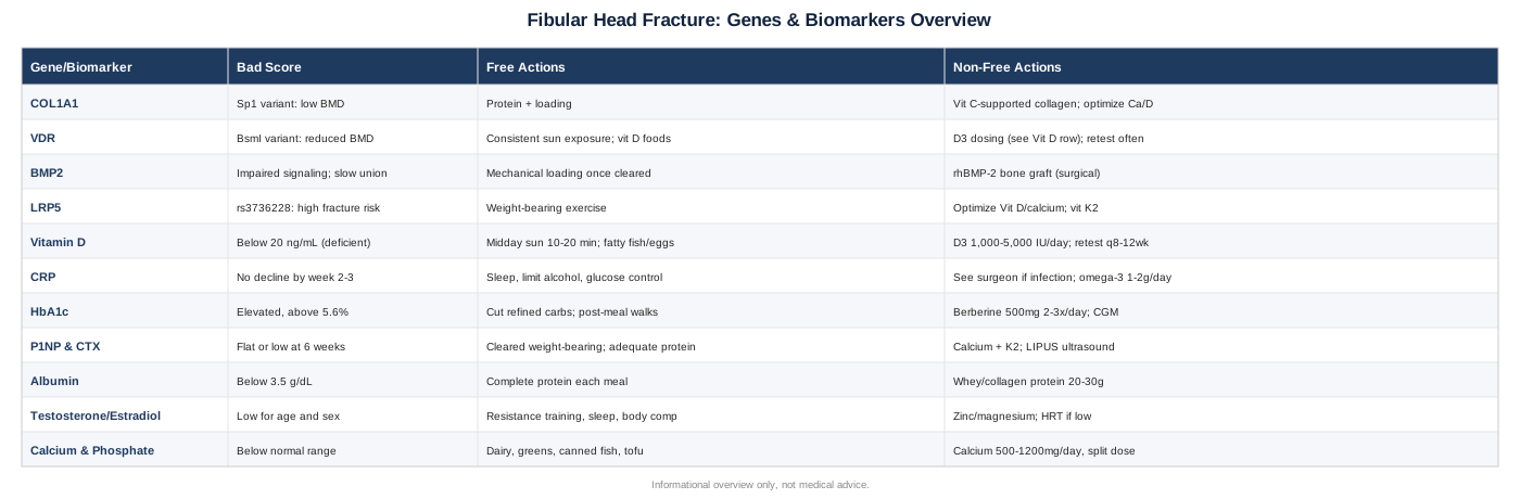
7 Biomarkers Worth Tracking After A Fibular Head Fracture
Fracture healing runs through overlapping biological stages: an inflammatory phase in the first days, a soft-callus phase over the following weeks, hard-callus mineralization over one to three months, and remodeling that can continue for a year or more. Each stage leans on different biological inputs — inflammatory signaling, mineral availability, collagen synthesis, hormonal support — and each of those inputs leaves a trace in the blood. Peter Attia has built much of his practice around the idea that waiting for a symptom or an X-ray change is a lagging indicator; checking the underlying numbers is a leading one. That philosophy applies just as well to a fibular head fracture as it does to cardiometabolic health, even though the specific markers are different. The seven below are not markers your orthopedist will necessarily order by default, but each has a plausible, evidence-backed connection to how well and how fast this specific injury heals.
1. Vitamin D (25-Hydroxyvitamin D)
Vitamin D governs how efficiently your gut absorbs calcium and how your body regulates the calcium and phosphate that get deposited into new bone matrix. Low 25(OH)D is common — population studies routinely find a third or more of adults below sufficient levels — and it's one of the few fracture-relevant markers that's cheap, standardized, and easy to act on.
The evidence here is more nuanced than it's often presented. A randomized trial in fracture patients with confirmed low vitamin D found that a single high-dose vitamin D3 bolus did not significantly change union versus nonunion rates compared with placebo (Haines et al., Bone & Joint Journal, 2017). That doesn't mean vitamin D is irrelevant — chronic deficiency is still linked to poorer bone quality overall — but it does mean a single big dose isn't a fix, and correcting deficiency should be thought of as removing a headwind rather than adding a tailwind.
How to measure it
A basic serum 25(OH)D blood test, available through primary care, most direct-to-consumer lab services, or hospital pre-op panels. Cost typically runs $40–$100 out of pocket, less if covered by insurance or bundled into a broader panel.
If the score is low, the plan without supplements
Ten to twenty minutes of midday sun exposure on bare skin, several times a week, can meaningfully raise vitamin D synthesis in most skin tones, though this is slower and less reliable during winter months or at higher latitudes. Dietary sources — fatty fish, egg yolks, fortified dairy — contribute modestly but rarely close a real deficiency on their own.
If the score is low, the plan with supplements
Vitamin D3 (cholecalciferol) at 1,000–2,000 IU daily is a reasonable general maintenance dose for someone with mild insufficiency; more significant deficiency (below 20 ng/mL) is often addressed with 4,000–5,000 IU daily for 8–12 weeks under a clinician's guidance, followed by retesting. Taking it with a fat-containing meal improves absorption. Side effects are rare at these doses but very high, prolonged intake (10,000+ IU/day for months) can raise calcium to unsafe levels, so retesting every 2–3 months during correction, rather than supplementing indefinitely at high doses, is the safer pattern.
2. C-Reactive Protein (CRP)
CRP is a general inflammation marker, and after any fracture surgery it follows a predictable arc: a rise in the first 48–72 hours as the body mounts an inflammatory response to the injury and any surgical fixation, then a steady decline over the following two to three weeks. A systematic review of orthopedic trauma patients found this normal peak-then-decline pattern, and noted that a secondary rise or a failure to normalize is one of the more reliable early signals of a postoperative infection or healing complication (Kruidenier et al., Acta Orthopaedica Belgica, 2018).
How to measure it
A standard or high-sensitivity CRP blood draw, widely available and inexpensive — roughly $15–$40, often bundled into post-surgical monitoring already.
If the pattern looks abnormal, the plan without supplements
A CRP that isn't trending down by week two to three is a conversation for your surgeon, not a self-management project — it may reflect infection, hardware irritation, or another complication that needs direct evaluation, not lifestyle tweaks. That said, general anti-inflammatory habits (adequate sleep, minimizing alcohol, controlling blood sugar) support a normal inflammatory trajectory throughout recovery.
If the pattern looks abnormal, the plan with supplements or equipment
There's no supplement that reliably normalizes a fracture-related CRP elevation, and self-treating a possible infection signal with anti-inflammatory supplements can mask what your surgical team needs to see. Omega-3 fatty acids (1–2g EPA/DHA daily) have modest general anti-inflammatory evidence and are reasonable as a baseline habit, but they are not a substitute for follow-up imaging or bloodwork if CRP stays elevated.
3. HbA1c (Glycated Hemoglobin)
HbA1c reflects average blood sugar over roughly the past three months, and it turns out to matter a great deal for fracture healing. A meta-analysis of orthopedic trauma patients found that people with diabetes had more than double the risk of impaired fracture healing compared with non-diabetics (odds ratio 2.11) (Ding et al., ANZ Journal of Surgery, 2020). Elevated glucose impairs osteoblast function and collagen cross-linking, which slows the mineralization phase specifically.
How to measure it
A standard HbA1c blood test, roughly $15–$35, or included in most annual physical panels.
If the score is elevated, the plan without supplements
Reducing refined carbohydrate and added sugar intake, walking after meals (even seated ankle pumps and short walks are appropriate once weight-bearing is cleared), and prioritizing 7–9 hours of sleep all measurably improve glycemic control within weeks. Post-injury inactivity itself worsens insulin sensitivity, so maintaining whatever movement is medically permitted matters more than it might seem.
If the score is elevated, the plan with supplements or equipment
Berberine (500mg two to three times daily with meals) has trial evidence comparable to some oral glycemic agents for improving insulin sensitivity, though it can cause GI upset and interacts with several medications, so it warrants a conversation with a physician first, especially post-surgery. A continuous glucose monitor (roughly $50–$100/month) can help identify which specific foods spike blood sugar during a period when activity is restricted. These are adjuncts, not replacements for existing diabetes management if you're already diagnosed.
4. Bone Turnover Markers: P1NP And CTX
P1NP (procollagen type 1 N-terminal propeptide) reflects new bone formation, while CTX (C-telopeptide) reflects bone resorption — together they give a biochemical readout of the remodeling process that X-rays only show indirectly. A study following tibia and femur fracture patients found both markers rose and peaked around six weeks post-injury, and that higher 6-week levels correlated with better radiological healing by twelve weeks (Stewart et al., Bone & Joint Research, 2022) — meaning these markers may give you a readable signal weeks before it's visible on imaging.
How to measure it
These are more specialized tests, ordered less often in routine primary care but available through most major reference labs (Quest, LabCorp) or endocrinology practices. Combined cost typically runs $100–$250, and timing relative to time-of-day and fasting state matters (morning, fasted draws are more standardized), so ask your lab about protocol.
If turnover looks sluggish, the plan without supplements
Bone responds to mechanical load, and gentle, cleared weight-bearing or isometric loading (once your surgeon approves it) is one of the few interventions that directly stimulates both formation and remodeling. Adequate protein intake (roughly 1.2–1.6g/kg body weight daily) supports the collagen matrix that bone mineral deposits onto.
If turnover looks sluggish, the plan with supplements or equipment
Calcium citrate or carbonate (500–600mg twice daily with meals, rather than one large dose, for better absorption) paired with vitamin K2 (MK-7, ~100mcg daily) to support directing calcium into bone rather than soft tissue is a commonly used combination, though evidence for K2 specifically in fracture healing is still early-stage rather than definitive. Low-intensity pulsed ultrasound devices (prescription-grade units used 20 minutes daily) have RCT evidence for accelerating healing in certain fracture types and might be worth discussing with your surgeon if healing is confirmed to be slow, rather than self-purchasing a consumer device.
5. Serum Albumin
Albumin is a simple, often-overlooked marker of overall protein-energy nutritional status, and nutrition status has a surprisingly direct effect on surgical outcomes. A large study of geriatric hip fracture patients found that hypoalbuminemia (below 3.5 g/dL) was associated with significantly higher postoperative mortality (9.94% versus 5.53% in patients with normal albumin) (Bohl et al., Journal of Bone and Joint Surgery, 2017). While that study looked at hip fractures specifically, the underlying principle — that inadequate protein reserves impair surgical recovery and tissue repair broadly — applies to any fracture requiring fixation.
How to measure it
Included automatically in a standard comprehensive metabolic panel (CMP), so it often costs nothing extra beyond routine bloodwork, roughly $10–$30 if ordered alone.
If the score is low, the plan without supplements
Prioritizing complete protein at each meal — eggs, dairy, poultry, fish, legumes — rather than relying on carbohydrate-heavy convenience foods during a period of reduced mobility is the most direct lever. Low albumin in an older adult or anyone with reduced appetite post-injury is worth flagging to a physician, since it can also reflect liver, kidney, or inflammatory issues unrelated to diet.
If the score is low, the plan with supplements or equipment
A whey or collagen-based protein supplement (20–30g once or twice daily) can help close a gap when appetite is reduced post-surgery, particularly in older patients. Collagen peptides specifically pair well with vitamin C (see the connective tissue research below) for supporting the structural protein matrix. These are food-gap fillers, not substitutes for addressing why appetite or intake dropped in the first place.
6. Testosterone And Estradiol
Sex hormones influence osteoblast activity and bone matrix maintenance in both men and women. A study comparing elderly men with hip fractures to age-matched, fracture-free controls found the fracture group had significantly lower testosterone and estradiol levels (Kantharaj et al., Cureus). This doesn't mean low hormones caused the fracture, but it does suggest hormonal status is worth knowing, especially in older adults or anyone with symptoms of low testosterone or perimenopausal estrogen decline.
How to measure it
Total and free testosterone (men), or estradiol and FSH (women), via morning blood draw — morning timing matters for testosterone specifically, since levels are highest then. Cost is roughly $50–$120 per panel depending on how comprehensive it is.
If the score is low, the plan without supplements
Resistance training (once cleared for weight-bearing and upper-body work is always available even during lower-limb restriction), adequate sleep, and maintaining a healthy body composition all support endogenous hormone production over months, not weeks.
If the score is low, the plan with supplements or equipment
Vitamin D correction (see marker #1) and adequate zinc and magnesium intake support normal testosterone synthesis and are low-risk first steps. Hormone replacement therapy is a legitimate option for clinically low levels but is a decision for an endocrinologist or your physician, given the cardiovascular and other considerations involved — not something to self-initiate based on one blood draw during fracture recovery.
7. Calcium And Phosphate
These are the two minerals actually deposited into the bone matrix during mineralization, and while frank deficiency is less common than vitamin D insufficiency, both are cheap to check and relevant to confirm, especially if vitamin D or kidney function is also abnormal, since calcium and phosphate regulation are tightly linked to both.
How to measure it
Both are part of a standard comprehensive metabolic panel, typically $10–$20 if ordered outside of a broader panel.
If the score is low, the plan without supplements
Dairy, leafy greens, canned fish with bones, and tofu are strong dietary calcium sources; phosphate deficiency is rare outside of malnutrition or certain medication effects and usually resolves with a generally adequate diet.
If the score is low, the plan with supplements or equipment
Calcium supplementation (as above, split into smaller doses) is appropriate for a confirmed dietary shortfall, generally 500–1200mg daily depending on dietary intake, split across two doses for absorption. Very high calcium intake (above 2,000-2,500mg/day from food plus supplements combined) has been associated with kidney stone risk in some studies, so this is a "correct the gap, don't stack supplements indefinitely" marker — retest after 2-3 months rather than supplementing without a plan to reassess.
Tracking these seven numbers doesn't replace your surgeon's follow-up imaging, but it turns "wait and see" into something closer to "wait and know," which is a meaningfully different experience during a recovery that can otherwise feel like a black box.
The Genetics Behind Slow Or Fast Bone Healing
Biomarkers tell you what's happening right now; genetics offers a partial explanation for why some people's bones and connective tissue seem to knit faster or slower than others' even under similar conditions. Researchers like Ali Torkamani at Scripps Research, whose work focuses on how common genetic variants combine to shape disease and trait risk, have emphasized that most individual bone-related variants carry a small effect on their own — the value is in knowing the pattern, not in treating any single gene as destiny. Gary Brecka's popularized approach to biomarker-and-gene-informed health optimization follows a similar logic: know your variant, then compensate with the cheapest, lowest-risk intervention first. The four genes below are the ones with the most direct, human-study-backed relevance to fracture and bone healing.
COL1A1 (Collagen Type 1 Alpha 1)
COL1A1 codes for the main structural protein in bone and connective tissue. A specific variant in its Sp1 binding site has been shown in human studies to predispose people to reduced bone mineral density, altered bone quality, and higher osteoporotic fracture risk (Mann et al., Journal of Clinical Investigation, 2001). This variant affects collagen fiber quality rather than just quantity, which matters for a fracture site that depends on strong collagen scaffolding before mineralization can proceed properly.
If this variant is a known factor for you (through consumer genetic testing or a family history of fragility fractures), the plan without supplements centers on adequate, consistent protein intake and progressive mechanical loading once cleared, since loading is one of the few known stimuli for collagen remodeling regardless of genotype. The plan with supplements leans on vitamin C-supported collagen synthesis (detailed in the next section) and ensuring calcium and vitamin D status are optimized, since a collagen-quality disadvantage is partly compensated by making sure the mineralization side of the equation isn't also deficient.
VDR (Vitamin D Receptor)
The VDR gene determines how effectively your cells respond to the vitamin D circulating in your blood — it's the difference between having vitamin D and being able to use it. A study of postmenopausal women found the BsmI polymorphism in VDR showed a modest but significant association with reduced bone mineral density (Al-Barazenji et al., Pathophysiology, 2025), while other common VDR variants (ApaI, TaqI) did not show the same association in that cohort — a reminder that not every variant in a "bone gene" carries equal weight.
If you carry a less favorable VDR variant, the plan without supplements is simply more consistent sun exposure and dietary vitamin D sources, since the issue is receptor sensitivity rather than intake alone — meaning slightly higher, steady circulating levels may help compensate for reduced receptor efficiency. The plan with supplements involves the same vitamin D3 dosing discussed under biomarker #1, but with a stronger case for retesting 25(OH)D more frequently (every 8-12 weeks during active healing) since a less efficient receptor may mean your usual "normal" blood level isn't doing as much work at the tissue level.
BMP2 (Bone Morphogenetic Protein 2)
BMP2 sits at the center of the signaling pathway that tells stem cells to become bone-forming osteoblasts — it's one of the most-studied molecules in fracture biology. Its relevance to humans is best demonstrated not through a common polymorphism study but through direct clinical use: a meta-analysis of randomized trials found that adding recombinant human BMP-2 to the surgical fixation of open tibial fractures reduced rates of secondary intervention and treatment failure (Wei et al., Orthopedics, 2012). The foundational mechanism — that BMP2 signaling is essentially required for normal bone formation — comes primarily from animal knockout studies, so the human evidence here is strongest for BMP2's therapeutic use rather than for common genetic variation in the general population.
There's no simple consumer "fix" for BMP2 pathway efficiency, since it isn't a single well-characterized common variant the way COL1A1 or VDR are. The most realistic non-supplement approach is again mechanical loading, which upregulates BMP signaling naturally once bone is stable enough to bear it. If healing is confirmed to be unusually slow on imaging or via bone turnover markers, BMP2-based bone graft substitutes are a legitimate surgical option your orthopedic team may discuss — this is a clinical decision, not a supplement or lifestyle one.
LRP5 (LDL Receptor-Related Protein 5)
LRP5 is part of the Wnt signaling pathway, which regulates osteoblast activity and overall bone mass. A meta-analysis of case-control studies found a common LRP5 variant (rs3736228) associated with increased osteoporosis and fracture risk across multiple populations (Xu, Qiu, and Mao, BioMed Research International, 2014).
If this variant applies to you, the plan without supplements again comes back to mechanical loading — Wnt signaling is itself activated by mechanical strain on bone, so consistent, cleared weight-bearing exercise is one of the more direct ways to support this exact pathway. The plan with supplements has less direct evidence specific to LRP5, but ensuring adequate vitamin D and calcium status removes at least one compounding disadvantage, and some early research has explored vitamin K2's role in Wnt-related bone signaling, though this remains an area of ongoing rather than settled research.
Genetic testing isn't necessary to recover well from a fibular head fracture, and none of these variants are destiny — they're modifiers, not verdicts. Where genetics gives you a plausible "why," the biomarkers above give you a real-time "how's it going," and together they're more useful than either alone.
Ten Ideas From The Huberman Lab Episode On Tissue Repair That Push Back On Standard Recovery Advice
The Huberman Lab podcast episode featuring Dr. Keith Baar, a physiologist who studies connective tissue and collagen biology at UC Davis, is one of the more specific, evidence-referenced discussions available on how tendon, ligament, and bone-adjacent tissue actually repairs itself — and several of its central points cut against the traditional "just rest it" advice still common in general practice. Below are ten of the most useful, condensed for anyone recovering from a fibular head fracture (where the surrounding ligament and tendon tissue matters as much as the bone itself).
1. Connective Tissue Has Far Less Blood Flow Than Muscle
Tendons, ligaments, and the periosteum around bone receive a fraction of the blood supply that muscle does, which is a core reason connective tissue injuries and healing generally take longer than muscle injuries of similar severity — it isn't just "toughness," it's circulation.
2. Timing Of Loading Matters More Than Total Rest
Collagen-producing cells respond to mechanical signals with a delay; a landmark human study found collagen synthesis in tendon tissue rose after a single bout of loading exercise and stayed elevated for up to 72 hours afterward (Miller et al., Journal of Physiology, 2005), which is the basis for structured, cyclical loading rather than either total immobilization or constant strain.
3. Vitamin C Timing Around Loading May Matter
The most directly applicable human study here found that vitamin C-enriched gelatin, taken about one hour before intermittent exercise, roughly doubled blood markers of collagen synthesis compared with placebo (Shaw et al., American Journal of Clinical Nutrition, 2017) — the specific protocol used was 15g of gelatin with vitamin C, consumed roughly an hour before movement.
4. Total Immobilization Slows Recovery More Than It Protects It
Complete rest reduces the mechanical signal that connective tissue needs to remodel; the more current framing (once a fracture is surgically or clinically stable enough) favors early, controlled, cleared movement over prolonged full immobilization, which mirrors current best-practice guidance in fracture rehabilitation more broadly.
5. Gelatin, Not Just Any Collagen Source, Was The Specific Protein Studied
The Shaw et al. study specifically used gelatin (hydrolyzed collagen) rather than whey or other proteins, since gelatin's amino acid profile — high in glycine and proline — mirrors the amino acids connective tissue itself is built from.
6. Heat Exposure May Support Local Blood Flow To Repairing Tissue
Because connective tissue is blood-flow-limited, anything that reasonably and safely increases local circulation (warm compresses, contrast bathing once cleared by a clinician) is a plausible, low-risk adjunct — though this is a supportive idea drawn from broader physiology rather than a fracture-specific trial.
7. Recovery Nutrition Windows Aren't Just For Muscle
The same "nutrient timing around activity" logic long applied to muscle protein synthesis appears to apply to collagen synthesis as well, which is a relatively newer and less mainstream idea in general rehab advice.
8. Cyclical Loading Beats Static Stretching For Connective Tissue Adaptation
Repeated, moderate loading and unloading cycles appear to stimulate collagen remodeling more effectively than sustained static stretches, reinforcing why supervised, incremental rehab exercises tend to outperform passive stretching protocols once weight-bearing is permitted.
9. Individual Recovery Timelines Genuinely Vary
Given the blood-flow and collagen-turnover differences discussed above, along with the genetic variation covered earlier in this article (COL1A1 in particular), expecting a fixed, universal timeline for connective tissue and bone-adjacent healing isn't well supported by the underlying biology.
10. None Of This Replaces Structural Healing Time For Bone Itself
These ideas are specifically about connective tissue (ligament, tendon, periosteum) rather than the mineralization of bone itself, which still depends on the biomarkers and timeline discussed earlier in this article — connective tissue and bone healing run in parallel, not as substitutes for one another.
Taken together, these ideas suggest that the passive, purely time-based model of fracture recovery is outdated for the tissues surrounding the bone, even if the bone itself still needs its biological remodeling time. That distinction — between what can be actively supported and what simply needs to run its course — is a useful frame for the complementary approaches that follow.
Complementary Approaches Worth Considering
None of the following replace surgical or orthopedic management of a fibular head fracture, and the evidence quality varies — but each has at least one human study specific enough to be worth knowing about, rather than being a generic wellness recommendation stretched to fit.
Low-Level Laser Therapy (Photobiomodulation)
Low-level laser therapy uses specific wavelengths of red or near-infrared light to stimulate cellular activity in healing tissue, and it has more direct fracture-specific evidence than most complementary approaches on this list, making it relevant for someone specifically interested in supporting bone union rather than general wellness.
A randomized controlled trial of 50 patients with closed wrist and hand fractures found that 830nm laser therapy significantly improved pain, grip strength, function, and radiographic healing compared with sham treatment (Chang et al., Photomedicine and Laser Surgery, 2014). The fracture type differs from a fibular head fracture, but the mechanism — stimulating cellular metabolism at the fracture site — is not location-specific.
In practice, this means asking your physical therapist or orthopedic clinic whether they offer photobiomodulation as an adjunct once the wound (if surgical) has closed, typically administered in short sessions two to three times weekly for several weeks. It should be treated as a possible accelerant alongside standard care, not a substitute for surgical fixation or the standard healing timeline.
Massage Therapy
Massage therapy is relevant here less for the bone itself and more for the swelling, pain, and anxiety that commonly accompany fracture surgery, all of which can slow down early mobilization if left unmanaged.
A randomized clinical trial of 66 patients who had undergone tibial shaft fracture surgery found that a simple ten-minute foot massage significantly reduced both postoperative pain intensity and anxiety compared with standard care alone (Pasyar, Rambod, and Kahkhaee, Journal of Orthopaedic Trauma, 2018).
For a fibular head fracture, this looks like gentle massage above and below the injury site and cast or brace boundary (never directly over unhealed fracture sites or surgical incisions without clinician approval), a few times a week during the early post-operative period, primarily as a comfort and anxiety-reduction measure rather than a healing accelerant.
Tai Chi
Tai chi is most relevant to fibular head fracture recovery in the rehabilitation phase, where regaining balance and proprioception matters, particularly if the peroneal nerve was affected and ankle or foot control feels less reliable.
A meta-analysis of 24 randomized controlled trials in older adults found tai chi reduced fall risk by 24% and produced measurable improvements in balance (Chen et al., Frontiers in Public Health, 2023) — directly relevant given that impaired balance and fall risk are common concerns after any lower-limb fracture, especially one near a major nerve.
Realistically, this fits best once weight-bearing and basic mobility have been cleared by your surgeon or physical therapist, starting with seated or supported forms and progressing to standing sequences as confidence and strength return — a low-impact, low-risk way to rebuild the balance and proprioceptive control that this specific injury can compromise.
Mindfulness Meditation / MBSR
An acute fracture, especially a traumatic one, carries a real psychological load — pain, disrupted routine, and uncertainty about recovery — and that stress response can itself interfere with sleep, appetite, and rehab adherence.
A study of 135 trauma-fracture patients found that mindfulness-based stress reduction combined with conventional treatment significantly lowered the incidence of acute stress disorder (8.22% versus 24.19% in the conventional-treatment-only group) and improved anxiety, depression, sleep, and quality of life measures (Chen et al., Actas Españolas de Psiquiatría, 2025).
In practice, this means a structured MBSR program (many are available free or low-cost through hospital systems or apps) started soon after injury, with even 10–15 minutes of daily guided practice showing benefit in the studied population — a reasonable, low-risk complement to standard fracture care, particularly for anyone noticing anxiety or sleep disruption during recovery.
Conclusion
A fibular head fracture heals through a fairly specific sequence of biology — inflammation, mineral deposition, collagen remodeling, and nerve and soft-tissue recovery — and each stage leaves measurable clues. Tracking vitamin D, CRP, HbA1c, bone turnover markers, albumin, sex hormones, and calcium and phosphate turns a vague waiting period into something you can actually monitor and, in several cases, meaningfully influence. Understanding genetic factors like COL1A1, VDR, BMP2, and LRP5 adds context for why your healing pace might differ from someone else's with the same injury, and the research on connective tissue timing and loading offers a more active, evidence-grounded alternative to simply waiting it out.
None of this changes the fundamentals: follow your surgeon's weight-bearing timeline, attend your follow-up imaging, and treat any of the complementary approaches above as additions to that plan rather than replacements for it. The most useful next step is a practical one — ask your orthopedic team which of these biomarkers they'd be willing to check at your next visit, or request them independently if you want a clearer picture between appointments. Better numbers won't heal the bone for you, but they will tell you, with more confidence than a calendar ever could, how the healing is actually going.
Musculoskeletal: Bone Conditions Tendon & Ligament Conditions
Neurological: Nerve Conditions