This article contains AI generated content.
· UpdatedFibular Hemimelia Genes and Biomarkers — 6 Genes And 7 Biomarkers To Track
Introduction
Living with fibular hemimelia — whether as the person born with this condition or as a parent navigating care decisions for a child — means dealing with a medical reality that doesn't fit standard health frameworks. Most clinical conversations center on surgical timelines, prosthetic fitting, and rehabilitation milestones. What receives far less attention is the internal biochemistry that shapes how the body develops, heals, and sustains skeletal health across decades. That gap matters, and this is where a more precise approach begins.
Generic bone health advice — "get enough calcium," "take vitamin D," "do weight-bearing exercise" — is not wrong, but it misses the specificity that fibular hemimelia demands. Someone with this condition carries a unique combination of structural asymmetry, surgical history, altered biomechanics, and in some cases underlying developmental gene variants that no general prevention message was designed to address. A child going through tibial osteotomy or distraction osteogenesis has physiological needs that differ sharply from a healthy adult, and an adult managing compensatory knee and hip strain has priorities no standard guideline accounts for.
Two angles tend to be underused in clinical settings. The first is targeted biomarker tracking — a focused panel of blood markers that can reveal bone formation and resorption rates, growth factor availability, calcium-regulating hormones, and systemic inflammation levels. The second is the growing body of research on the developmental genes involved in limb formation, which increasingly points toward actionable nutritional and lifestyle implications for people carrying specific variants. Together, they offer a more precise map of what is happening biologically, beyond what imaging alone can capture.
This article explores both angles. The main focus covers seven biomarkers chosen for their direct relevance, measurability, and actionability in the context of fibular hemimelia. A genetics section examines six of the most relevant developmental genes and what current research suggests about supporting their downstream pathways. Two further sections draw on external expert frameworks and evidence-based complementary approaches. The goal is consistent: better information leads to better questions, and better questions lead to better care decisions over a lifetime.
Summary
This article takes a precision-health approach to fibular hemimelia, moving beyond surgery and rehabilitation to the biological variables you can actually measure and improve. Seven key biomarkers are covered in depth: 25-OH Vitamin D, IGF-1, PTH, CTX, P1NP, hsCRP, and alkaline phosphatase. For each, you will find out why it matters for this specific condition, how to measure it with cost ranges, and exactly what to do if your score is off — with and without supplements. Six developmental genes — PITX1, TBX4, FGF10, SHH/GLI3, WNT5A, and SALL4 — are then reviewed for what their variants may signal and how to support those pathways nutritionally and biomechanically. A section drawing on Peter Attia's Outlive applies that longevity framework directly to decades of living with fibular hemimelia. Finally, five evidence-supported complementary approaches close out the article. If you have been told there is nothing to track beyond surgical follow-ups, what follows will give you a meaningfully different view.
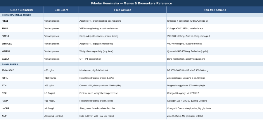
7 Biomarkers Worth Tracking With Fibular Hemimelia
Most monitoring for fibular hemimelia focuses on imaging: X-rays, leg-length discrepancy measurements, joint alignment assessments. Blood-based biomarkers rarely enter the conversation unless a specific clinical concern arises. That is a missed opportunity. These markers can detect problems early, guide nutrition and supplementation decisions, and provide objective feedback during critical growth windows and post-surgical recovery phases. The seven below are selected because they are directly tied to bone quality, growth factor signaling, calcium regulation, and systemic inflammation — all of which have compounding effects in this condition.
Biomarker 1: 25-OH Vitamin D
Vitamin D operates as a steroid hormone, regulating calcium absorption, bone mineralization, immune function, and gene expression across hundreds of target tissues. For fibular hemimelia, optimizing vitamin D matters because bone quality affects not only the absent or hypoplastic fibula, but the compensating tibia, knee, and hip joint that carry disproportionate load across a lifetime. Suboptimal vitamin D accelerates bone loss, impairs post-surgical healing, and reduces muscle strength — all of which create secondary complications in an already asymmetric musculoskeletal system.
The standard clinical reference range is 20–50 ng/mL, but Peter Attia and other precision medicine practitioners target 40–60 ng/mL for optimal musculoskeletal and immune function. Levels below 30 ng/mL are associated with elevated PTH, reduced bone mineral density, and slowed healing. Levels above 100 ng/mL carry toxicity risk, making monitoring essential when supplementing at higher doses.
How to measure it: Standard blood test (serum 25-hydroxyvitamin D). Cost: $30–80 at independent labs; often included in comprehensive metabolic or hormonal panels. Retest 8–12 weeks after any supplement change. The NIH Office of Dietary Supplements vitamin D fact sheet provides authoritative reference on dosing and safety thresholds.
If the score is low — plan without supplements: Ten to twenty minutes of midday sun exposure (UV index 3 or above) with arms and legs exposed is the most natural correction. Dietary sources include wild-caught salmon (600–1000 IU per 3 oz), sardines, mackerel, egg yolks, and cod liver oil (approximately 400–1000 IU per teaspoon). This approach alone is rarely sufficient for correcting significant deficiency but builds a meaningful baseline. Frequency: daily sun exposure where possible; dietary sources three to four times per week.
If the score is low — plan with supplements: Vitamin D3 at 4000–5000 IU/day for correction when levels are below 30 ng/mL, paired with vitamin K2 in the MK-7 form at 100–200 mcg/day. K2 activates osteocalcin and matrix Gla protein, directing calcium toward bone rather than soft tissue — this pairing matters most when correcting severe deficiency. Once levels reach 40–60 ng/mL, a maintenance dose of 2000–4000 IU/day is appropriate. No cycling required. Side effects: toxicity requires sustained doses above 10,000 IU/day for extended periods without monitoring; K2 at these doses has an excellent safety profile.
Biomarker 2: IGF-1 (Insulin-like Growth Factor 1)
IGF-1 is the primary downstream mediator of growth hormone signaling. It drives bone growth, muscle protein synthesis, and tissue repair. In children with fibular hemimelia undergoing leg lengthening procedures, IGF-1 levels directly influence how well regenerate bone fills in during distraction osteogenesis — the process by which new bone is laid down as the two bone ends are gradually separated. In adults, IGF-1 continues to regulate bone turnover and muscle maintenance. Both low and high IGF-1 carry risks: low levels are associated with poor bone density and slow healing; chronically elevated levels are associated with increased cancer risk. The goal is optimal range, not maximum.
Age-adjusted adult targets are approximately 130–220 ng/mL, with Attia suggesting the upper third of the age-adjusted range for musculoskeletal optimization. In children, IGF-1 should be interpreted against growth velocity references. Low IGF-1 in a child undergoing bone lengthening may contribute to slower regenerate consolidation and longer external fixation time.
How to measure it: Fasting blood test (serum IGF-1). Cost: $50–150 at specialty labs; may require a physician's order. Always interpret against age- and sex-adjusted reference ranges — raw numbers without age context are not meaningful.
If the score is low — plan without supplements: Resistance training is the most potent lifestyle activator of the GH/IGF-1 axis. Compound movements adapted for the affected limb — upper body pressing and pulling, seated resistance, aquatic resistance training — performed three to four times per week produce measurable IGF-1 improvements. Adequate protein intake (1.6–2.2g per kilogram of body weight per day) is required because IGF-1 synthesis depends on amino acid availability. Sleep quality is equally critical: growth hormone secretes primarily in the first two hours of deep sleep, driving liver IGF-1 production. Seven to nine hours of quality sleep is non-negotiable for IGF-1 optimization.
If the score is low — plan with supplements: Zinc deficiency independently suppresses IGF-1. Zinc picolinate at 15–25mg/day with food addresses this cofactor. For long-term zinc use, add 1–2mg copper to prevent depletion from competition. Glycine at 3–5g before bed supports sleep architecture and collagen synthesis. Creatine monohydrate at 3–5g/day has emerging evidence for supporting bone and muscle anabolism through IGF-1-related pathways, with an excellent long-term safety record and no cycling required. Side effects: zinc at doses above 40mg/day can cause GI upset; at 15–25mg it is well-tolerated.
Biomarker 3: PTH (Parathyroid Hormone)
Parathyroid hormone regulates calcium and phosphate balance in the blood, primarily by controlling bone resorption and renal calcium retention. Chronically elevated PTH — secondary hyperparathyroidism — is a direct driver of bone loss. The most common reversible cause is vitamin D deficiency, which is why PTH and vitamin D should always be measured together. In fibular hemimelia, elevated PTH undermines bone quality in the compensating structures — tibia, knee joint, contralateral hip — and can slow healing after any surgical intervention.
The intact PTH reference range is 15–65 pg/mL. Values consistently above 65 pg/mL warrant investigation and intervention. The target for bone health optimization is the lower half of the normal range — chronically elevated PTH within "normal" limits still represents a meaningful bone loss driver over years.
How to measure it: Intact PTH blood test, ideally fasting and in the morning. Cost: $50–120. Best measured alongside serum calcium, phosphate, and 25-OH vitamin D for a complete mineral metabolism picture.
If the score is elevated — plan without supplements: Correcting vitamin D deficiency is the most effective first step — it addresses the most common reversible cause directly. Adequate dietary calcium from food sources (1000–1200mg/day from dairy, leafy greens, fortified foods, canned fish with bones) reduces the stimulus for PTH secretion. Weight-bearing activity signals bones to retain calcium, reducing reliance on PTH-driven renal calcium retention. Reducing excess caffeine and alcohol, both of which increase urinary calcium loss, also contributes.
If the score is elevated — plan with supplements: Magnesium glycinate at 300–400mg taken at night is the highest-priority supplement here. Magnesium deficiency directly elevates PTH and is significantly underdiagnosed in the general population. Correcting both magnesium and vitamin D simultaneously will in most cases normalize elevated PTH within 8–12 weeks. The NIH ODS magnesium fact sheet provides comprehensive dosing and safety reference data. No cycling required for magnesium at these doses. Side effects: glycinate form is better tolerated than oxide; loose stools are possible above 400mg.
Biomarker 4: CTX (C-terminal Telopeptide of Type I Collagen)
CTX, also referred to as β-CrossLaps, is a bone resorption marker — it reflects how actively osteoclasts are breaking down bone collagen. Elevated CTX means bone is being degraded faster than it is being rebuilt, which in the context of fibular hemimelia can compound existing structural vulnerabilities, accelerate compensatory joint wear, and delay post-surgical bone healing. CTX is highly sensitive to nutritional status, cortisol load, and exercise habits, making it a dynamic marker that responds relatively quickly to interventions.
A critical pre-analytical requirement: CTX follows a strong diurnal rhythm and is suppressed significantly by food intake. A valid CTX result requires a fasting morning sample drawn between 7 and 9 am, with nothing consumed since midnight except water. An improperly collected sample cannot be interpreted. In adults, fasting CTX above 0.7 ng/mL is generally considered elevated in the bone health monitoring context.
How to measure it: Fasting morning serum draw. Cost: $60–100. Some labs offer home blood collection kits with cold-chain packaging. Measure alongside P1NP (below) for the complete bone turnover picture.
If the score is elevated — plan without supplements: The four most common reversible drivers are inadequate protein intake, chronic sleep disruption (elevated cortisol directly drives osteoclast activity), insufficient weight-bearing exercise, and nutritional deficiencies including vitamin D, calcium, and magnesium. Address all four before adding supplements: target 1.6–2.0g protein per kilogram bodyweight daily, prioritize 7–9 hours of sleep, include three to four days per week of adapted resistance or weight-bearing exercise, and address any identified dietary deficiencies.
If the score is elevated — plan with supplements: Omega-3 fatty acids (EPA+DHA) at 2–4g/day have well-documented anti-resorptive properties — they reduce osteoclast activity and suppress bone-resorbing cytokines including IL-6 and TNF-α. The NIH ODS omega-3 fact sheet summarizes the evidence base. Vitamin K2 (MK-7 form, 100–200mcg/day) activates carboxylated matrix Gla protein, which directly suppresses osteoclast-mediated bone loss. This combination can meaningfully reduce elevated CTX over 12–16 weeks. No cycling required for either. Side effects: high-dose omega-3 (above 4g/day) can have mild anticoagulant effects — discuss with a physician if on blood-thinning medication.
Biomarker 5: P1NP (Procollagen Type 1 N-terminal Propeptide)
If CTX tells you how fast bone is being broken down, P1NP tells you how fast new bone is being built. P1NP is released as a byproduct when type 1 collagen — the primary structural protein in bone — is synthesized by osteoblasts. It is widely considered the gold-standard bone formation marker, preferred over older alternatives like bone-specific alkaline phosphatase for its sensitivity and precision. Tracking P1NP alongside CTX gives the full picture of bone turnover balance: you need both the resorption and formation sides to understand whether the net result is bone gain, maintenance, or loss. Low P1NP paired with elevated CTX is the most clinically concerning pattern.
For adults in bone health monitoring, P1NP in the range of 15–80 mcg/L is typical, with higher values expected during active growth phases in children, post-fracture healing, and the distraction osteogenesis phase of leg lengthening. Values below 15 mcg/L in a context where bone formation should be active suggest an inadequate building response.
How to measure it: Blood test (serum). Cost: $80–150 at most endocrinology or specialist labs. Best measured in the morning at the same time as CTX for within-person consistency across retests. No fasting requirement, though morning collection is conventional.
If the score is low — plan without supplements: Resistance exercise is the most reliable non-pharmacological stimulator of bone formation. Adapted resistance training — pool resistance, upper body compound movements, seated leg press as structurally tolerated — produces measurable increases in P1NP within 8–12 weeks. Adequate protein intake is foundational: bone matrix is collagen, and without sufficient amino acids the anabolic signal from exercise cannot translate into actual new bone. Sleep is the third non-supplement lever — bone remodeling is predominantly nocturnal, and sleep deprivation measurably suppresses bone formation markers.
If the score is low — plan with supplements: Collagen peptides at 10g/day taken alongside 50–100mg of vitamin C have growing evidence for supporting bone matrix formation — the vitamin C is required as a cofactor in collagen cross-linking and cannot be omitted. Silicon (as orthosilicic acid or horsetail/bamboo extract) supports collagen matrix organization. Creatine monohydrate at 3–5g/day has been studied in the context of bone outcomes combined with resistance training and shows modest but consistent support for P1NP. Cycling: all three can be taken daily without requiring breaks. Side effects: all three have excellent safety profiles at these doses. Note that collagen peptides are derived from animal sources — relevant for dietary preference considerations.
Biomarker 6: hsCRP (High-sensitivity C-reactive Protein)
High-sensitivity CRP is the most widely available marker of systemic low-grade inflammation. Chronic elevation signals that the immune system is in a sustained state of low-level activation — a state that impairs bone formation, delays tissue healing, accelerates cartilage degradation, and increases long-term cardiovascular risk. For fibular hemimelia, hsCRP monitoring is particularly relevant in three contexts: post-surgical recovery (elevated CRP is expected acutely but should normalize within four to six weeks), management of chronic compensatory joint overload, and general long-term cardiovascular health monitoring given that sedentary periods during recovery can affect metabolic health in ways that accumulate over time.
Optimal hsCRP is below 1.0 mg/L. Values of 1.0–3.0 mg/L reflect moderate risk; above 3.0 mg/L is elevated (excluding acute illness or recent surgery, which can transiently drive CRP far higher). Thomas Dayspring and Allan Sniderman both emphasize hsCRP as a cardiovascular risk modifier that operates independently of standard lipid markers — a dimension that becomes relevant if prolonged recovery or limited mobility has affected metabolic health.
How to measure it: Standard blood test. Cost: $20–50 — one of the most affordable markers on this list. Widely available through any general practitioner or lab. Must be measured during a stable health period, at minimum four to six weeks post-surgery, and in the absence of active infection.
If the score is elevated — plan without supplements: The most powerful lifestyle interventions are sleep optimization (poor sleep is among the strongest drivers of elevated CRP), reduction of excess visceral body fat (metabolically active and producing pro-inflammatory cytokines), and a dietary shift toward whole foods and away from refined vegetable oils and processed carbohydrates. Regular zone 2 cardio — conversational-pace aerobic exercise for 30–45 minutes, four to five times per week — reduces CRP measurably over 8–12 weeks. Addressing periodontal disease is often overlooked but is a significant and reversible driver of systemic CRP.
If the score is elevated — plan with supplements: Omega-3 fatty acids (EPA+DHA, 2–4g/day) represent the best-evidenced anti-inflammatory supplement with a strong safety profile. Curcumin as a standardized extract with piperine at 500–1000mg/day curcumin with 5–10mg piperine has demonstrated CRP-lowering effects across multiple randomized trials. Magnesium glycinate at 300–400mg/night supports both CRP reduction and sleep quality simultaneously. Cycling: omega-3 can be taken continuously; curcumin benefits from cycling (8 weeks on, 4 weeks off) to maintain responsiveness. Side effects: omega-3 above 4g/day has mild anticoagulant potential; curcumin can cause GI discomfort in sensitive individuals at higher doses.
Biomarker 7: Alkaline Phosphatase (ALP)
Alkaline phosphatase is an enzyme present in bone, liver, and kidney tissues. The bone-specific isoform is produced by osteoblasts — the cells that build new bone — making it a useful bone formation indicator in the right context. In fibular hemimelia, ALP monitoring helps interpret whether elevated values reflect healthy healing (such as active distraction osteogenesis or fracture repair) or a more concerning process. Critically, ALP must always be interpreted with context: elevated ALP in a 10-year-old during active bone lengthening is expected and reassuring; the same value in a stable 40-year-old without recent surgery warrants further investigation.
Standard total ALP reference ranges: adults 44–147 U/L; children 100–400 U/L (naturally higher during growth phases). Bone-specific ALP testing is available where disambiguation from liver-origin ALP is needed.
How to measure it: Included in the standard comprehensive metabolic panel (CMP). Cost: $20–50 — often already part of routine bloodwork. Bone-specific ALP (bALP) costs $80–150 as a separate test and requires an explicit lab order.
If the score is unexpectedly low: Persistently low ALP outside of known growth suppression should prompt evaluation for hypophosphatasia — a rare enzyme deficiency with direct skeletal implications. This condition requires specialist management and is not addressed by general supplementation. If ALP is simply at the low end of normal without pathology, the approach mirrors low P1NP: resistance exercise, adequate protein, vitamin D optimization, and sleep quality.
If the score is elevated outside a known healing context — plan without supplements: First rule out liver disease by checking ALT, AST, and GGT concurrently. If liver markers are normal, elevated ALP is bone-origin. Ensure vitamin D and calcium are adequate — high bone turnover states drive ALP elevation. Reduce excess preformed vitamin A (retinol form), which at high intake increases bone resorption and can elevate ALP.
If the score is elevated — plan with supplements: Zinc at 15–25mg/day supports ALP enzyme activity as a direct cofactor and helps normalize abnormal bone turnover in the context of zinc deficiency. Magnesium glycinate at 300–400mg/night supports broader mineral homeostasis. Vitamin D3 paired with K2 provides the bone-signaling hormones needed for normalized turnover. For long-term zinc use, pair with 1–2mg copper daily. Side effects: zinc at these doses is well-tolerated; copper supplementation at 1–2mg is safe.
Tracking these seven biomarkers consistently — ideally every six months during active growth or recovery, annually during stable phases — builds a meaningful longitudinal picture of bone and metabolic health. The next layer of understanding is genetic: what the developmental biology of limb formation reveals about individual variation in this condition.
The Genetic Landscape of Fibular Hemimelia: What Research Currently Suggests
Most cases of fibular hemimelia are sporadic — occurring without a clear family history or identified genetic mutation. However, the developmental biology of limb formation is now well-mapped, and specific genes in critical signaling pathways have been linked to lower limb deficiency spectrums. Genetic testing cannot change an underlying variant, but understanding which developmental pathway was affected can usefully guide nutritional support decisions, exercise priorities, and where to direct preventive attention. The six genes below represent the most relevant ones based on current limb development research. This area is still evolving, and clinical genetic testing for fibular hemimelia is not yet routine practice — these genes represent research-informed context, not diagnostic certainties.
Gene 1: PITX1 — Master Regulator of Lower Limb Identity
PITX1 (Paired-like homeodomain transcription factor 1) is one of the few transcription factors expressed specifically in the hindlimb rather than the forelimb. It functions as a master regulator of lower extremity identity, operating upstream of TBX4 and multiple other lower-limb patterning genes. Heterozygous deletions at the PITX1 locus have been documented in isolated lower limb malformations, including cases with fibular hypoplasia and clubfoot. Its lower-limb specificity makes it one of the most directly relevant genes in this condition.
If the gene variant is present — plan without supplements: PITX1 variants affect how the lower limb was patterned during fetal development — this cannot be altered postnatally. The focus shifts entirely to optimizing functional outcomes: adapted physical therapy targeting proprioception and neuromuscular balance, progressive resistance loading of the affected limb as structurally tolerated, and proactive gait analysis to prevent compensatory hip and lumbar strain from becoming habitual. Biofeedback-assisted gait training is particularly well-matched to the proprioceptive challenges this variant creates.
If the gene variant is present — plan with supplements or equipment: No supplement directly modulates PITX1 expression in clinically meaningful ways after birth. The most impactful equipment intervention is a well-fitted orthotic or prosthetic with systematic adjustment tracking — the biomechanical compensation pattern around a PITX1-related lower limb anomaly creates secondary joint loading that accumulates silently if not monitored. A comprehensive bone health support stack (vitamin D3/K2 + magnesium + omega-3) provides nutritional foundation for the compensating skeletal structures that bear disproportionate load.
Gene 2: TBX4 — Knee, Patella, and Lower Leg Development
TBX4 (T-box transcription factor 4) is expressed selectively in the hindlimb bud and regulates knee, patella, and lower leg development. Heterozygous loss-of-function variants cause small patella syndrome, characterized by absent or hypoplastic patella, ischio-pubic hypoplasia, and lower limb anomalies that can include fibular deficiency. TBX4 sits directly downstream of PITX1 in the lower-limb transcription factor hierarchy, making these two genes functionally linked.
If the gene variant is present — plan without supplements: Patella insufficiency creates specific quadriceps mechanism challenges that, if unaddressed, accelerate cartilage wear. Targeted physiotherapy focusing on vastus medialis oblique (VMO) strengthening and hip abductor stability reduces compensatory patellar stress. Aquatic resistance training provides the mechanical stimulus without the compressive load of land-based exercise on a structurally compromised knee. Regular imaging follow-up — particularly MRI when symptoms develop — can detect early cartilage changes before they become structural.
If the gene variant is present — plan with supplements or equipment: Collagen peptides (10g/day with vitamin C) support the connective tissue matrix around a structurally vulnerable knee. MSM (methylsulfonylmethane) at 1000–3000mg/day has modest but consistent evidence for joint matrix support in clinical trials. Patellar tracking braces during high-activity periods reduce mechanical stress on an improperly tracking patella. Frequency: daily collagen peptides without cycling; MSM can be taken continuously. Side effects: both supplements have excellent safety profiles.
Gene 3: FGF10 — Limb Bud Outgrowth and Tissue Repair Capacity
FGF10 (Fibroblast Growth Factor 10) signals through FGFR2b to drive limb bud outgrowth and progenitor cell proliferation in early embryonic development. Beyond its developmental role, the FGF signaling axis continues to regulate adult tissue repair, wound healing, and bone regeneration — making FGF10 relevant not just for understanding the origin of fibular hemimelia, but for understanding individual variation in surgical recovery. People with variants that reduce FGF10 signaling efficiency may show slower tissue regeneration following surgery.
If the gene variant is present — plan without supplements: Optimize the conditions for tissue regeneration around every surgical procedure: maintain adequate caloric intake (undereating suppresses growth factor signaling broadly), allow full healing intervals between staged operations when clinically possible, and prioritize sleep as the primary tissue-repair window. Protein timing matters — a protein-rich meal or shake within two hours of a physiotherapy session or minor procedure supports FGF-driven repair signaling when it is most active.
If the gene variant is present — plan with supplements or equipment: Vitamin C at 500–1000mg/day is required as a cofactor for collagen synthesis downstream of FGF signaling — this is rarely sufficient alone but is non-negotiable as a foundation. Zinc at 15–25mg/day supports growth factor receptor function and cell proliferation. Omega-3 fatty acids at 2–4g/day reduce the excessive post-surgical inflammation that can interfere with the tissue proliferation phase. Red light therapy (photobiomodulation) has emerging evidence for supporting FGF-related tissue repair mechanisms and is discussed further in the complementary approaches section.
Gene 4: SHH and GLI3 — Anterior-Posterior Limb Patterning
SHH (Sonic Hedgehog) is one of the most critical morphogens in limb development, establishing the anterior-posterior axis of the limb bud. Its downstream effector GLI3 (chromosome 7p14.1) acts as both an activator and repressor of hedgehog target genes depending on context. GLI3 loss-of-function causes Greig cephalopolysyndactyly syndrome and Pallister-Hall syndrome, both of which involve digit and limb abnormalities. More broadly, SHH pathway dysregulation is associated with pre- and postaxial polydactyly and axial limb deficiency — patterns that overlap with the fibular hemimelia spectrum.
If the gene variant is present — plan without supplements: SHH/GLI variants that affect limb patterning are structural — the developmental outcome is established at birth and cannot be reversed. Management focuses on adaptive physical therapy, gait optimization, and proactive monitoring of digit and joint function as growth continues. Digit abnormalities associated with fibular hemimelia (often preaxial ray deficiency, absent or fused toes) may require separate orthopedic management coordinated with the fibular deficiency treatment plan.
If the gene variant is present — plan with supplements or equipment: Vitamin D functions as a partial modulator of hedgehog pathway activity in postnatal tissues — maintaining optimal levels (40–60 ng/mL) supports healthy SHH pathway signaling in bone cells and the immune system. This makes vitamin D doubly relevant for individuals with SHH/GLI variants: directly relevant for bone quality and secondarily relevant as a pathway modulator. Custom footwear and orthotics to accommodate digit variants and distribute plantar load appropriately are the most impactful equipment intervention.
Gene 5: WNT5A — Skeletal Patterning and Bone Remodeling
WNT5A operates through non-canonical Wnt signaling to regulate skeletal segment proportions, joint development, and limb axis elongation. Loss-of-function mutations cause Robinow syndrome, characterized by shortened limb segments and vertebral anomalies. More common low-function variants in the broader WNT pathway contribute to reduced bone mineral density and impaired bone remodeling. The canonical Wnt/β-catenin branch — activated by mechanical loading in osteoblasts — is the most important bone-formation signaling cascade identified to date, and it is directly stimulated by weight-bearing exercise.
If the gene variant is present — plan without supplements: Weight-bearing activity is the most direct activator of canonical Wnt bone formation signaling. Even partial weight-bearing — standing with support, walking with orthotic assistance, seated resistance exercise — generates the mechanical signals that activate Wnt target genes and drive osteoblast proliferation. This is one of the strongest mechanistic arguments for maintaining weight-bearing activity as a life-long priority in fibular hemimelia, not just during active rehabilitation phases.
If the gene variant is present — plan with supplements or equipment: Quercetin at 500–1000mg/day has early in-vitro evidence for activating Wnt signaling in osteoblast cell lines. Berberine at 500mg twice daily with food activates AMPK and has emerging evidence for supporting Wnt-driven bone metabolism. Both have broader metabolic benefits that justify use even beyond the Wnt pathway rationale. Cycling for berberine is recommended at 8 weeks on, 4 weeks off, due to microbiome and enzymatic adaptation. Side effects: quercetin is well-tolerated; berberine can cause GI effects in some individuals and may interact with certain medications — check interactions if on prescriptions.
Gene 6: SALL4 — Transcriptional Networks in Limb Patterning
SALL4 (Sal-like protein 4) is a zinc-finger transcription factor participating in the transcriptional networks that govern limb, kidney, and eye development during embryogenesis. Mutations cause Okihiro syndrome (Duane-radial ray syndrome), primarily characterized by radial ray defects and thumb aplasia; in some cases lower limb involvement has also been reported. SALL4 participates in developmental networks shared with SALL1, which causes Townes-Brocks syndrome. While more commonly associated with upper limb and radial defects, its role in the broader limb patterning gene regulatory network makes it relevant to the genetic landscape of fibular hemimelia, particularly in cases presenting with multi-limb involvement.
If the gene variant is present — plan without supplements: For individuals presenting with both upper and lower limb variants suggesting SALL4 involvement, occupational therapy for fine motor and upper extremity function should be coordinated with physical therapy for lower limb management rather than addressed in isolation. The biomarker monitoring framework described throughout this article applies — SALL4 variants do not change the approach to bone quality optimization.
If the gene variant is present — plan with supplements or equipment: No SALL4-specific nutritional interventions with meaningful clinical evidence currently exist. The foundational bone health stack — vitamin D3/K2, magnesium glycinate, omega-3 — remains appropriate as broad metabolic support. A comprehensive adaptive equipment assessment covering both upper and lower limb functional needs is the most impactful practical intervention for individuals with multi-limb involvement.
The genetic picture does not change the structural reality of fibular hemimelia — but it can inform how aggressively to support particular biological pathways. The perspective from longevity medicine adds another complementary layer.
What Peter Attia's Outlive Reveals About Bone and Musculoskeletal Health — 10 Things Worth Knowing
Peter Attia's Outlive: The Science and Art of Longevity (2023) is primarily structured around the four leading causes of chronic disease mortality. But threaded through the book is a rigorous, evidence-based framework for musculoskeletal health, bone density management, and performance longevity that maps directly onto the challenges of living with fibular hemimelia across a full lifespan. Attia's emphasis on measurable biology, early intervention, and functional outcomes over decades makes his framework one of the most practically applicable for a structural condition managed over a lifetime.
1. Bone Density Loss Begins Earlier Than Most People Expect
Attia emphasizes that peak bone mass is established in the third decade of life, and that the window between ages 20 and 30 is often the last opportunity to add meaningful density. After that, the goal becomes preservation. For fibular hemimelia, this reframes the teenage and early adult years as critical investment periods for bone quality — not only in the affected limb, but across the entire compensating skeleton that will carry asymmetric load for decades.
2. DEXA Scans Should Be Part of Routine Monitoring
Attia advocates for regular DEXA (dual-energy X-ray absorptiometry) scanning as a baseline and longitudinal tracking tool for bone mineral density. He recommends establishing a baseline by age 25–30 for anyone with risk factors for bone loss — a category that fibular hemimelia clearly occupies. Cost: $100–300 at most imaging centers. T-scores and Z-scores should be tracked across time, not evaluated as single measurements in isolation.
3. Resistance Training Is the Non-Negotiable Bone Intervention
No supplement, dietary strategy, or medication produces the bone formation signal as reliably as mechanical load. Attia is explicit that resistance training — not cardio, not stretching alone — is required to preserve and build bone mineral density. For fibular hemimelia, this means identifying the adapted resistance protocol that works within individual biomechanical constraints, rather than using those constraints as a reason to avoid resistance-based exercise entirely.
4. Protein Is Chronically Under-Consumed
Attia recommends 1g of protein per pound of body weight per day for active adults — significantly above the official RDA. Bone matrix is primarily collagen, which is protein. Muscle is protein. Healing tissue is protein. Anyone consuming below 1g/kg/day and wondering why their bone markers or surgical recovery are suboptimal is likely missing a foundational input before any supplement consideration is warranted.
5. IGF-1 Requires Calibration, Not Maximization
Unlike simplistic "more is better" thinking about anabolic hormones, Attia discusses the nuanced role of IGF-1: essential for bone and muscle maintenance, but chronically elevated IGF-1 is associated with accelerated cellular aging and cancer risk. The goal is achieving the optimal range through exercise and nutrition — not through pharmaceutical elevation of growth factors.
6. Zone 2 Cardio Delivers Metabolic Benefits Without Joint Destruction
Zone 2 — conversational-pace aerobic exercise sustained for 30–60 minutes — supports mitochondrial health, reduces visceral fat, and lowers systemic CRP without the joint loading that high-impact training creates. For fibular hemimelia, swimming, cycling, or walking at low intensity can deliver significant metabolic health benefits while protecting structurally vulnerable joints from additional wear.
7. Sleep Is When Bone Remodeling Primarily Occurs
Attia treats sleep not as a lifestyle preference but as a non-negotiable physiological process. Growth hormone secretion, bone remodeling, tissue repair, and immune regulation all peak during deep sleep. Seven to nine hours of quality sleep is a prerequisite for anyone managing an active bone remodeling or post-surgical healing process — not a bonus.
8. Grip Strength Is a Reliable Proxy for Musculoskeletal Health
Attia highlights grip strength as one of the most reliable predictors of overall musculoskeletal health and longevity outcomes. For fibular hemimelia, this is an easily trackable bilateral metric that captures overall musculoskeletal investment across the body. It can be measured with a hand dynamometer ($30–50) regardless of lower limb status, and tracked over years as an objective indicator of physical decline or improvement.
9. Asymmetric Compensation Creates Silent Secondary Risks
Attia's broader framework of proactive medicine — anticipating problems before they become symptomatic — maps directly onto fibular hemimelia. Contralateral knee, hip, and lumbar strain from years of asymmetric loading are predictable, preventable consequences. These can be monitored through biomarkers, periodic imaging, and pain pattern tracking, and mitigated through targeted exercise and orthotic optimization before structural damage accumulates.
10. ApoB Matters Even in Musculoskeletal-Focused Care
Attia's emphasis on ApoB as a cardiovascular risk marker is a reminder that bone and joint health does not exist in metabolic isolation. Periods of reduced activity during recovery, chronic pain-related compensation, and sedentary phases affect metabolic health broadly. Adding ApoB alongside the bone biomarkers in this article gives a more complete picture of long-term health trajectory — a dimension that orthopedic-focused care rarely addresses.
Complementary Approaches With Clinical Support
The approaches below have meaningful human evidence for supporting bone healing, pain management, neuromuscular function, or quality of life in musculoskeletal conditions. They are selected for their clinical plausibility in fibular hemimelia, and none of them require abandoning standard medical care — they are additive, not alternative.
Low-level Laser Therapy / Photobiomodulation
Photobiomodulation (PBM) uses red and near-infrared light (typically 630–850nm) to stimulate cytochrome c oxidase in mitochondria, increasing cellular ATP production and reducing oxidative stress in target tissues. In the context of bone healing, PBM has been studied for its ability to accelerate osteoblast activity and collagen matrix organization. For fibular hemimelia, it is most relevant in two specific windows: post-surgical soft tissue and bone healing, and scar tissue management around operative sites that can create secondary adhesion and movement restriction.
A systematic review examining LLLT in bone and soft tissue repair across multiple controlled studies found statistically significant improvements in bone formation markers and healing time, with consistent results across multiple tissue types. Human clinical trials have examined effects on fracture healing and post-surgical tissue repair. The evidence for soft tissue outcomes is stronger than for large structural bone defects, but the risk profile is minimal.
Practically: three to five sessions per week, five to fifteen minutes per session on target areas, using devices delivering 630–850nm at appropriate power density (10–50mW/cm²). Sessions are available at physiotherapy clinics (approximately $50–100 per session) or through quality home devices ($200–600 one-time). Begin two to three weeks post-surgery once incisions are fully closed. Side effects are minimal; avoid directing light at the eyes.
Massage Therapy
Fibular hemimelia creates predictable compensatory loading patterns: the contralateral leg carries surplus force, the hip abductors work asymmetrically, the lumbar paraspinals adapt to structural pelvic tilt, and the plantar fascia of the affected foot often bears altered pressure distribution. Massage therapy targeting these compensation zones reduces chronic muscle guarding, improves local circulation, and supports the neuromotor relaxation response — all of which contribute to better gait quality, reduced chronic pain, and improved range of movement over time.
Multiple systematic reviews support manual therapy for musculoskeletal pain management. A Cochrane review of massage for chronic low back pain — among the most common compensatory consequences of lower limb asymmetry — found short-term clinically significant improvements in pain intensity and functional outcomes compared to inactive controls. Condition-specific RCTs for fibular hemimelia do not exist, but the mechanistic rationale and evidence base for compensatory musculoskeletal patterns are solid.
Practically: monthly deep-tissue massage sessions focusing on the contralateral calf and hamstring, ipsilateral hip flexors, and bilateral hip external rotators represents a sustainable maintenance protocol. Self-myofascial release using a foam roller daily (five to ten minutes targeting the IT band, TFL, and calf) extends the benefit cost-effectively between professional sessions. Ensure therapists are briefed on the orthopedic history to avoid inappropriate joint manipulation.
Biofeedback
Biofeedback uses electronic sensors to measure physiological signals — muscle activation, plantar pressure distribution, surface EMG — and display them in real time, allowing conscious optimization of patterns that would otherwise operate below the threshold of awareness. For fibular hemimelia, gait biofeedback is particularly relevant: real-time feedback on step symmetry, force distribution, and muscle firing sequence improves gait quality and reduces compensatory biomechanical strain in ways that verbal instruction alone cannot achieve. Every orthotic or prosthetic change creates a brief window where habitual patterns are in flux and most responsive to retraining.
Studies in lower limb prosthetic users and patients with limb length discrepancy have demonstrated that real-time plantar pressure biofeedback improves gait symmetry and reduces compensatory trunk lean during rehabilitation. Force plate and EMG biofeedback are available through specialist sports medicine and rehabilitation centers.
Practically: an initial course of 8–12 biofeedback-assisted gait training sessions (approximately $80–150 per session) during any significant change in orthotic or prosthetic fitting represents high-return timing for this intervention. Home pressure-sensing insole systems ($150–400) extend feedback into daily life after the initial clinical course. The goal is retraining automatic gait patterns — not creating ongoing dependence on the device.
Mindfulness Meditation / MBSR
Mindfulness-Based Stress Reduction is an 8-week structured program that trains sustained, non-judgmental present-moment attention. For fibular hemimelia, its relevance extends in two directions: reducing the pain catastrophizing and surgical anxiety that commonly accompany a condition involving multiple procedures across childhood, and modulating the HPA (hypothalamic-pituitary-adrenal) axis in a way that measurably reduces cortisol — which, as covered in the CTX biomarker section, directly drives osteoclast-mediated bone resorption.
A 2014 meta-analysis in JAMA Internal Medicine covering 47 randomized controlled trials of mindfulness-based interventions found significant reductions in pain intensity, anxiety, and depression, with effects persisting at follow-up. MBSR-specific trials have demonstrated reductions in salivary cortisol and hsCRP — two biomarkers with direct relevance to bone health in the fibular hemimelia context.
Practically: the formal 8-week MBSR program — available in-person or online through MBSR-certified teachers, approximately $300–600 — is the most evidence-based entry point. For those not ready for the full commitment, a structured daily 10-minute breath-focused meditation practice using a well-designed app (Insight Timer, Waking Up) produces measurable HPA axis benefits within 4–8 weeks of consistent practice. Zero side effects, low cost, and direct biomarker relevance make this one of the most practical additions to a fibular hemimelia management plan.
Breathing-based Therapies
Slow, controlled breathing at 4–6 breaths per minute activates the parasympathetic nervous system via the vagus nerve, reduces cortisol output, and improves heart rate variability (HRV). HRV is increasingly used as a recovery and readiness marker in athletic and clinical contexts. For someone managing chronic compensatory pain, periodic surgery, and the associated physiological stress, the autonomic nervous system balance that breathing practices support has measurable downstream effects on CRP, cortisol, and subjective pain tolerance — connecting directly to several of the biomarkers discussed earlier in this article.
Meta-analyses of slow breathing interventions consistently demonstrate improvements in HRV and reductions in perceived stress and autonomic sympathetic dominance in clinical populations. HRV improvement is associated with better immune regulation and faster physiological recovery from stressors including surgical procedures. The intervention requires no equipment, no cost, and no side effects.
Practically: a daily 10–15 minute practice of 5-second inhale through the nose followed by 5-second exhale produces the core autonomic benefit. The 4-7-8 method (4 seconds inhale, 7 seconds hold, 8 seconds exhale) is useful for pre-sleep practice and sleep onset support. A HRV tracking device (Polar H10 paired with the Elite HRV app, or a WHOOP band) at $50–250 provides objective feedback on whether practice is improving recovery readiness over time — connecting breathing practice to measurable physiological data.
Conclusion
Fibular hemimelia is a structural condition, but the quality of life over decades depends on biological variables that extend far beyond what imaging captures. The seven biomarkers covered here — vitamin D, IGF-1, PTH, CTX, P1NP, hsCRP, and alkaline phosphatase — provide a measurable, actionable window into the bone metabolism, growth factor signaling, and inflammatory status that shape healing, compensation, and long-term joint health. The six developmental genes — PITX1, TBX4, FGF10, SHH/GLI3, WNT5A, and SALL4 — do not change the structural reality, but they can inform smarter decisions about where to focus nutritional and biomechanical support.
The most useful next step is not to act on everything at once. Start with the highest-yield, most accessible biomarkers: 25-OH vitamin D, hsCRP, and PTH. Bring those results to a physician or functional medicine practitioner who can contextualize them against your specific surgical history. Build the lifestyle foundations first — resistance exercise adapted to your biomechanics, adequate protein, sleep optimization — before adding targeted supplements. Then layer in additional monitoring and interventions as the picture becomes clearer. Better information, applied systematically and revisited over time, is the most reliable path toward better long-term outcomes in this condition.
Musculoskeletal Endocrine & Metabolic
Musculoskeletal: Bone Conditions Joint Conditions
Autoimmune: Inflammatory Conditions