This article contains AI generated content.
· UpdatedFixed Drug Eruption — 5 Genes and 6 Biomarkers to Track
If you have experienced a fixed drug eruption, you know the pattern: a round, darkening patch appears in the exact same spot every time you take a particular medication. It might be on your lip, your forearm, your groin, or your torso. The dermatologist tells you to avoid the drug. And while that advice is technically correct, it leaves the most important question completely unanswered — why you, and why there?
Fixed drug eruption is one of the few hypersensitivity reactions defined by its anatomical memory. That specificity is not random. It reflects something deeply biological: a cluster of immune cells permanently residing in that patch of skin, waiting. Understanding why those cells are there — and why they react so intensely to a particular drug — requires looking at genetic predisposition, drug metabolism capacity, and immune regulation in a more granular way than most clinical conversations allow.
What research published over the last two decades has made increasingly clear is that specific genetic variants — particularly in the HLA system and in drug-metabolizing enzyme genes — significantly influence who develops FDE and to which medications. At the same time, several measurable biomarkers can track inflammatory activity, immune activation, and detoxification burden in ways that create a more complete picture than symptom observation alone.
This article covers both angles in practical depth. The first section examines five genes most strongly implicated in FDE susceptibility, with a concrete plan for each regardless of whether you have access to genetic testing. The second explores six biomarkers worth tracking if recurrent or unexplained drug reactions are a concern. Beyond those two frameworks, the article also draws on a book that reframes genetic risk in actionable terms, and three evidence-grounded complementary approaches relevant to this condition. The goal is not to replace a dermatologist — it is to help you arrive at the conversation better prepared.
Summary
FDE is not just a skin reaction. It is the visible surface of a layered biological process involving immune memory, enzyme variants, inflammatory gene expression, and detoxification capacity. Most treatment conversations stop at drug avoidance. This article starts there and goes much further.
Here is what is covered:
Five genetic variants most strongly associated with FDE risk: HLA-B*22:01 (a key driver in antibiotic-triggered FDE), CYP2C9 (the enzyme governing NSAID clearance speed), GSTM1 and GSTT1 (deleted in roughly half the population, leaving reactive drug metabolites unscavenged), TNFA -308G>A (a promoter variant that cranks up inflammatory output), and MTHFR C677T (the methylation gene that affects how your body handles folate antagonists like trimethoprim). For each gene, you will find a plan for what to do with supplements and without.
Six biomarkers worth tracking: including IFN-γ as the central cytokine of the FDE response, serum tryptase for differential diagnosis, eosinophil count as an activity signal, hsCRP as an inflammatory baseline, liver function markers as proxies for drug processing capacity, and the lymphocyte transformation test for identifying the culprit drug with precision.
A summary of Dr. Ben Lynch's book Dirty Genes: ten insights about how genetic variants in detoxification, methylation, and inflammation pathways can be targeted practically — content that maps directly onto the gene-level discussion in this article.
Three complementary approaches: mindfulness-based stress reduction, low-level laser therapy for post-FDE hyperpigmentation, and microbiome-directed strategies that influence drug metabolism from the gut up.
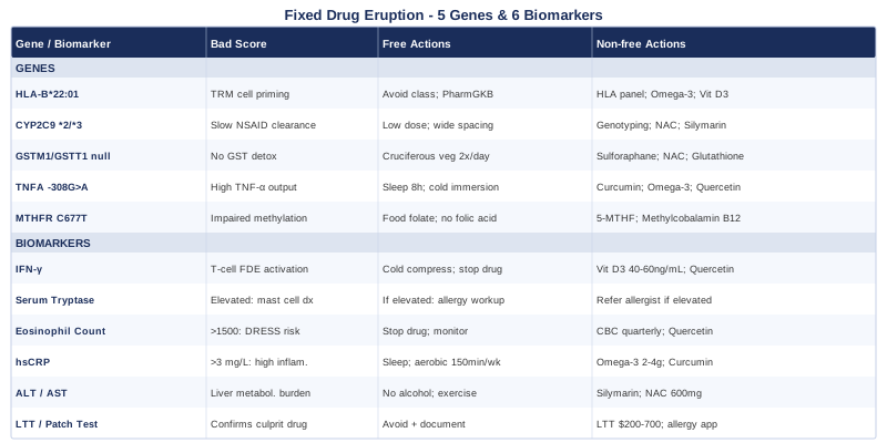
5 Genes That May Be Driving Your Fixed Drug Eruption Risk
The idea that pharmacogenomics — the study of how genes affect drug response — could explain who develops FDE and who does not is no longer speculative. Researchers like Ali Torkamani at the Scripps Research Translational Institute have been among the loudest voices making the case that individual genomic profiles should inform prescribing decisions long before reactions occur. Gary Brecka, who has built a public platform around methylation genetics and functional genomics, has helped bring concepts like MTHFR and detoxification gene variants into conversations that previously stayed in academic journals. Both perspectives are useful here.
What follows is not a genomics primer. It is a focused breakdown of the five variants most relevant to FDE — what each one does, what happens when it is working against you, and what you can do about it.
Gene 1: HLA-B Alleles — The Immune Recognition Lock
What it is and why it matters for FDE
Human Leukocyte Antigen (HLA) genes encode proteins that sit on the surface of your cells and present fragments — including fragments of drugs and their metabolites — to circulating T cells. The specific shape of your HLA binding groove determines which drug fragments get "flagged" for immune attention. HLA-B*22:01 is the most well-studied allele in the context of FDE, with particular evidence linking it to trimethoprim-sulfamethoxazole (TMP-SMX) and mefenamic acid-induced reactions in Asian populations. Other HLA-B alleles have been implicated in reactions to specific NSAIDs and anticonvulsants depending on ethnic background.
The mechanism connects directly to the defining feature of FDE: CD8+ tissue-resident memory T cells (TRM) that establish permanent residence in previously affected skin sites. These cells were primed during the initial reaction, and HLA-B alleles determined how efficiently the drug or its metabolite was presented to them. Studies available through PubMed on HLA-B and FDE consistently show that these TRM cells express high levels of IFN-γ upon drug re-exposure, which drives keratinocyte apoptosis and the characteristic erosion.
If the gene is working against you — the plan without supplements
The most practical step is pharmacogenomics testing, available through services like GeneSight, Color Genomics, or through a clinical geneticist. Knowing your HLA-B allele profile before medications are prescribed enables anticipatory avoidance. This is already standard of care in some contexts — HLA-B*57:01 testing before abacavir (HIV medication) is now routine — and the logic applies here. Beyond testing, cross-referencing your HLA alleles with drug databases like the PharmGKB database can flag medications with known allele-specific risk before you are exposed.
Practically, if you know your HLA-B allele confers risk for TMP-SMX reactions, you and your physician can substitute trimethoprim-free antibiotics for urinary tract infections. For mefenamic acid risk, the substitution is any NSAID outside the mefenamic acid (anthranilic acid) class. Drug class alternatives almost always exist; the challenge is knowing which class to avoid.
If the gene is working against you — the plan with supplements or equipment
There are no supplements that change your HLA-B allele. This is a structural genetic variant. However, there is evidence that reducing overall immune activation load — through anti-inflammatory nutritional strategies — can reduce the threshold at which TRM cells fire. Omega-3 fatty acids (2–4 g EPA+DHA daily, from fish oil) have demonstrated reductions in T cell-mediated skin inflammation in clinical trials. Cycling is generally 3 months on, 1 month off to avoid habituation and monitor tolerability. Vitamin D3 (2,000–4,000 IU daily, with K2) has demonstrated immune-modulatory effects on TRM cell populations in multiple studies. Check serum 25-OH vitamin D before starting; target 40–60 ng/mL. Side effects at standard doses are minimal, but hypercalcemia risk increases above 10,000 IU daily.
The more important equipment consideration is pharmacogenomics reporting tools. Services that produce HLA genotype reports paired with actionable drug-interaction databases give you a practical decision-support layer that most prescribers cannot currently provide from memory.
Gene 2: CYP2C9 — The NSAID Processing Gate
What it is and why it matters for FDE
Cytochrome P450 2C9 (CYP2C9) is the primary enzyme responsible for metabolizing a significant portion of commonly used NSAIDs — including ibuprofen, naproxen, diclofenac, meloxicam, and piroxicam. The gene encoding this enzyme exists in several functionally distinct variants. CYP2C9*2 (R144C substitution) and CYP2C9*3 (I359L substitution) both result in significantly reduced enzyme activity. Individuals who carry two copies of these reduced-function alleles are classified as poor metabolizers, meaning they clear NSAIDs much more slowly than the general population.
This matters for FDE in two ways. First, slower drug clearance means higher and more prolonged drug concentrations in tissue, including skin. Second, some NSAIDs produce reactive metabolites during incomplete hepatic processing. Poor metabolizers may generate a different metabolite profile than extensive metabolizers, potentially presenting novel drug fragments to HLA molecules and priming or re-stimulating TRM cells. Studies in pharmacogenomics consistently identify CYP2C9 poor metabolizers as having elevated adverse reaction risk across multiple drug classes. Research on this topic is searchable at PubMed: CYP2C9 and NSAID skin reactions.
If the gene is working against you — the plan without supplements
CYP2C9 genotyping is widely available through clinical pharmacogenomics panels and direct-to-consumer services. Knowing your metabolizer status allows several practical adjustments: first, selecting NSAIDs with minimal CYP2C9 dependence (nabumetone, for example, is metabolized differently); second, using the lowest effective dose and shortest duration for any CYP2C9-metabolized NSAID; third, spacing doses wider than standard recommendations to allow longer clearance windows. Your prescriber should be aware of your metabolizer status because it also affects anticoagulants (warfarin), oral hypoglycemics (glipizide), and certain antihypertensives — CYP2C9 is not NSAID-specific.
Acetaminophen is metabolized primarily through glucuronidation and sulfation, not CYP2C9, which is one reason it is sometimes used as an alternative in CYP2C9 poor metabolizers — though it carries its own limitations. Aspirin at low dose is also not meaningfully CYP2C9-dependent. These alternatives reduce reliance on the impaired pathway.
If the gene is working against you — the plan with supplements or equipment
Supplements that support liver phase I and phase II metabolism can help reduce the burden of incomplete drug clearance. Milk thistle (silymarin) at 140–300 mg three times daily has demonstrated hepatoprotective effects and mild CYP2C9 modulation in human trials. Cycle 6 weeks on, 2 weeks off. It is generally well-tolerated; rare gastrointestinal effects reported. N-acetylcysteine (NAC) at 600 mg twice daily supports glutathione reserves that neutralize reactive drug metabolites generated during CYP2C9-dependent processing. Take on an empty stomach; note that NAC has a brief odor due to sulfur content. Avoid in individuals with asthma (can trigger bronchospasm at high doses). For equipment, home-use laboratory services (like LabCorp's patient portal) allow you to track ALT and AST quarterly if you use NSAIDs regularly — early hepatic stress signals faster than symptoms.
Gene 3: GSTM1 and GSTT1 Null Genotypes — The Detoxification Gap
What they are and why they matter
Glutathione S-transferases M1 and T1 (GSTM1 and GSTT1) are detoxification enzymes that conjugate reactive compounds — including toxic drug metabolites — to glutathione for safe excretion. Both genes can exist in a "null" genotype, meaning the entire gene is deleted and no functional enzyme is produced. This is not rare: approximately 50% of European and East Asian populations carry the GSTM1 null genotype, and roughly 15–25% carry the GSTT1 null genotype. Double-null individuals have no functional copies of either enzyme.
For FDE, the relevance is significant. Many drugs that commonly cause FDE — including sulfonamides, NSAIDs, and certain anticonvulsants — generate electrophilic metabolites during hepatic processing. In individuals with GSTM1/GSTT1 null genotypes, these reactive metabolites cannot be efficiently conjugated to glutathione and are therefore available longer to form covalent bonds with proteins. When this happens in the skin, it creates modified self-proteins — neoantigens — that can prime the very CD8+ T cell responses characteristic of FDE. Research connecting GST null genotypes to drug hypersensitivity is searchable at PubMed: GSTM1 null drug hypersensitivity.
If the genotype is null — the plan without supplements
The dietary approach here is well-supported. Cruciferous vegetables (broccoli, Brussels sprouts, cauliflower, kale) are rich in glucosinolates that upregulate Nrf2, the transcription factor that drives expression of other phase II enzymes including NQO1 and heme oxygenase — partially compensating for absent GSTM1/GSTT1 activity. Aiming for at least two servings of cruciferous vegetables daily, lightly cooked or raw, provides measurable benefit without any supplementation. Avoiding long-fasting periods before taking reactive drugs may also be helpful, since hepatic glutathione is lower in the fasted state.
Avoiding medications with known reactive metabolite profiles when alternatives exist is the most direct intervention. Discussing this openly with prescribers using the language of "reactive metabolite burden" will often prompt consideration of alternatives they might not have volunteered.
If the genotype is null — the plan with supplements or equipment
Sulforaphane (from broccoli sprout extract, standardized to 10–30 mg sulforaphane equivalent) is the most evidence-backed Nrf2 activator available without a prescription. It compensates for null GST activity by inducing alternative phase II pathways. Take daily with food; cycling 5 days on, 2 days off can prevent adaptation. Heat destroys the myrosinase enzyme that activates sulforaphane — active supplements should use cold-processed standardized extract or add mustard seed powder to activate sulforaphane in broccoli-containing meals. N-acetylcysteine (NAC) at 600–1200 mg daily replenishes cysteine for glutathione synthesis, directly addressing the substrate deficit that null GST creates. Liposomal glutathione (250–500 mg daily) provides more direct support; standard oral glutathione is poorly absorbed, so liposomal form or sublingual delivery matters here. No significant cycling is required for NAC or glutathione at standard doses, though high-dose NAC above 1,800 mg daily warrants medical supervision. Equipment: at-home urine tests that measure 8-OHdG (oxidative DNA damage marker) can serve as indirect proxies for reactive metabolite burden and help gauge whether these interventions are having effect.
Gene 4: TNFA -308G>A — The Inflammatory Volume Dial
What it is and why it matters
The TNFA gene encodes tumor necrosis factor alpha (TNF-α), one of the most potent pro-inflammatory cytokines in the immune system. A well-studied single nucleotide polymorphism (SNP) at position -308 in the gene's promoter region — specifically the G-to-A substitution — creates what is called a "high-producer" variant. Individuals carrying the A allele at this position transcribe significantly more TNF-α in response to immune stimulation than those with the GG genotype. The heterozygous (GA) form affects roughly 30–40% of most populations.
For FDE, TNF-α plays a dual role: it amplifies the local inflammatory cascade during active reactions and contributes to the recruitment of immune cells that sustain skin damage. High-producer individuals are more likely to experience intense erythema, more blistering, and more pronounced post-inflammatory hyperpigmentation from FDE events. This SNP has been studied in multiple inflammatory and drug reaction contexts; relevant research is available at PubMed: TNF-α -308 polymorphism and drug reactions.
If the gene is working against you — the plan without supplements
Chronic background inflammation raises the TNF-α baseline that any immune trigger — including a drug metabolite — must overcome. Lowering that baseline through lifestyle gives high-producer allele carriers more buffer. Cold water immersion (10–15 minutes in water below 15°C, 3–5 times weekly) has demonstrated acute reductions in circulating TNF-α in controlled human studies. Sleep is non-negotiable: TNF-α circadian rhythm means that chronic sleep restriction consistently elevates baseline TNF-α; targeting 7.5–9 hours is a structural anti-inflammatory intervention. Refined carbohydrate reduction — specifically removing sugar-sweetened beverages and ultra-processed snack foods — is among the most evidence-supported dietary strategies for lowering chronic TNF-α output.
If the gene is working against you — the plan with supplements or equipment
Curcumin (from turmeric root, as BCM-95 or Meriva form for bioavailability; 500–1,000 mg twice daily) has the strongest supplement evidence for TNF-α modulation. Multiple randomized controlled trials show significant reductions in circulating TNF-α at these doses. Take with a fat-containing meal; absorption is substantially improved. Cycle 8 weeks on, 2–4 weeks off. Note curcumin inhibits CYP3A4 at high doses — if taking medications metabolized by CYP3A4, check for interactions. Omega-3 fatty acids (2–4 g combined EPA+DHA) consistently reduce TNF-α in meta-analyses of RCTs across multiple inflammatory conditions. Cycle similar to curcumin; monitor for blood-thinning effects if combined with anticoagulants. Quercetin (500 mg twice daily) has modest but consistent evidence for TNF-α inhibition and synergizes with curcumin at lower doses of each. Equipment: high-sensitivity CRP home testing or quarterly lab draws provide a cheap proxy for TNF-α inflammatory activity and can track whether interventions are reducing baseline inflammation over time.
Gene 5: MTHFR C677T — The Methylation Link to Folate Antagonists
What it is and why it matters for FDE
MTHFR (methylenetetrahydrofolate reductase) is the enzyme that converts folic acid into its active form, 5-methyltetrahydrofolate (5-MTHF), which is essential for the methylation cycle. The C677T polymorphism reduces enzyme activity by approximately 30% in heterozygotes (CT genotype) and 60–70% in homozygotes (TT genotype). This variant is extremely common — roughly 10–15% of most populations are TT homozygotes.
The MTHFR-FDE connection becomes particularly relevant with trimethoprim, the antibiotic component of TMP-SMX and the drug most consistently linked to FDE in published literature. Trimethoprim is a folate antagonist — it blocks dihydrofolate reductase, the enzyme that works downstream of MTHFR in the folate pathway. In individuals with MTHFR TT genotype, folate processing is already impaired. Adding a drug that further blocks the folate pathway creates a compounding deficit that affects cellular methylation, DNA repair capacity, and potentially antigen presentation dynamics in immune cells. Gary Brecka has emphasized in his widely viewed content that the TT genotype, when combined with environmental folate antagonists (medications, alcohol, processed foods), creates a "methylation debt" that affects systemic immune and inflammatory regulation. The relevant research on methylation and immune function is accessible through PubMed: MTHFR C677T immune function.
If the gene is working against you — the plan without supplements
The immediate practical intervention is eliminating supplemental folic acid (the synthetic form that MTHFR cannot efficiently convert) and replacing it with naturally occurring folate from food — dark leafy greens, lentils, black beans, avocado, asparagus. The distinction between folic acid and food-based folate is significant in MTHFR TT carriers because unmetabolized folic acid may actually compete with active 5-MTHF for cellular uptake. Reading supplement labels and avoiding products containing folic acid (rather than methylfolate) is a first-step intervention that requires no testing to implement.
More directly: if trimethoprim-containing medications have triggered FDE for you, informing your physician of MTHFR TT carrier status provides a clinical rationale for switching to alternative antibiotics that do not interfere with the folate cycle — nitrofurantoin, fosfomycin, or fluoroquinolones (depending on the indication), for example.
If the gene is working against you — the plan with supplements or equipment
5-methyltetrahydrofolate (5-MTHF) — also labeled as methylfolate, Metafolin, or Quatrefolic — bypasses the impaired MTHFR enzyme entirely, delivering active folate directly. Doses of 400–1,000 mcg daily are appropriate for most carriers; TT homozygotes may benefit from the higher end of this range under physician guidance. Start low (200 mcg) to monitor for "start-up" reactions including headache or irritability that some sensitive individuals experience. Methylcobalamin (B12 in active form, 500–1,000 mcg daily sublingually) is the essential cofactor for the methionine synthase step that requires methylfolate — the two should not be taken without each other. Riboflavin (B2) at 10–30 mg daily is the required cofactor for MTHFR enzyme itself and can modestly increase residual enzyme activity even in TT carriers; clinical trials specifically examining this are available through PubMed: MTHFR C677T and riboflavin. These three should be cycled every 3–4 months with a 2–4 week break to reassess baseline methylation markers (homocysteine is the most accessible proxy — target below 10 µmol/L).
6 Biomarkers to Track If Fixed Drug Eruption Is a Recurring Problem
Genetics tells you about predisposition. Biomarkers tell you about current biological state. The two are not interchangeable — a high-risk genotype does not mean a reaction is inevitable, and a low-risk genotype does not guarantee immunity. What biomarkers offer is a dynamic snapshot that can shift with lifestyle, medication exposure, and immune load. For FDE specifically, the following six markers span inflammatory activity, immune activation, drug-processing capacity, and differential diagnosis — each serving a distinct purpose.
Biomarker 1: Interferon-Gamma (IFN-γ)
Why it matters
IFN-γ is the central cytokine in FDE pathophysiology. When CD8+ tissue-resident memory T cells in the affected skin patch are activated by the offending drug, IFN-γ is the primary signal they release — and it directly drives the apoptosis of surrounding keratinocytes that creates the clinical lesion. Elevated IFN-γ during an active FDE episode confirms T cell-mediated immune activation and distinguishes FDE from other mechanisms (mast cell-driven reactions do not generate significant IFN-γ). Research on IFN-γ and FDE is indexed at PubMed: IFN-γ and fixed drug eruption.
How to measure it
In clinical settings, IFN-γ is most relevantly measured through the lymphocyte transformation test (LTT) or ELISpot assay — specialized laboratory tests that expose peripheral blood lymphocytes to the suspected drug and measure cytokine release (including IFN-γ). These tests are not universally available; they are offered by specialty dermatology and allergy centers. Cost typically ranges from $200–$600 USD depending on the number of drugs tested. Serum IFN-γ ELISA is available at most clinical laboratories ($30–$80) but is less specific to the skin-resident mechanism. More practically, a skin biopsy of an active FDE lesion with immunohistochemical staining for CD8+ cells and IFN-γ is the gold standard for confirming FDE diagnosis and its immune mechanism.
If the marker is elevated — the plan without supplements
Active FDE requires identifying and eliminating the culprit drug. Beyond that, cooling the inflammatory response through cold compresses (15–20 minutes, 3 times daily on active lesions), avoiding re-exposure, and keeping the affected skin clean with gentle non-irritating emollients while the T cell response subsides. Phototherapy (narrowband UVB) has evidence for accelerating resolution of FDE lesions by modulating local T cell activity — this is typically managed through dermatology.
If the marker is elevated — the plan with supplements or equipment
No supplement directly neutralizes IFN-γ, but modulating the upstream T cell activation that drives its release is achievable. Vitamin D3 has demonstrated consistent ability to downregulate IFN-γ production in CD8+ T cells across several clinical studies — targeting serum 25-OH vitamin D above 40 ng/mL is worth pursuing. Quercetin (500 mg twice daily) has shown IFN-γ suppression in cell studies and some human trials involving immune-mediated skin conditions. These are supportive measures during remission, not treatments for active reactions.
Biomarker 2: Serum Tryptase
Why it matters
Serum tryptase is the diagnostic marker for mast cell activation. In FDE, tryptase is typically normal — and that is precisely the point. If a patient with suspected FDE shows elevated tryptase (above 11.4 ng/mL), mast cell-mediated reactions (allergic urticaria, anaphylaxis, mastocytosis) become the priority differential, and management changes substantially. Confirming normal tryptase in the context of a drug reaction helps narrow the diagnosis toward T cell-mediated reactions including FDE.
How to measure it
Serum tryptase is a routine clinical laboratory test available through LabCorp, Quest Diagnostics, and most hospital systems. Cost is typically $50–$150. Optimal timing is within 1–3 hours of an acute reaction episode for maximum sensitivity. A baseline (non-reaction) tryptase level is also useful for comparison. Peter Attia has noted in his work on personalized medicine that establishing personal baselines for immune markers is more informative than a single reading compared to population norms.
If tryptase is elevated — the plan
Elevated tryptase redirects the workup toward mast cell disorders and true IgE-mediated allergy. This warrants referral to an allergist-immunologist for comprehensive evaluation including skin prick testing and possibly bone marrow assessment if systemic mastocytosis is suspected. This path moves away from FDE as the diagnosis and toward a different management algorithm entirely. Normal tryptase, in contrast, supports FDE and closes that differential efficiently.
Biomarker 3: Eosinophil Count (CBC with Differential)
Why it matters
Peripheral eosinophilia — an elevated eosinophil count in circulating blood — can accompany FDE, particularly in the acute phase of a reaction. While eosinophilia in FDE is typically mild (below 1,500 cells/µL), its presence signals allergic or hypersensitivity activation and can correlate with reaction severity. More importantly, marked eosinophilia (above 1,500 cells/µL) shifts concern toward DRESS syndrome (Drug Reaction with Eosinophilia and Systemic Symptoms), a far more dangerous drug reaction requiring urgent management — making eosinophil count a critical safety filter.
How to measure it
A complete blood count with differential (CBC w/ diff) is one of the most affordable laboratory tests available — typically $20–$50 at any clinical lab. It directly provides absolute eosinophil count and percentage. For ongoing monitoring, quarterly CBCs during periods when you are taking medications with known FDE or drug reaction risk profiles represent an inexpensive safety net. Thomas Dayspring and other clinical lipidologists who advocate for comprehensive lab panels routinely include CBC as a baseline panel component precisely because of its low cost and broad diagnostic utility.
If eosinophils are elevated — the plan without supplements
Mild eosinophilia in the context of a recent drug reaction is expected and typically resolves within weeks of stopping the offending drug. Persistent eosinophilia beyond 6 weeks warrants re-evaluation. High eosinophilia (above 1,500 cells/µL) with systemic symptoms (fever, lymphadenopathy, organ involvement) requires urgent medical evaluation to rule out DRESS.
If eosinophils are elevated — the plan with supplements or equipment
Quercetin (500 mg twice daily) has evidence for reducing eosinophilic inflammation in conditions like allergic rhinitis and eosinophilic gastrointestinal disease, with plausible mechanisms relevant to drug hypersensitivity. Omega-3 fatty acids have also demonstrated modest eosinophil-modulating effects. These are maintenance strategies during remission, not interventions during active reactions.
Biomarker 4: High-Sensitivity CRP (hsCRP)
Why it matters
C-reactive protein (CRP) is a general-purpose inflammatory marker produced by the liver in response to cytokine signaling — particularly IL-6 and TNF-α. High-sensitivity CRP (hsCRP) measures the same protein but at lower concentrations, allowing detection of subclinical chronic inflammation. For FDE, hsCRP is most useful as a baseline tracker: elevated CRP between drug reaction episodes suggests ongoing systemic inflammatory load that lowers the threshold for T cell re-activation and may contribute to more intense reactions when they occur.
How to measure it
hsCRP is available as a standalone test at most labs for $15–$40, or is often included in expanded metabolic panels. Optimal levels are below 1.0 mg/L; 1.0–3.0 mg/L is moderate risk; above 3.0 mg/L suggests high systemic inflammation. Peter Attia considers hsCRP one of the most actionable preventive biomarkers precisely because it is cheap, responsive to lifestyle change, and provides a proxy for inflammatory biology across multiple systems simultaneously.
If hsCRP is elevated — the plan without supplements
The most potent non-pharmacological interventions for elevated hsCRP are sleep optimization (7.5–9 hours nightly), aerobic exercise (150+ minutes moderate intensity weekly), smoking cessation, and dietary Mediterranean or whole-food pattern with reduced ultra-processed food intake. These interventions, in combination, can reduce hsCRP by 30–50% within 8–12 weeks in people with elevated baselines.
If hsCRP is elevated — the plan with supplements or equipment
Omega-3 fatty acids (2–4 g EPA+DHA daily) have the most consistent RCT data for hsCRP reduction in populations without active infection. Curcumin (BCM-95 or Meriva form, 500 mg twice daily) provides complementary anti-inflammatory signaling through NF-κB inhibition. A wearable heart rate variability (HRV) tracker (Oura Ring, WHOOP) provides indirect daily feedback on systemic inflammatory load — low HRV correlates with elevated inflammatory cytokines — and serves as a motivational proxy between lab draws.
Biomarker 5: Liver Function Tests (ALT and AST)
Why it matters
The liver is the primary organ of drug metabolism. Hepatocyte health — measured most accessibly through alanine aminotransferase (ALT) and aspartate aminotransferase (AST) — directly determines how efficiently drugs are processed, how long reactive metabolites circulate, and how much oxidative stress drug processing generates. In individuals with impaired drug metabolism (CYP2C9 variants, GSTM1/GSTT1 null genotypes), baseline liver function is especially relevant. Subclinical hepatic stress — even mild ALT elevation — impairs the efficiency of phase I and phase II metabolism and may increase reactive metabolite burden.
How to measure it
ALT and AST are standard components of any comprehensive metabolic panel (CMP), costing $20–$80. Optimal ALT is below 25 U/L in women and below 30 U/L in men according to updated liver health guidelines — the conventional upper limit of normal (40–55 U/L) is now considered too permissive by hepatologists including those Allan Sniderman has cited in discussions of cardiovascular and metabolic risk. Quarterly testing is appropriate for individuals regularly taking CYP2C9-metabolized or GSTM1-dependent drugs.
If liver enzymes are elevated — the plan without supplements
Alcohol elimination (even moderate consumption elevates ALT) is the single most impactful intervention for most individuals with mild elevation. Fructose reduction (eliminating sugar-sweetened beverages, fruit juice, high-fructose corn syrup) directly reduces hepatic lipid accumulation — a common cause of elevated liver enzymes in non-alcoholic populations. Moderate-intensity aerobic exercise 5 times weekly has demonstrated ALT reductions equivalent to pharmaceutical interventions in several NAFLD trials.
If liver enzymes are elevated — the plan with supplements or equipment
Silymarin (milk thistle, 140–300 mg standardized extract three times daily) has the strongest human evidence for hepatoprotection and modest enzyme reduction. Cycle 6–8 weeks with 2–3 week breaks. NAC (600 mg twice daily) supports hepatic glutathione reserves. Berberine (500 mg twice daily with meals) has shown meaningful ALT reductions in NAFLD trials and may complement milk thistle synergistically; however, berberine is a moderate CYP3A4 inhibitor — check for drug interactions before combining.
Biomarker 6: Lymphocyte Transformation Test (LTT) / Drug-Specific Patch Testing
Why it matters
Identifying the specific drug responsible for FDE is not always straightforward, especially in individuals on polypharmacy. The lymphocyte transformation test (LTT) measures proliferation of peripheral blood lymphocytes in response to in vitro drug exposure — a positive result confirms drug-specific T cell sensitization, which is the immunological hallmark of FDE. Epicutaneous patch testing on a previously affected skin site using the suspected drug (a technique pioneered in the FDE context) is a complementary clinical tool; it provokes a localized reaction at the pre-sensitized site, confirming both the diagnosis and the causative drug without systemic drug challenge.
How to measure it
LTT is available through specialized allergy and immunology centers; cost ranges from $200–$700 depending on the number of drugs tested. Sensitivity is moderate (approximately 50–70% depending on the drug class) and specificity is generally high. Provocation patch testing on the old lesion site is performed by trained dermatologists and costs significantly less (typically $150–$400) but requires appropriate timing (test during remission, at least 4 weeks after the active lesion). False negatives occur with both tests; a negative result does not fully exclude a drug as the cause. Research on these diagnostic approaches is indexed at PubMed: LTT and fixed drug eruption.
If the test confirms a causative drug — the plan without supplements
Permanent avoidance of the confirmed drug and documentation in your medical record (with notation in pharmacy records and allergy alert systems) is the direct next step. Equally important is identifying structurally similar drugs that might cross-react through the same HLA-mediated recognition pathway — a clinical pharmacologist or allergist with pharmacogenomics training is best positioned to advise on this cross-reactivity mapping.
If the test confirms a causative drug — the plan with supplements or equipment
Allergy management apps (AllergyEats, Orca Health's drug allergy tracker) and pharmacy-connected allergy-alert systems (some offer API integration with prescribing systems) can serve as technology guardrails against inadvertent re-exposure. Medical ID wristbands or phone apps that list confirmed drug allergies function as critical safety redundancy. There are no supplements that prevent re-sensitization or eliminate the resident TRM population in affected skin sites.
Dirty Genes — 10 Insights That Reframe How You Think About Drug Metabolism
Dr. Ben Lynch's Dirty Genes (2018) starts from a premise that challenges most conventional medical framing: genetic variants in your detoxification, methylation, and inflammatory pathways are not permanent sentences. They are tendencies — and those tendencies can be meaningfully influenced by what you eat, how you sleep, what you're exposed to, and which supplements you take. The book's thesis maps directly onto what is relevant for FDE, and several of its central points deserve emphasis here.
1. Genes Are Not Destiny — They Are Dimmer Switches
Lynch opens with the distinction between genetic variants that are "born dirty" (true loss-of-function mutations) and those that are "soiled" by environmental inputs — poor diet, toxin exposure, poor sleep. Most of the variants relevant to FDE, including MTHFR and GSTM1, fall into the soiled category. This reframe matters because it suggests intervention is possible even without genetic testing: cleaning up inputs reduces gene expression of harmful variants even when you cannot change the gene itself.
2. MTHFR Is the Master Regulator — and Most Doctors Miss It
Lynch dedicates significant attention to MTHFR, emphasizing that its effects ripple across neurotransmitter synthesis, antioxidant production, DNA repair, and immune regulation — not just homocysteine levels. Trimethoprim's mechanism as a folate antagonist is directly relevant here. A physician who screens only for obvious symptoms of folate deficiency (macrocytic anemia) will miss the subtler immune dysregulation that MTHFR TT carriers may experience when placed on folate-blocking antibiotics.
3. Glutathione Is the Missing Piece — and You Can Build It
Lynch emphasizes glutathione as the body's master antioxidant and its central role in detoxifying reactive metabolites — exactly the function that is impaired in GSTM1/GSTT1 null carriers. His recommended approach mirrors what is described in this article: NAC as the precursor, glycine (3–5 g daily) as the second building block, and glutathione-rich food sources (avocado, spinach, asparagus) as dietary foundation.
4. Synthetic Folic Acid May Be Making Things Worse
One of Lynch's most practically actionable points is the distinction between folic acid (synthetic, added to fortified foods and most supplements) and methylfolate (the bioavailable form). MTHFR TT carriers do not convert folic acid efficiently. Unmetabolized folic acid accumulates and may actually block folate receptors, impairing active 5-MTHF uptake. Checking every supplement and fortified food label for the presence of folic acid — and replacing it with methylfolate — is a concrete step that requires no prescription.
5. COMT Affects Drug-Related Mood and Stress Response
COMT (catechol-O-methyltransferase) breaks down dopamine, adrenaline, and estrogens. Slow-COMT individuals (Val158Met Met/Met genotype) clear catecholamines slowly and tend toward heightened stress reactivity. This matters for FDE because stress elevates cortisol, which dysregulates immune cell trafficking and can lower the threshold for skin T cell activation. Lynch recommends magnesium glycinate (300–400 mg nightly), gentle aerobic exercise, and avoiding COMT-inhibiting polyphenols (like EGCG from green tea at high doses) in slow-COMT individuals.
6. NQO1 — The Backup Detoxifier That Needs Riboflavin
NAD(P)H:quinone oxidoreductase 1 (NQO1) is a phase II enzyme that Lynch calls the "backup quarterback" for detoxification when GST enzymes are absent. The C609T polymorphism in NQO1 reduces this enzyme's activity significantly. The intervention is riboflavin (B2), which is both an MTHFR cofactor and NQO1 support. This connection — a single B vitamin with dual relevance to methylation and detoxification — is the kind of convergence Lynch's framework highlights as the most efficient intervention targets.
7. Inflammation Genes Respond to Daily Choices Faster Than Expected
Lynch reviews research showing that pro-inflammatory gene expression can shift measurably within days of dietary change. His protocols for high TNF-α producers (the TNFA -308 A allele discussed in this article) center on removing inflammatory dietary triggers — sugar, refined seed oils, alcohol — before adding anti-inflammatory supplements. This sequence matters: supplements added to an inflammatory diet show blunted effects in intervention trials.
8. Testing Before Supplementing — the Lynch Rule
A recurring caution throughout Dirty Genes is the risk of blindly supplementing without knowing your specific variant profile. Some interventions (high-dose methylfolate, high-dose B12) can cause anxiety, insomnia, or overmethylation in individuals who do not need them. Lynch recommends starting at the lowest effective dose, monitoring symptoms for the first two weeks, and using homocysteine, organic acid panels, and symptom tracking as response indicators before escalating.
9. The Sneak Peek Genes — MTR and MTRR
Lynch dedicates a chapter to MTR and MTRR — two enzymes in the methylation cycle that work directly upstream and downstream of MTHFR. MTR is methionine synthase; MTRR is its regenerator. Variants in either gene can cause methylation cycle stalling independent of MTHFR, and they often co-occur. For FDE patients investigating the MTHFR-trimethoprim connection, knowing MTR and MTRR status adds precision to the intervention — some individuals with stalled methylation despite correct methylfolate supplementation have a MTR or MTRR variant as the actual bottleneck.
10. Epigenetic Momentum — Why Changes Take Weeks, Not Days
Lynch closes with the concept of "epigenetic momentum" — the idea that gene expression patterns established over years of inflammatory dietary and lifestyle input do not reset overnight. His clinical observations suggest 3–6 months of consistent intervention before lab markers and symptoms reflect the full benefit of gene-targeted strategies. This matches what the pharmacogenomics research suggests: reducing reactive metabolite burden and inflammatory background through MTHFR support, GST pathway compensation, and TNF-α modulation is a cumulative intervention, not a quick fix. Patience with the protocol is built into the biology.
Complementary Approaches With Meaningful Evidence
The following three approaches are selected from the evidence-supported modality list because each has specific relevance to either the underlying immune mechanism of FDE, its skin-level consequences, or the drug metabolism pathways that influence its severity. None is a replacement for drug avoidance or medical management — each represents a complementary layer worth considering.
Mindfulness-Based Stress Reduction (MBSR)
MBSR is an 8-week structured program developed by Jon Kabat-Zinn at the University of Massachusetts Medical School, combining body scan meditation, mindfulness sitting practice, and gentle movement. Its relevance for FDE centers on the well-established connection between psychological stress and immune dysregulation — specifically, stress-induced cortisol fluctuations that alter trafficking and activation thresholds of skin-resident T cells. Individuals under chronic stress have demonstrably elevated baseline inflammatory cytokines, including IFN-γ, making the T cell response in FDE sites more volatile.
A meta-analysis published in PubMed examining MBSR and inflammatory markers found significant reductions in circulating CRP and IL-6 across randomized controlled trials. The Dusek et al. (2008) genomic study showed that relaxation response training — the physiological mechanism MBSR activates — produced measurable changes in gene expression pathways governing inflammation and oxidative stress within 8 weeks.
Practically, the MBSR protocol can be accessed through structured online courses (Palouse Mindfulness offers a free, evidence-based version), hospital-based programs, or certified instructors. The weekly time commitment is approximately 45 minutes of formal practice daily. For FDE specifically, the application is preventive rather than acute: MBSR is unlikely to abort an active reaction but may reduce the frequency and intensity of future reactions by lowering the chronic stress baseline that primes T cell reactivity.
Low-Level Laser Therapy (LLLT) / Photobiomodulation
Low-level laser therapy uses wavelengths of light — typically in the 630–1000 nm range — at non-thermal intensities to stimulate cellular energy production via cytochrome c oxidase, reduce oxidative stress, and modulate inflammatory cytokine expression in skin cells. Its most directly applicable role in FDE is the treatment of post-inflammatory hyperpigmentation (PIH), the darkening that persists at FDE sites long after the active lesion resolves. PIH at FDE sites can persist for months to years and causes significant cosmetic concern for many patients.
Multiple randomized controlled trials have examined LLLT for PIH, with generally positive outcomes. A double-blind RCT found that low-fluence 1064 nm Nd:YAG laser treatment significantly reduced melanin index at PIH sites compared to sham treatment, with minimal adverse effects. Research on photobiomodulation and skin pigmentation is indexed at PubMed: LLLT and post-inflammatory hyperpigmentation. The evidence base specifically for FDE-related PIH is limited, extrapolated from general PIH literature.
Realistically, LLLT for PIH is most accessible through dermatology or medical aesthetics clinics offering laser pigmentation treatments. Home-use LLLT devices are available (typically 630–850 nm LEDs) for $50–$300, but evidence for their efficacy on established PIH is weaker than clinic-based devices. For FDE PIH specifically, patience is key: the hyperactive melanocyte response at repeatedly inflamed FDE sites tends to be more persistent than PIH from other causes, and multiple sessions (6–10) are typically needed before meaningful pigment reduction is observed.
Microbiome-Directed Therapies
The gut microbiome is now recognized as a significant modulator of drug metabolism — through direct bacterial enzymatic transformation of drugs and metabolites, and indirectly through microbial modulation of host CYP450 enzyme expression in the liver. Emerging research shows that gut microbiome composition influences the activity of CYP2C9, CYP3A4, and other drug-metabolizing enzymes. Specific bacterial species produce β-glucuronidase enzymes that reactivate drug metabolites that the liver has already conjugated for excretion — effectively recycling drugs and their metabolites back into systemic circulation and potentially increasing reactive metabolite exposure.
A growing body of clinical evidence supports targeted microbiome intervention for inflammatory skin conditions. Research on the gut-skin axis and drug metabolism is accessible through PubMed: gut microbiome and drug metabolism. A randomized trial examining Lactobacillus rhamnosus and inflammatory skin reactions found modulation of both systemic inflammatory markers and skin barrier function. The connection to FDE specifically is indirect but mechanistically plausible: reducing the microbiome-driven reactivation of drug metabolites, and supporting the mucosal immune environment that shapes systemic immune tone, may lower reactive metabolite burden at skin sites.
The most practically accessible microbiome-directed approach for FDE management combines: a high-fiber, diverse-plant diet (30+ different plant foods weekly, as recommended by the British Gut Project data); avoidance of unnecessary antibiotic courses that deplete protective commensal species; and targeted probiotic supplementation with strains that have demonstrated specific β-glucuronidase-reducing activity (Lactobacillus acidophilus and Bifidobacterium longum in particular). Fermented foods (yogurt, kefir, kimchi, sauerkraut, miso) provide direct bacterial seeding. A 16S rRNA gut microbiome test (Viome, Thryve, or clinical-grade through a gastroenterologist) can identify specific dysbiosis patterns that warrant targeted correction. Cost ranges from $100–$400 for consumer-grade testing.
Conclusion
Fixed drug eruption has a cleaner biological story than most chronic skin conditions, and that story — told in HLA alleles, metabolizing enzyme variants, and cytokine patterns — is increasingly legible through genetics and biomarker tracking. The five genes covered in this article are not obscure research curiosities. They are testable, actionable variants that explain why some people react and others do not, and for each one there is a practical response path — with or without supplements. The six biomarkers are not exotic panels available only in academic medical centers; most are accessible through standard clinical labs for under $100 per draw.
The most useful next step depends on where you currently are. If you have never identified the causative drug definitively, a lymphocyte transformation test or structured provocation patch test under dermatologic supervision is the priority. If the drug is known, a pharmacogenomics panel covering HLA-B alleles and CYP2C9 will sharpen your understanding of why you are susceptible and which drug classes to approach with additional caution. If recurrent reactions are a concern, tracking hsCRP and liver function quarterly, supporting methylation with methylfolate and methylcobalamin, and optimizing glutathione pathways through diet and NAC gives your biology more capacity to process reactive compounds before they reach the immune threshold.
None of this replaces a conversation with a dermatologist or clinical pharmacologist who knows your history. But arriving at that conversation with a clearer understanding of your own biology — your HLA alleles, your metabolizer status, your inflammatory baseline — changes what becomes possible in that room.
Digestive: Liver & Gallbladder Conditions
Skin: Inflammatory Skin Conditions Pigmentation Conditions
Autoimmune: Inflammatory Conditions