This article contains AI generated content.
· UpdatedFlexion Contracture of the Knee: 6 Genes and 7 Biomarkers to Track
Introduction
Living with a knee that will not fully straighten changes more than your range of motion. It reshapes how you walk, how you sleep, how you climb stairs, and eventually how you think about your own body. If you have been through physical therapy, diligent stretching, or even surgical intervention and still hit a wall, it is not necessarily because you have not tried hard enough. It is more likely that the biology underneath the contracture has not been fully addressed.
Flexion contracture is not a single problem with a single solution. It is the visible end result of a biological cascade — chronic inflammation driving fibrosis in the posterior joint capsule, collagen crosslinking that stiffens connective tissue, muscle imbalances maintained by neurological patterns, and nutrient deficiencies that impair tissue repair. When you treat the symptom without addressing those root drivers, progress tends to plateau.
The question worth asking is: what is your internal environment actually doing? Specific blood markers can tell you whether inflammation is actively feeding tissue fibrosis, whether your collagen metabolism is out of balance, or whether a simple deficiency is quietly blocking recovery. And if you have access to genetic data from a consumer DNA test, certain gene variants can explain why your connective tissue, inflammation response, or muscle fiber composition makes you more prone to this condition — and what to do about it.
This article covers both. You will find seven biomarkers worth testing, each with a practical plan for when the score is abnormal. You will also find six gene variants that meaningfully influence joint health, along with specific protocols tailored to each risk profile. Beyond that, the article covers what leading researchers in flexibility science have identified as the most overlooked drivers of chronic stiffness — and four complementary approaches with real clinical support. No miracle claims. Just a more complete picture that you can act on.
Summary
This article covers the internal biology that most standard knee contracture treatments never address. Here is what you will find:
Seven blood biomarkers that directly reflect the biological environment driving or sustaining your contracture — including inflammatory markers, a fibrosis signal, two joint-specific proteins, and a mineral deficiency that contributes to muscle tension. For each one: how to test it (with cost ranges), and precise plans — with and without supplements — for when results are abnormal.
Six genetic variants that influence how your body builds and remodels connective tissue, responds to inflammation, processes B vitamins, and activates vitamin D. These do not determine your fate, but they explain why standard protocols work for some people and not others — and they point to specific compensating strategies.
A deep dive into the neuroscience of flexibility, drawing on research synthesized by Andrew Huberman's lab, including ten findings that most physio protocols have not yet caught up to.
Four evidence-backed complementary approaches — including yoga, photobiomodulation, massage therapy, and progressive muscle relaxation — with specific protocols and supporting clinical trials.
If you have been doing everything right and still not seeing the results you expected, the answer is almost certainly somewhere in this article.
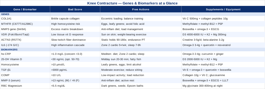
Understanding your biomarker profile is arguably the fastest shortcut to knowing which part of the biological cascade to target first. That is where this article begins.
7 Biomarkers Worth Testing If Your Knee Will Not Fully Straighten
Biomarkers are measurable signals in your blood that reflect what is happening inside your joints and connective tissue right now. They are not diagnostic in the traditional sense — they will not tell you definitively why your contracture developed. But they do tell you what your body's biology is currently doing, which is exactly the information needed to make smarter decisions about treatment and recovery.
The seven biomarkers below were selected because they each reflect a distinct mechanism relevant to flexion contracture: inflammation, fibrosis, connective tissue turnover, cartilage stress, and mineral status. Several of them can be ordered through a standard GP visit or a direct-to-consumer lab. Others require specialty panels. All of them are actionable.
1. High-Sensitivity C-Reactive Protein (hs-CRP)
Why it matters. CRP is produced by the liver in response to inflammatory signaling, primarily interleukin-6. In the context of knee contracture, systemic inflammation accelerates fibroblast activation in the joint capsule — the mechanism that causes excess collagen to accumulate and the capsule to tighten. Chronic low-grade inflammation, even at levels that cause no obvious symptoms, sustains this process quietly. Peter Attia consistently includes hs-CRP in his longevity panels precisely because it tracks background inflammation that standard CRP misses at low levels. For anyone with a persistent joint stiffness issue, this marker is non-negotiable. Research linking CRP elevation to joint capsule fibrosis processes.
How to measure it. Standard blood draw. Available through most general practitioners or direct-to-consumer labs (LabCorp, Quest). Cost: $15–$50 USD. Optimal target: below 1.0 mg/L. Concerning: above 3.0 mg/L. Anything above 1.0 mg/L warrants investigation even if the clinical threshold is set higher.
If the score is bad — the plan without supplements. Prioritize a Mediterranean-style dietary pattern (olive oil, fatty fish, vegetables, legumes, minimal ultra-processed food) with a target of at least 5 servings of vegetables per day. Add 150–200 minutes per week of Zone 2 cardio (conversational pace), which is one of the most reliably anti-inflammatory interventions known. Reduce sleep debt — even partial sleep deprivation acutely elevates CRP. Retest every 8–12 weeks.
If the score is bad — the plan with supplements or equipment. Omega-3 fatty acids (EPA+DHA): 2–4 g/day with meals; safe long-term at this dose, mild blood-thinning effect at higher doses — cycle to 2 g/day after 3 months if INR is a concern. Curcumin with piperine (BioPerine): 500–1000 mg/day, taken with fat; cycle off every 8 weeks. Ginger extract (standardized): 250–500 mg/day. Retest hs-CRP at 90 days. If still elevated despite interventions, investigate underlying drivers (gut permeability, sleep apnea, dental infection).
2. 25-OH Vitamin D
Why it matters. Vitamin D receptors (VDR) are expressed in skeletal muscle, connective tissue, and immune cells throughout the knee. Deficiency is associated with muscle weakness, impaired satellite cell activation, and increased inflammatory cytokine production — all of which worsen the functional deficits and tissue environment that sustain contracture. Gary Brecka has emphasized that standard reference ranges for vitamin D (often 30 ng/mL) are set to prevent bone disease, not to optimize muscle function and tissue repair. Functional optimization requires levels closer to 50–70 ng/mL. Research on vitamin D and musculoskeletal function.
How to measure it. Standard blood draw (25-hydroxyvitamin D). Available everywhere. Cost: $30–$80. Optimal range: 50–70 ng/mL. Deficiency: below 30 ng/mL. Note: ask specifically for 25-OH Vitamin D, not 1,25-OH (which measures active form and is a different test).
If the score is bad — the plan without supplements. Twenty to thirty minutes of direct midday sun exposure on arms and legs (not through glass), five or more days per week. Increase dietary sources: wild salmon, sardines, mackerel, egg yolks, liver. These are insufficient alone for severely deficient individuals but meaningful as a foundation. Retest in 3 months.
If the score is bad — the plan with supplements or equipment. Vitamin D3 (cholecalciferol): 2000–5000 IU/day depending on starting level. Always pair with vitamin K2 (MK-7 form, 100–200 mcg/day) to direct calcium appropriately. Magnesium glycinate or malate (300–400 mg/day) is also needed for vitamin D conversion — deficiency in magnesium impairs D3-to-25OH conversion. Retest in 12 weeks and adjust dose to maintain 50–70 ng/mL. Side effects of excess (above 100 ng/mL): hypercalcemia — do not supplement blindly above 5000 IU without testing.
3. Homocysteine
Why it matters. Homocysteine is an amino acid that, when elevated, disrupts normal collagen crosslinking and impairs the structural integrity of connective tissue. Abnormal crosslinking produces stiffer, less organized collagen fibers — which is precisely the tissue problem at the core of joint capsule fibrosis. Gary Brecka has built much of his work around homocysteine as a master marker that most clinicians systematically underscreen, despite being associated with a wide range of musculoskeletal and cardiovascular pathologies. In the context of knee contracture, elevated homocysteine suggests that the collagen forming in and around the joint capsule may be structurally disorganized. Research on homocysteine and collagen structure.
How to measure it. Standard blood draw. Available through GP or direct-to-consumer labs. Cost: $20–$60. Optimal: below 8 µmol/L. Concerning: above 10 µmol/L. Elevated: above 15 µmol/L (hyperhomocysteinemia). Note: fasting for at least 8 hours before the draw for accurate measurement.
If the score is bad — the plan without supplements. Increase B-vitamin-rich foods: leafy green vegetables (folate), eggs and meat (B12 and B6), legumes. Reduce alcohol consumption, which impairs B-vitamin absorption and metabolism. Limit coffee to 2 cups/day if above normal range, as high caffeine intake can mildly raise homocysteine. Retest at 8 weeks.
If the score is bad — the plan with supplements or equipment. The key intervention is the methylation trio: Methylfolate (L-5-MTHF, 400–800 mcg/day), Methylcobalamin B12 (500–1000 mcg/day sublingually), and Pyridoxal-5-Phosphate (P5P, active B6, 25–50 mg/day). Note: use methyl forms — standard folic acid is ineffective in people with MTHFR variants (see genetics section). Add riboflavin (B2, 50 mg/day) if MTHFR C677T is confirmed. Cycle after 3 months — reduce to a maintenance dose of methylfolate 400 mcg and B12 500 mcg once homocysteine normalizes. No serious side effects at these doses; very high B6 (>200 mg/day for months) can cause peripheral neuropathy — stay well below that.
4. TGF-β1 (Transforming Growth Factor Beta-1)
Why it matters. TGF-β1 is arguably the most direct biological driver of joint capsule fibrosis. It activates fibroblasts to produce excessive collagen, inhibits matrix metalloproteinases (the enzymes that break down old collagen), and promotes the differentiation of fibroblasts into myofibroblasts — contractile cells that physically shorten the joint capsule tissue. Multiple studies have found elevated TGF-β1 in posterior knee capsule tissue from patients with persistent post-surgical or idiopathic contracture. Research on TGF-β1 in knee joint capsule fibrosis. This is the marker that most directly measures whether the fibrotic process is actively driven.
How to measure it. Serum TGF-β1 via ELISA assay. Available through specialty labs (Boston Heart Diagnostics, MDLAB, some university hospitals). Cost: $100–$300. Not typically available through standard GP panels — you may need to order through a functional medicine physician or a direct-to-consumer specialty service. Reference ranges vary by lab; generally: normal < 2500 pg/mL, elevated > 3000 pg/mL.
If the score is bad — the plan without supplements. Regular moderate-intensity exercise is one of the most potent known reducers of circulating TGF-β1 over time — both aerobic training and resistance training appear to down-regulate the signal through mechanotransduction pathways. Reduce sedentary time: prolonged immobility in flexion (sitting) is itself a TGF-β1 driver. Manage sleep, which regulates circadian immune cycling. Reduce alcohol and ultra-processed food intake. Retest at 12 weeks minimum — this marker moves slowly.
If the score is bad — the plan with supplements or equipment. Vitamin E (mixed tocopherols, 200–400 IU/day): research has shown it can modulate TGF-β1 signaling in fibrotic tissues — cycle to 3 months on, 1 month off. Omega-3 EPA+DHA (3–4 g/day): suppresses TGF-β1 indirectly via prostaglandin modulation. Quercetin (500 mg/day with fat): inhibits fibroblast activation downstream of TGF-β1 — cycle 8 weeks on, 4 weeks off. Boswellia serrata extract (standardized to 65% boswellic acids, 300–400 mg twice daily): significant anti-fibrotic and anti-inflammatory evidence, especially for joint conditions. Side effects: mild GI discomfort at higher doses; not recommended during pregnancy.
5. COMP (Cartilage Oligomeric Matrix Protein)
Why it matters. COMP is a structural protein in cartilage and tendon. It leaks into serum when these tissues are under abnormal mechanical stress or undergoing degeneration. Elevated serum COMP indicates that the joint's cartilage or periarticular connective tissue is being progressively damaged — a condition that both contributes to contracture and predicts its worsening. Thomas Dayspring has noted COMP as an underutilized marker in musculoskeletal medicine. It is particularly informative when considering whether a contracture is damaging the joint over time, which in turn affects treatment urgency and target range of motion. Research on COMP as a knee biomarker.
How to measure it. Serum COMP, via specialty labs. Cost: $100–$250. Not widely available in standard clinical panels — functional medicine physicians and sports medicine specialists are most likely to order it. Normal serum COMP: approximately 5–10 U/L (lab-dependent). Elevated signals active tissue breakdown.
If the score is bad — the plan without supplements. Load management is the primary intervention: reduce high-impact activities (running, jumping, stairs with heavy load) while maintaining joint movement through low-impact options (pool walking, cycling with full extension, supported ROM exercises). Unloading the joint through temporary use of a walking aid during peak activity reduces acute COMP spikes. Continue physical therapy focused on improving extension ROM to reduce abnormal contact stress patterns.
If the score is bad — the plan with supplements or equipment. Hydrolyzed collagen peptides (10 g/day with vitamin C 50 mg): supported by emerging RCT evidence for joint cartilage health — take 30–60 minutes before a loading activity for best tissue delivery. Vitamin C (500 mg/day): required cofactor for collagen synthesis. Glucosamine sulfate (1500 mg/day): moderate evidence for knee cartilage; most effective in early to moderate degeneration — allow 12 weeks to assess response. Side effects: minimal; mild GI discomfort for some. Undenatured type II collagen (UC-II) (40 mg/day): more recent evidence suggests immune tolerance induction to slow autoimmune cartilage attack; cycle continuously at low dose.
6. MMP-3 (Matrix Metalloproteinase-3)
Why it matters. MMP-3, also called stromelysin-1, is an enzyme responsible for breaking down extracellular matrix components including proteoglycans, fibronectin, and collagen types II, III, IV, and IX. In a healthy joint, MMPs and their inhibitors (TIMPs) are in balance. In joints undergoing contracture, this balance is often disrupted — either too much matrix degradation (driven by MMP-3) or too little (driven by TGF-β1 suppressing MMPs), depending on the phase of the condition. Elevated serum MMP-3 indicates active joint matrix breakdown and heightened inflammatory remodeling. This marker is regularly used in rheumatology to track disease activity. Research on MMP-3 and knee joint remodeling.
How to measure it. Serum MMP-3, available through standard rheumatology panels and some functional medicine labs. Cost: $80–$200. Normal range: approximately 3–13 ng/mL (men), 3–9 ng/mL (women), though this varies significantly by lab methodology. A value substantially above the upper limit is clinically meaningful.
If the score is bad — the plan without supplements. Anti-inflammatory dietary approach is foundational. Critically: if MMP-3 is elevated, this suggests active joint inflammation — returning to high-impact loading during this phase risks accelerating damage. Prioritize active rest, controlled ROM work, and addressing the inflammatory triggers (diet, sleep, psychological stress). Retest after 8 weeks of intervention.
If the score is bad — the plan with supplements or equipment. Boswellia serrata (as above, 300–400 mg twice daily) has specific MMP-3 inhibitory activity in joint tissue. Omega-3 EPA+DHA (3 g/day): inhibits NF-κB pathway upstream of MMP production. Green tea extract (EGCG) (400–800 mg/day with food): inhibits MMP-3 gene expression; cycle 8 weeks on, 4 off; avoid on an empty stomach. Low-level laser therapy applied to the knee (see Complementary section) has shown measurable MMP-3 modulation in joint tissue studies.
7. RBC Magnesium
Why it matters. Magnesium is a cofactor in over 300 enzymatic reactions and is particularly critical for muscle relaxation, nerve conduction, and ATP production. When intracellular magnesium is low, muscles have difficulty fully releasing from contraction — a physiological reality that directly worsens the hamstring and posterior chain tension that sustains and deepens knee flexion contracture. Standard serum magnesium is notoriously unreliable because the body maintains serum levels at the expense of intracellular stores. RBC magnesium (red blood cell magnesium) is the accurate test. Peter Attia consistently emphasizes this distinction — serum magnesium can appear normal while RBC magnesium is deficient. Research on intracellular magnesium and muscle function.
How to measure it. RBC magnesium blood test (not standard serum magnesium). Available through most labs including LabCorp and Quest on request; some GPs will need guidance to order the RBC-specific version. Cost: $30–$80. Optimal RBC magnesium: 5.5–6.5 mg/dL. Below 5.5 mg/dL warrants intervention. Below 5.0 is significantly deficient.
If the score is bad — the plan without supplements. Increase dietary magnesium: dark leafy greens (spinach, Swiss chard), pumpkin seeds, sunflower seeds, black beans, almonds, avocado, and dark chocolate. This is the most sustainable long-term strategy. Reduce dietary depleters: excessive caffeine and alcohol impair magnesium absorption and increase urinary excretion. Epsom salt baths (magnesium sulfate, 2 cups in warm water, 20 minutes, 3× per week): while transdermal absorption is debated, this provides localized muscle relaxation and may support RBC levels modestly.
If the score is bad — the plan with supplements or equipment. Magnesium glycinate (300–400 mg elemental magnesium/day, at night): best tolerated form, crosses the blood-brain barrier, minimal laxative effect. Alternatively: magnesium L-threonate if cognitive benefits are also desired (200–300 mg/day). Avoid magnesium oxide — poorly absorbed. Allow 6–8 weeks before retesting RBC levels. Side effects: loose stools if dose is too high (reduce by half if this occurs). Use magnesium bisglycinate powder if capsule doses are insufficient.
Taken together, these seven markers give you a complete functional picture: inflammation level, vitamin status, connective tissue quality, fibrosis drive, joint stress load, matrix remodeling activity, and muscle mineral status. With this data in hand, targeted interventions become far more rational than standard protocols. The next dimension to consider is genetic — because your DNA determines the baseline efficiency of nearly every pathway these biomarkers measure.
What Your Genetic Profile Reveals About Knee Contracture Risk and Recovery
Consumer DNA testing through services like 23andMe, AncestryDNA, or more clinically oriented platforms like SelfDecode or StrateGene gives access to SNP (single nucleotide polymorphism) data that can explain a great deal about individual variability in musculoskeletal health. The gene variants below are relevant specifically because they influence connective tissue structure, inflammatory response, B-vitamin metabolism, vitamin D utilization, and muscle fiber composition — all of which intersect directly with the mechanisms behind knee flexion contracture.
This is not determinism. Having a risk variant does not mean you will develop a severe contracture, and not having these variants does not make you immune. What gene data offers is a map: it shows you where your biology is likely under-resourced, and where targeted compensation is worth the effort.
COL1A1 — Collagen Type I Alpha Chain
What it does. COL1A1 encodes the primary structural protein of tendons, ligaments, joint capsules, and bone. Variants like the Sp1 polymorphism (rs1800012) alter the ratio of collagen alpha-1 to alpha-2 chains, producing collagen fibers with modified mechanical properties — often stiffer, less resilient, and prone to abnormal remodeling under stress. In the context of knee contracture, an unfavorable COL1A1 variant can mean that the joint capsule forms more brittle, less extensible collagen in response to injury or inflammation.
If the gene is bad — the plan without supplements. Emphasize eccentric loading of the hamstrings and posterior chain at 3–4× per week — eccentric contractions promote type I collagen remodeling and tendon reorganization in a way that concentric loading does not. Proprioceptive training (balance board, single-leg work) should be a daily fixture to reinforce joint stability. Avoid rapid passive overstretching, which in COL1A1-variant tissue can cause micro-trauma rather than extensibility gains.
If the score is bad — the plan with supplements or equipment. Vitamin C (500 mg, 30–60 min before collagen-loading activity): vitamin C is the rate-limiting cofactor for lysyl hydroxylase, the enzyme that crosslinks collagen properly. This timing protocol (now supported by multiple exercise-collagen trials) significantly improves collagen quality. Hydrolyzed collagen peptides (10–15 g/day, paired with vitamin C): provides the proline and hydroxyproline building blocks your COL1A1 pathway needs. Research on COL1A1 variants and collagen remodeling.
MTHFR — Methylenetetrahydrofolate Reductase
What it does. MTHFR is the enzyme that converts dietary folate into its active usable form (5-MTHF), which drives the methylation cycle and the conversion of homocysteine to methionine. Two common variants — C677T and A1298C — reduce enzyme activity by 30–70%. Gary Brecka has extensively discussed MTHFR as arguably the most clinically underrecognized genetic variant affecting systemic health, noting that both variants impair homocysteine clearance and thus disrupt collagen quality. As noted in the biomarker section, elevated homocysteine directly impairs collagen crosslinking. Research on MTHFR and connective tissue health.
If the gene is bad — the plan without supplements. Prioritize dietary methyl donors: eggs (choline), leafy greens, beets, liver. Avoid folic acid in its synthetic form (found in most fortified foods and standard supplements) — in MTHFR carriers, unconverted folic acid accumulates and can paradoxically worsen methylation by blocking folate receptors.
If the score is bad — the plan with supplements or equipment. This is the case where supplementation has particularly strong rationale. Switch all B-vitamin supplements to activated (methylated) forms: methylfolate (400–800 mcg/day), methylcobalamin (500–1000 mcg sublingually), P5P (25–50 mg/day). For C677T homozygotes, add riboflavin B2 (50–100 mg/day) which significantly improves MTHFR enzyme function at this variant. Retest homocysteine at 8 weeks. Side effects: very rare, but some individuals experience anxiety or overstimulation from methylated B12 — switch to hydroxocobalamin if this occurs.
MMP3 — Matrix Metalloproteinase-3 Gene
What it does. The MMP3 gene promoter carries the rs679620 polymorphism (5A/6A variant), which determines how much MMP-3 enzyme is produced in response to inflammatory stimuli. The 5A allele drives substantially higher MMP-3 expression — meaning that in carriers, joint inflammation triggers more aggressive matrix breakdown than in non-carriers. This creates a paradox: while MMP-3 elevation contributes to joint destruction, MMP-3 suppression by TGF-β1 contributes to capsule fibrosis. MMP3 variants affect which failure mode you are more prone to. Research on MMP3 polymorphism in joint conditions.
If the gene is bad — the plan without supplements. For 5A carriers (high-expression phenotype): prioritize joint load management and anti-inflammatory diet above all else. Early treatment of any joint inflammation is essential — passive acceptance of chronic low-grade joint symptoms risks accelerated matrix degradation. For 6A carriers (low-expression phenotype): the capsule fibrosis risk may be higher; prioritize passive stretching protocols and TGF-β1 management.
If the score is bad — the plan with supplements or equipment. Boswellia serrata and omega-3 (as above) address the MMP-3 pathway directly. For 5A carriers, limiting pro-inflammatory triggers (seed oils, refined carbohydrates, alcohol) has outsized impact. EGCG at 400 mg/day specifically inhibits MMP-3 transcription. Consider low-level laser therapy (see complementary section) as it has shown MMP modulation in joint studies.
VDR — Vitamin D Receptor
What it does. VDR encodes the receptor protein that binds vitamin D3 inside cells to produce its downstream effects. Several common variants — including FokI, BsmI, and TaqI — alter the receptor's function, reducing tissue responsiveness to vitamin D even when serum levels appear adequate. In musculoskeletal terms, VDR variant carriers may experience reduced muscle strength, impaired satellite cell activation, and blunted immune modulation despite having normal 25-OH vitamin D levels. Research on VDR variants and muscle function.
If the gene is bad — the plan without supplements. Supplement sun exposure on skin not just face and forearms, but larger body surface areas (torso, legs) — VDR variant carriers likely need higher serum D levels to achieve the same tissue effect. Weight-bearing exercise up-regulates VDR expression in muscle tissue.
If the score is bad — the plan with supplements or equipment. VDR carriers typically need to target serum 25-OH D at the higher end: 65–80 ng/mL rather than 50–60 ng/mL. This may require 4000–6000 IU/day D3 with K2 (200 mcg MK-7). Test every 3 months to avoid overshooting. Also: magnesium (300–400 mg/day) is essential for D3 conversion — VDR inefficiency compounds quickly with magnesium deficiency.
ACTN3 — Alpha-Actinin-3
What it does. The ACTN3 gene codes for alpha-actinin-3, a structural protein found exclusively in fast-twitch (type IIx) muscle fibers. The R577X variant, present in approximately 18% of the population as a homozygous XX genotype, results in complete absence of alpha-actinin-3. This profoundly shifts muscle fiber composition toward slow-twitch dominance. For knee contracture, this matters because the muscle groups sustaining the contracture — the hamstrings and gastrocnemius — rely on a balance of fiber types for both active tension and extensibility. XX genotype individuals tend to have less explosive power but greater fatigue resistance. Research on ACTN3 variants and exercise adaptation.
If the gene is bad — the plan without supplements. XX genotype individuals respond better to endurance-style physical therapy — higher rep, lower load eccentric stretching maintained over longer durations. Static stretch holds of 90–180 seconds leverage the slow-twitch endurance capacity. Rapid PNF techniques may be less effective — prefer longer, sustained contract-relax cycles.
If the score is bad — the plan with supplements or equipment. Creatine monohydrate (3–5 g/day, continuous, no need to load): partially compensates for reduced fast-twitch capacity and may support overall neuromuscular recovery. Beta-alanine (3.2 g/day, split doses to reduce paresthesia): improves buffering capacity in type I-dominant muscles during prolonged therapeutic exercise sessions. These are long-term supplements with good safety profiles — creatine particularly well-documented. No cycling required at these doses.
IL6 — Interleukin-6 Promoter Variants
What it does. The IL6 gene promoter region carries several SNPs (notably rs1800795, the -174 G/C variant) that determine basal and stimulated IL-6 production. The G allele is associated with higher IL-6 output under inflammatory conditions. Since IL-6 is one of the primary drivers of both hs-CRP elevation and TGF-β1 activation, IL6 promoter variants represent an upstream genetic risk for the entire inflammatory-fibrotic cascade that sustains joint contracture. It also explains why some people develop severe post-surgical contracture while others with similar procedures do not. Research on IL-6 polymorphism and inflammatory response.
If the gene is bad — the plan without supplements. For GG carriers (high IL-6 expressers), the anti-inflammatory lifestyle interventions carry disproportionately higher returns. Specifically: Zone 2 cardio (30–45 min, 5× per week) is the most potent lifestyle intervention for IL-6 down-regulation. Adequate sleep (7–9 hours) is not optional — IL-6 is significantly elevated after sleep deprivation. Thermal stress management: avoid prolonged exposure to excessive heat or cold in acute phases, which can transiently spike IL-6.
If the score is bad — the plan with supplements or equipment. Omega-3 EPA+DHA (3–4 g/day) suppresses NF-κB and AP-1 transcription factors upstream of IL-6 gene expression. Quercetin (500 mg/day) and resveratrol (250–500 mg/day with fat) have shown IL-6 modulating effects. Resveratrol: cycle 8 weeks on, 4 weeks off; mild blood-thinning effect. Consider testing hs-CRP and IL-6 serum levels concurrently to track whether genetic predisposition is being expressed or successfully managed.
With an understanding of both your biomarkers and genetic tendencies, it becomes possible to build a recovery approach that fits your specific biology rather than a statistical average. The next section goes deeper into the neuroscience of flexibility — because the nervous system, it turns out, is often the largest bottleneck.
What the Science of Flexibility Really Says: Key Insights From Huberman Lab Research
Andrew Huberman and his collaborators at Stanford have synthesized a substantial body of peer-reviewed research into practical, evidence-based protocols for improving joint range of motion and connective tissue extensibility. His work challenges several deeply held assumptions in physical therapy — particularly the idea that tight muscles are the primary limitation in chronic stiffness. What the neuroscience actually shows is considerably more nuanced.
1. Most Range of Motion Limitations Are Neurological, Not Structural
The nervous system imposes a protective ceiling on range of motion that is entirely separate from the physical length or stiffness of the tissue. This neural governor can be released through specific techniques — which is why some people gain full extension in a single session when fear or pain is removed. Addressing this requires a different intervention than stretching more aggressively.
2. Static Stretching Is Most Effective After, Not Before, Activity
Pre-activity static stretching reduces power output and may temporarily impair proprioception. For contracture recovery, static stretching should be performed after 10–15 minutes of low-load, blood-flow-enhancing movement. This pre-warms connective tissue and reduces neurological resistance, making each stretch more effective.
3. Duration Matters More Than Intensity
Huberman's synthesis of the stretching literature points to a minimum of 30 seconds per hold to produce any neural adaptation, and 90–180 seconds per hold for durable connective tissue change. For joint contracture specifically — where connective tissue is the primary target — shorter holds are largely ineffective. The daily total time under appropriate stretch appears to be more important than any single session.
4. PNF (Contract-Relax) Stretching Is Neurologically Superior for Chronic Stiffness
Proprioceptive neuromuscular facilitation works by briefly contracting the muscle being stretched (activating the Golgi tendon organ), then releasing — which produces a brief window of reduced neural resistance. For hamstring and posterior capsule stiffness driving knee contracture, contract-relax protocols consistently outperform passive static stretching in clinical trials. The technique: contract the target muscle against resistance for 5–8 seconds at 50% effort, then relax and move into the stretch. Repeat 3–5 times per session.
5. Heat Before Stretching Significantly Improves Tissue Extensibility
Collagen viscosity decreases substantially with temperature. Tissue heated to 39–41°C (warm bath, heat pack applied 10–15 minutes before stretching) is significantly more extensible than tissue at resting temperature. This is not trivial — the research suggests 15–20% improved range of motion gains from the same stretching protocol when the tissue is properly warmed first.
6. Inflammation Is Actively Working Against You
Elevated inflammatory cytokines (particularly IL-6 and TNF-α) directly suppress neurological flexibility by sensitizing nociceptors in joint tissues. This creates a pain-tension feedback loop that makes stretching less effective and more painful. Huberman's synthesis emphasizes that managing inflammation is a prerequisite to effective flexibility training, not an optional addition. This is exactly where the biomarker section becomes relevant — if your hs-CRP is elevated, stretching harder will not compensate.
7. Consistency Beats Volume — Daily Brief Sessions Outperform Weekly Long Ones
Neurological adaptation to range of motion requires frequent signaling. A daily 8–10 minutes of targeted stretching produces greater improvements over 12 weeks than a single 60-minute session once or twice a week, even when total time is equalized. For contracture recovery, this principle suggests structuring a short daily routine is more valuable than infrequent intensive physical therapy sessions.
8. Zone 2 Cardio Improves Tissue Elastic Properties Through Oxygenation
Regular aerobic exercise at Zone 2 intensity (conversational pace, 55–70% max heart rate) improves capillary density in connective tissue, enhancing oxygen and nutrient delivery to structures that are largely avascular. Over 8–12 weeks, this measurably improves the compliance of tendons, ligaments, and joint capsule tissue — the very structures involved in flexion contracture.
9. Sleep Is When Connective Tissue Repairs
Collagen synthesis and remodeling peak during slow-wave sleep. This is not metaphorical — the anabolic signaling cascades that reorganize joint capsule tissue are largely GH-dependent, and GH secretion is sleep-stage-dependent. Chronically poor sleep quality or duration directly impairs the tissue remodeling that flexibility training is trying to accomplish. Prioritizing 7–9 hours of uninterrupted sleep is a structural intervention, not a lifestyle luxury.
10. Breathing Patterns Directly Modulate Neural Tension in Muscles
Slow diaphragmatic breathing during a stretch activates the parasympathetic nervous system, reduces the defensive neural response in the muscle being stretched, and directly lowers perceived pain. The practical protocol: inhale for 4 counts through the nose, exhale slowly for 6–8 counts through the mouth, specifically during the peak of the stretch position. This is not relaxation theater — it has measurable electromyographic effects on muscle tone.
These principles collectively describe a fundamentally different approach to flexibility than most clinical settings currently practice. Andrew Huberman Lab continues to update its protocols as the research evolves.
Complementary Approaches With Real Clinical Support
The modalities below were selected specifically for their clinical evidence base in conditions involving joint stiffness, musculoskeletal pain, and connective tissue dysfunction. They are not replacements for medical care but well-researched adjuncts that can meaningfully augment a recovery program.
Yoga
Yoga is relevant to knee flexion contracture because it combines sustained passive stretching, eccentric loading through bodyweight positions, and systematic attention to posterior chain flexibility — all applied in a manner that also addresses the neurological tension component. Unlike isolated stretching exercises, yoga integrates hip flexor and posterior chain work in functional patterns that more closely match the demands of normal gait, which matters for functional recovery. Yin yoga in particular, with its emphasis on 3–5 minute passive holds in connective tissue-targeting positions, is particularly aligned with the evidence on stretch duration.
A randomized controlled trial on yoga and musculoskeletal range of motion found significant improvements in hamstring extensibility and knee ROM in participants practicing yoga 3× per week for 8 weeks versus a stretching control. Supine hamstring stretches (Supta Padangusthasana) and reclined hero pose (Supta Virasana, with blocks as needed) are particularly well-matched to the posterior capsule and hamstring shortening pattern in knee contracture.
Practically: begin with a daily 15–20 minute yin or restorative yoga routine focused on posterior chain and hip flexor release. Use props (blocks, bolsters) to allow full relaxation into positions rather than forced tension. Avoid deep flexion poses (child's pose, deep squat) in phases where range is severely limited. Progress to 30–45 minute sessions as ROM improves. If any sharp pain occurs in the joint (as distinct from muscular discomfort), stop and reassess with a clinician.
Massage Therapy
Massage therapy addresses the myofascial component of knee contracture directly: the posterior compartment soft tissue — hamstrings, gastrocnemius, and posterior joint capsule — is frequently hypertonic and contains trigger points that actively restrict knee extension. Manual therapy to this region can break adhesion patterns, reduce neurological guarding, and transiently improve ROM in a way that facilitates more effective stretching afterward. It is most useful when combined with active stretching within 24 hours of treatment — the window when neurological resistance is reduced.
Research on massage and knee ROM improvement supports manual therapy's effectiveness for hamstring flexibility and posterior knee tissue compliance. Cross-fiber friction technique applied to the posterior capsule and hamstring-tendon junction has shown the most consistent results in reducing myofascial restriction contributing to joint contracture. Sessions of 30–45 minutes at 1–2× per week for 6–8 weeks is a reasonable therapeutic trial.
Realistically, deep tissue massage and myofascial release by a qualified therapist is the safest approach. Self-massage using a foam roller (posterior thigh, calf) can provide meaningful adjunct benefit between sessions — 5–10 minutes per session, 5–7× per week. Apply moderate pressure, pause on tender areas for 30–60 seconds rather than rolling continuously. Avoid direct pressure on the posterior joint line in acute inflammatory phases.
Low-Level Laser Therapy (Photobiomodulation)
Photobiomodulation (PBM) uses specific wavelengths of red and near-infrared light (typically 630–850 nm) to stimulate mitochondrial activity in cells, reduce oxidative stress, and modulate inflammatory cytokine production in target tissues. For knee contracture, PBM is relevant because it can reach the joint capsule and periarticular connective tissue, where it appears to down-regulate fibrogenic signaling and promote more balanced collagen remodeling. It is also one of the few interventions with direct evidence for joint capsule fibrosis in a clinical setting. Research on LLLT and knee joint stiffness.
Several RCTs and meta-analyses of LLLT in knee osteoarthritis and post-surgical stiffness have documented meaningful improvements in ROM and pain reduction compared to sham irradiation. A typical protocol used in studies: 630–850 nm, 4–8 J/cm², applied to 4–6 points around the joint, 3–5× per week for 4–8 weeks. Clinical LLLT devices (class 3B or class 4 lasers) deliver more reliable dosing than consumer devices, but consumer-grade 630–850 nm LED panels have shown efficacy in some trials for superficial joint applications.
Practically: if using a professional device, 8–10 sessions over 4 weeks is a common initial trial, with reassessment of ROM. Consumer near-infrared panels (660 nm + 850 nm combination) used 10–15 minutes daily over the anterior and posterior knee at close proximity (5–10 cm) are a lower-cost option. PBM is considered very safe at standard doses — avoid direct eye exposure to the light source, and consult a clinician if there is any active infection or cancer in the treatment area.
Progressive Muscle Relaxation
Progressive muscle relaxation (PMR), developed by Edmund Jacobson, is a structured technique involving deliberate sequential tension and release of muscle groups throughout the body. For knee contracture, PMR is relevant because chronic muscle hypertonicity — particularly in the hamstrings, hip flexors, and calf — is both a cause and a consequence of the contracture pattern. Neurological muscle tension that is not fully under voluntary control can be gradually retrained through consistent PMR practice, which reduces baseline resting tension in the posterior chain.
Research on PMR and musculoskeletal tension reduction supports its use as a complement to physical rehabilitation in conditions with significant muscle guarding. A study involving patients with chronic musculoskeletal pain found that PMR practice 5× per week over 8 weeks significantly reduced muscle resting tone and pain intensity compared to controls.
Practically: perform a full-body PMR session nightly before sleep (15–20 minutes). Start with feet and calves, progress upward through the posterior chain, paying particular attention to hamstrings (contract firmly for 7–10 seconds, then relax completely for 30 seconds; repeat 3× per muscle group). Over time — typically 3–4 weeks of consistent practice — baseline muscle resting tone in the target groups measurably decreases. This makes daily stretching sessions more effective and sleep-based connective tissue recovery more productive. No equipment required; several free audio guides are available through evidence-based mindfulness and rehabilitation programs.
Conclusion
Flexion contracture of the knee is rarely explained by a single cause, and rarely resolved by a single intervention. The evidence reviewed here points consistently to an overlapping web of drivers — chronic inflammation sustaining joint capsule fibrosis, connective tissue metabolism impaired by nutrient deficiencies or genetic variants, and neurological tension patterns that no amount of passive stretching will fully overcome without addressing the underlying biology.
The most useful next step is not necessarily adding another stretch to your routine. It is understanding which of these drivers is actually active in your case. A basic panel — hs-CRP, 25-OH Vitamin D, homocysteine, and RBC magnesium — costs under $200 at most direct-to-consumer labs and provides an immediately actionable snapshot. If you have consumer DNA data already, reviewing the six gene variants covered here takes an afternoon and may explain years of inexplicable variability in your response to treatment.
Work with a qualified clinician — ideally one who integrates functional medicine or sports medicine alongside standard orthopedic care — to interpret your results and tailor interventions safely. The information in this article is a starting framework, not a prescription. But better information genuinely does lead to better decisions, and the gap between generic and personalized is often where lasting progress finally happens.
Musculoskeletal: Joint Conditions Muscle Conditions Tendon & Ligament Conditions
Autoimmune: Inflammatory Conditions Connective Tissue Conditions