This article contains AI generated content.
· UpdatedFocal Cartilage Defect of the Knee — 6 Genes and 6 Biomarkers to Track
Introduction
A focal cartilage defect in the knee is not simply a wear-and-tear story. It is a localized failure of a highly specialized tissue — articular cartilage — that has almost no capacity for spontaneous self-repair. If you have received this diagnosis, you may already know the structural picture from imaging. What is far less likely is that anyone has explained why your cartilage is behaving the way it is right now, or what your body's molecular environment is actually doing in response to the injury.
The standard clinical conversation tends to stay at the symptom level: pain, function, surgical thresholds. That is understandable, but it leaves out the variables that most directly determine whether a focal defect stabilizes, progresses slowly, or accelerates. People with nearly identical MRI findings can have vastly different trajectories. The difference is often not about the size of the defect — it is about the biological environment surrounding it.
What drives that environment? Inflammatory load, cartilage matrix turnover rate, chondrocyte survival signaling, and the genetic architecture that sets the baseline for all of the above. These are measurable. Blood tests, urine tests, and genetic data can give you a metabolic and molecular picture that transforms the clinical conversation from "wait and see" into something more targeted and more actionable.
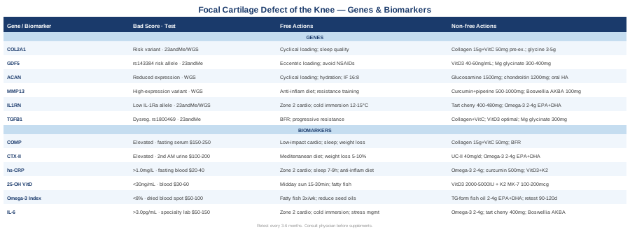
This article takes two complementary approaches to that problem. The first covers six biomarkers measurable through standard and specialty lab tests, reflecting how much cartilage degradation is actively occurring and how much inflammation is fueling it. The second examines six genes whose variants influence how cartilage is built, maintained, and broken down over a lifetime. Together, they offer a framework for making better decisions — one grounded in data rather than generalities.
6 Biomarkers to Track for Focal Cartilage Defect of the Knee
Biomarkers offer something structural imaging cannot: a window into what is happening in the joint right now, biochemically. While an MRI captures damage that has already occurred, metabolic and inflammatory markers reflect the rate of ongoing degradation and the adequacy of repair signaling. For a focal cartilage defect, the following six markers form the most actionable and evidence-supported panel available.
1. COMP — Cartilage Oligomeric Matrix Protein
Why it matters
COMP is a non-collagenous glycoprotein that plays a structural role in assembling the collagen network within articular cartilage. When chondrocytes are mechanically stressed or the extracellular matrix begins to degrade, COMP is released into the synovial fluid and eventually enters systemic circulation. Serum COMP is currently one of the most specific and well-validated blood markers for articular cartilage catabolism. Multiple longitudinal studies have shown that elevated serum COMP correlates with imaging-confirmed cartilage loss rates and predicts disease progression in both osteoarthritis and focal defect contexts. As a trend marker — measured repeatedly over months — it offers meaningful signal about whether interventions are working.
How to measure it
COMP is measured via a fasting serum blood draw. In the United States it is available through specialty labs including Boston Heart Diagnostics, certain academic medical centers, and functional medicine panels. Cost ranges from $150 to $250 depending on the lab. Reference ranges vary by assay method; consistently elevated values above the lab's stated upper limit, or a rising trend across repeated measurements, warrant investigation. Retesting every three to six months provides the most clinically useful trend data.
If the score is elevated — the plan without supplements
The most direct modifiable driver of high COMP is excessive or aberrant mechanical loading on a compromised joint surface. If you are carrying excess body weight, each additional kilogram adds approximately three to four times that force to the knee during walking. Prioritizing low-impact aerobic activity — cycling, swimming, aquatic resistance training — maintains metabolic health and quadriceps strength without amplifying COMP release. Neuromuscular training through balance board work and proprioceptive drills reduces peak impact loads on the cartilage. Sleep quality also plays a direct role: collagen synthesis peaks during slow-wave sleep, and poor sleep acutely elevates systemic inflammation while increasing COMP release even in joints that are not structurally compromised.
If the score is elevated — the plan with supplements or equipment
The most evidence-backed nutritional protocol for slowing cartilage matrix degradation involves hydrolyzed collagen peptides combined with vitamin C. Research from Dr. Keith Baar's lab at UC Davis, published in the American Journal of Clinical Nutrition, demonstrated that 15g of gelatin or collagen plus 50mg of vitamin C, consumed approximately 45 to 60 minutes before brief joint-loading exercise (6–10 minutes), significantly increased collagen synthesis markers compared to placebo. Vitamin C is the cofactor for prolyl and lysyl hydroxylases — the enzymes that create the cross-links responsible for mature collagen fiber tensile strength. Frequency: before each exercise session, or once daily if not exercising. Cycling: continuous use is appropriate long-term. Side effects: gastrointestinal discomfort is possible at higher collagen doses in sensitive individuals.
Blood flow restriction (BFR) training — using an occlusion cuff at 60–80% limb occlusion pressure during low-load resistance exercise at 20–30% of 1RM — builds quadriceps strength and generates local anabolic growth factor signaling with minimal compressive joint forces. A stronger quadriceps directly reduces tibio-femoral contact stress, which in turn reduces COMP release over time.
2. CTX-II — Urinary C-Terminal Crosslinking Telopeptide of Type II Collagen
Why it matters
CTX-II is arguably the most specific urinary marker for type II collagen degradation — the protein that forms the load-bearing scaffold of hyaline cartilage. When type II collagen fibers are enzymatically cleaved, CTX-II fragments pass into the synovial fluid, enter circulation, and are excreted in urine. Elevated urinary CTX-II has been associated with faster joint space narrowing on radiographs, greater cartilage defect progression on MRI, and worse functional outcomes in longitudinal cohort studies. Research on CTX-II consistently supports its role as a dynamic surrogate for active cartilage catabolism that is invisible on standard imaging until structural damage is already advanced.
How to measure it
CTX-II is measured from a second morning urine void — the first void is discarded to reduce overnight concentration effects, and the second sample is collected and normalized to urinary creatinine. Specialty rheumatology or sports medicine labs offer this test at a cost of $100 to $200. It is not routinely available at standard primary care labs in the United States but can be ordered through European specialty diagnostics or academic hospital rheumatology programs. Consistent collection timing (same time of day, same hydration status) significantly improves the reproducibility of serial measurements.
If the score is elevated — the plan without supplements
Elevated CTX-II reflects a combination of excessive mechanical loading, systemic inflammation, and insufficient anabolic signaling to the chondrocytes. A Mediterranean-pattern diet — high in colorful vegetables, olive oil, fatty fish, legumes, and low in refined carbohydrates and ultra-processed foods — consistently reduces the inflammatory drivers of type II collagen breakdown. Weight management is the highest single-leverage intervention for the knee: even a five to ten percent reduction in body weight produces measurable improvements in articular cartilage biomarkers. Replacing high-impact activities (running, jumping) with cyclical low-impact loading (cycling, swimming, walking on soft surfaces) reduces the peak enzymatic stress on type II collagen while preserving joint nutrition.
If the score is elevated — the plan with supplements or equipment
Undenatured type II collagen (UC-II) at 40mg per day operates through a fundamentally different mechanism than hydrolyzed collagen. UC-II acts on gut-associated lymphoid tissue (Peyer's patches) to induce oral tolerance against type II collagen fragments, thereby reducing the immune-mediated component of cartilage matrix destruction. Several randomized trials have found UC-II to be superior to glucosamine and chondroitin combination for both pain and function in knee OA. Omega-3 fatty acids at 2–4g EPA+DHA per day reduce prostaglandin E2 and leukotriene B4, the eicosanoids that upregulate MMP-13, the primary enzyme responsible for type II collagen cleavage. Retest CTX-II after 90 days of sustained intervention to assess response; adjust omega-3 dose based on concurrent Omega-3 Index testing (target above 8%).
3. hs-CRP — High-Sensitivity C-Reactive Protein
Why it matters
CRP is synthesized by the liver in response to interleukin-6 (IL-6) signals originating from inflamed tissues. In a focal cartilage defect, damaged chondrocytes and activated synoviocytes generate a local inflammatory environment that elevates systemic hs-CRP. While hs-CRP is not tissue-specific, it is an accessible and affordable marker that reflects the inflammatory load your cartilage is operating under daily. Peter Attia, who tracks this marker as a cornerstone of metabolic and longevity profiling, targets values below 0.5 mg/L in his patients. Values consistently above 1.0 mg/L in a person without active infection or injury indicate a pro-inflammatory metabolic state that will accelerate cartilage matrix degradation regardless of other interventions.
How to measure it
hs-CRP is a standard fasting blood test available at any laboratory. Cost: $20 to $40, often covered by insurance with appropriate diagnostic codes. It should not be interpreted during or immediately after an acute illness, viral infection, or significant injury, as values will be misleadingly elevated. Retesting every three to six months while implementing lifestyle changes provides actionable trend data. A single measurement is less meaningful than a pattern across three or more tests.
If the score is elevated — the plan without supplements
Sleep quality is the most underestimated driver of systemic CRP. Even one night of poor sleep acutely elevates CRP in otherwise healthy individuals. Prioritizing seven to nine hours of sleep in a cool, dark, consistent environment is foundational. Zone 2 aerobic exercise — sustained low-intensity cardio at a pace where you can hold a full conversation — performed three to four times per week for 30 to 45 minutes consistently lowers systemic inflammatory markers over eight to twelve weeks. Removing ultra-processed foods, refined seed oils, and high-glycemic carbohydrates from the diet, while increasing polyphenol-rich vegetables, berries, and olive oil, produces measurable hs-CRP reductions within weeks. Stress management through structured practices addresses cortisol-driven CRP amplification that purely dietary approaches cannot reach.
If the score is elevated — the plan with supplements or equipment
Omega-3 fatty acids (EPA+DHA, 2–4g/day) remain the most consistently evidence-supported supplement for lowering hs-CRP across multiple randomized trials. Curcumin with piperine (500–1000mg/day, phospholipid-bound or liposomal formulation for adequate bioavailability) has demonstrated anti-inflammatory effects in OA and general inflammation trials. Vitamin D3 combined with K2 (MK-7) — typically 2000–5000 IU D3 with 100–200mcg K2 taken with a fat-containing meal — is important when 25-OH vitamin D is below 40 ng/mL, as vitamin D deficiency is independently associated with elevated CRP and impaired chondrocyte survival. Regular sauna use (15–20 minutes at approximately 80°C, three times per week) has evidence from Finnish population studies and small RCTs for reducing cardiovascular and systemic inflammatory markers including CRP over six to eight weeks. Side effects: omega-3 at high doses can mildly thin blood — relevant to discuss with a clinician before surgery.
4. 25-OH Vitamin D
Why it matters
Vitamin D receptors (VDRs) are expressed directly on chondrocytes, and vitamin D signaling plays an active role in regulating chondrocyte survival, extracellular matrix production, and the cellular response to mechanical load. Population studies consistently show that vitamin D insufficiency below 30 ng/mL is associated with accelerated cartilage loss on MRI, reduced proteoglycan content, and worse outcomes following cartilage repair procedures. Beyond the joint, vitamin D regulates calcium homeostasis, neuromuscular function, and sleep architecture — all of which influence cartilage indirectly. VDR activation suppresses NF-κB, the master transcription factor governing inflammatory gene expression in synoviocytes and chondrocytes alike. Research on vitamin D and cartilage makes it one of the few nutritional variables with a mechanistically plausible and clinically replicated connection to joint tissue health.
How to measure it
25-OH Vitamin D is a routine fasting blood test available at any laboratory. Cost: $30 to $60, often covered by insurance. Testing twice per year — once at the end of summer (typically your seasonal peak) and once at the end of winter (typically your nadir) — is sufficient for most people. Target range for joint health and longevity: 40–60 ng/mL. Note that conventional labs typically flag deficiency at below 20 ng/mL, which reflects the minimum for bone health but not optimal tissue function. Testing should be 25-OH vitamin D, not 1,25-OH (calcitriol), which reflects different physiology and is not appropriate for routine status assessment.
If the score is low — the plan without supplements
Midday sun exposure on large skin surface areas — arms, legs, and torso — for 15 to 30 minutes without sunscreen is the most physiologically natural way to raise vitamin D for lighter skin tones. Darker skin tones require substantially longer exposure times. Fatty fish (salmon, sardines, mackerel), egg yolks, and fortified foods contribute meaningfully but rarely suffice to raise levels into the optimal range on their own. If you live above the 40th parallel latitude and spend most daylight hours indoors, food and sun exposure alone will be insufficient through winter months.
If the score is low — the plan with supplements or equipment
Vitamin D3 (cholecalciferol) — not D2 — is the standard supplemental form with superior absorption and biological activity. Pairing it with vitamin K2 as MK-7 directs calcium metabolism toward bone rather than soft tissues, which becomes particularly relevant at higher D3 doses. Standard starting dose: 2000–4000 IU D3 with 100–200mcg MK-7 K2, taken with a fat-containing meal daily. Retest after 90 days and adjust. Very low baseline levels may require a higher supervised loading protocol. Side effects are rare below 10,000 IU/day; toxicity is possible with prolonged doses above that threshold without monitoring. Cycling: continuous year-round use with twice-yearly testing is appropriate.
5. Omega-3 Index
Why it matters
The Omega-3 Index measures EPA and DHA as a percentage of total red blood cell fatty acids, providing a three-month snapshot of omega-3 status that outperforms single-point serum measurements. A low Omega-3 Index tips the eicosanoid balance toward pro-inflammatory prostaglandins and leukotrienes that directly upregulate MMP-13 and other cartilage-degrading metalloproteinases. Research linking omega-3 status to inflammatory biomarkers, chondrocyte survival, and joint pain scores is substantial. Thomas Dayspring, the leading clinical lipidologist in the United States, routinely includes the Omega-3 Index in comprehensive metabolic panels, identifying it as one of the most modifiable inflammatory risk factors in clinical practice. Most Americans test in the 4–6% range — well below the cardioprotective and anti-inflammatory threshold.
How to measure it
The Omega-3 Index is assessed via a dried blood spot collected at home through a finger prick kit or at a lab. OmegaQuant is the reference laboratory developed by the researchers who first validated the index clinically. Cost: $50 to $100. Most conventional physicians do not routinely order this test, so patients typically self-order. Target: above 8% for anti-inflammatory and cardiovascular benefit. Values below 4% represent a significant inflammatory risk. Retest after 90–120 days of any intervention.
If the score is low — the plan without supplements
Eating fatty fish — salmon, sardines, mackerel, herring — three or more times per week meaningfully raises the Omega-3 Index over 12 weeks. Equally important is reducing competing omega-6 linoleic acid from seed oils (soybean, corn, canola, sunflower), which occupy the same enzymatic conversion pathways and membrane incorporation sites. Simply eliminating seed oils from cooking — replacing them with olive oil, avocado oil, or butter — while maintaining regular fatty fish intake often shifts the index by one to two percentage points within three months.
If the score is low — the plan with supplements or equipment
Triglyceride-form or re-esterified omega-3 fish oil at 2–4g combined EPA+DHA per day offers higher bioavailability than ethyl ester forms. For those who prefer plant-based sources, algae-derived EPA+DHA provides the same marine fatty acids without the fish supply chain. Take with the largest fat-containing meal of the day to maximize absorption. If the index fails to rise above 6% despite consistent supplementation, high-dose prescription omega-3 (icosapent ethyl, 4g/day) under physician supervision may be warranted. Retest at 90–120 days. Side effects: mild blood thinning at doses above 3g/day is relevant to communicate to a surgeon before any planned procedure. Cycling: continuous use is appropriate given the persistent inflammatory benefit.
6. IL-6 — Interleukin-6
Why it matters
IL-6 is the cytokine that bridges local joint inflammation to systemic CRP elevation and serves as a direct activator of the metalloproteinases that degrade cartilage. Within the knee joint, IL-6 produced by activated synoviocytes and stressed chondrocytes directly upregulates MMP-1, MMP-3, and MMP-13 — the enzymes responsible for cleaving type II collagen and aggrecan from the extracellular matrix. In a focal cartilage defect, the local IL-6 environment largely determines whether the defect edge remains stable or begins to expand. Chronically elevated circulating IL-6 also drives muscle wasting and impairs sleep quality — creating a negative feedback loop where reduced quadriceps strength increases cartilage contact stress, which worsens the defect and raises IL-6 further. The IL-6/MMP pathway in cartilage destruction is one of the most studied and pharmacologically targeted mechanisms in rheumatology.
How to measure it
IL-6 is available through specialty labs and academic medical centers, often within broader inflammatory cytokine panels. Cost: $50 to $150 depending on panel inclusion. Reference range: typically below 3.0 pg/mL in healthy adults; optimal is likely below 2.0 pg/mL for joint protection. Importantly, IL-6 is acutely elevated by intense exercise, active infection, and psychological stress. Testing should occur during a baseline period, at least 48 hours after significant physical exertion, and not during or immediately after illness.
If the score is elevated — the plan without supplements
Regular moderate-intensity aerobic exercise paradoxically lowers chronic resting IL-6 even though it transiently raises it during each session. This occurs through a myokine feedback loop: exercise-induced transient IL-6 release from working muscles stimulates anti-inflammatory IL-10 and IL-1Ra (the IL-1 receptor antagonist). Over weeks of consistent Zone 2 training, basal IL-6 falls measurably. Cold water immersion (10–15 minutes at 12–15°C post-exercise) has been shown to reduce acute post-exercise inflammatory signaling and may contribute to lower resting IL-6 over time. Chronic psychological stress drives cortisol-mediated IL-6 production from adipose tissue — making structured stress management practices a direct, if indirect, cartilage-protective intervention.
If the score is elevated — the plan with supplements or equipment
Omega-3 fatty acids (2–4g EPA+DHA/day) represent the most consistently replicated supplement for lowering resting IL-6 across multiple randomized trials. Tart cherry extract (standardized to 400–480mg anthocyanins per day, or 30mL concentrated juice twice daily) has RCT evidence for reducing IL-6 in athletes and patients with inflammatory joint conditions. Boswellia serrata extract standardized to AKBA (acetyl-keto-β-boswellic acid) at 100–200mg AKBA per day blocks 5-lipoxygenase, reducing leukotriene B4 production upstream of IL-6 amplification in the joint. Frequency: daily for all three. Cycling: Boswellia is typically cycled at 8–12 weeks on with 4 weeks off; omega-3 and cherry extract can be taken continuously. Reassess IL-6 after 90 days of sustained intervention.
With a clear picture of the inflammatory and catabolic signals active in your joint, the next logical layer is understanding the genetic architecture that sets the baseline for how your cartilage is built and how it responds to both load and injury.
6 Genes That Shape Your Cartilage — What the Research Suggests
Genetics does not determine destiny, but it shapes tendency. Knowing which gene variants you carry can explain why your cartilage behaves the way it does under load, why inflammation resolves slowly or not at all, and which of the interventions above are most likely to work for your specific biology. The field of cartilage genetics has progressed significantly over the past decade, with several genes now robustly replicated across multiple populations.
COL2A1 — The Structural Blueprint of Articular Cartilage
What this gene does
COL2A1 encodes type II procollagen, the foundational structural protein of articular cartilage. Type II collagen fibers form the tensile scaffold that entraps aggrecan and water molecules, providing both tensile strength and compressive resistance to the joint surface. Variants in COL2A1 alter fiber geometry, cross-linking density, and the tissue's inherent resistance to enzymatic degradation. In the most severe cases, COL2A1 mutations cause chondrodysplasias; in more common subtle variants, the effect is reduced baseline cartilage quality that accelerates under repetitive mechanical stress. Epigenetically, research has shown that the COL2A1 promoter becomes increasingly methylated as OA progresses, progressively silencing type II collagen production in affected chondrocytes — a reversible epigenetic change in principle.
Testing
Raw data from 23andMe or AncestryDNA can reveal several common COL2A1 SNPs. Whole genome sequencing through services such as Nebula Genomics provides broader coverage. Clinical genetic testing through a genetic counselor or rheumatology center is appropriate for those with suspected connective tissue disorders involving multiple joints or systemic signs.
If the gene is unfavorable — the plan without supplements
COL2A1 expression responds directly to cyclical mechanical load. Chondrocytes under appropriate compressive and tensile cycling — not extreme, not absent — upregulate COL2A1 transcription through integrin-mediated intracellular signaling. Aquatic resistance exercise, moderate-resistance cycling, and progressive low-impact strength training provide this stimulus without the destructive peak forces of running or jumping on a compromised surface. Prolonged immobilization should be avoided — it rapidly silences COL2A1 transcription and accelerates cartilage thinning. Prioritizing sleep quality is essential: growth hormone secretion during slow-wave sleep directly drives type II collagen synthesis.
If the gene is unfavorable — the plan with supplements or equipment
Hydrolyzed collagen peptides (15g) combined with vitamin C (50mg), taken 45 to 60 minutes before joint-loading exercise, directly increase the circulating substrate available for type II collagen synthesis at the moment of peak joint blood flow. Glycine — the most abundant amino acid in collagen, comprising approximately one-third of its sequence — is often rate-limiting. Supplemental glycine at 3–5g per day is inexpensive, safe for long-term use, and has essentially no meaningful side effects. Blood flow restriction training at the knee (60–80% limb occlusion, 20–30% of 1RM) creates a local IGF-1 and TGF-β1 surge in the periarticular tissue, stimulating chondrocyte anabolic pathways without high joint compressive loads. Frequency: collagen + vitamin C before each training session; BFR three times per week.
GDF5 — The Joint Formation Gene
What this gene does
GDF5 (Growth Differentiation Factor 5) is a member of the TGF-β/BMP superfamily with a critical role in joint morphogenesis during development and in maintaining chondrocyte differentiation and survival in adult tissue. The SNP rs143384 in the 5' untranslated region of GDF5 reduces the gene's transcriptional output and has been replicated as one of the most significant genetic risk factors for knee and hip osteoarthritis across European, Asian, and mixed-ancestry populations — making it one of the most robustly validated OA susceptibility genes identified to date. Carriers of the risk allele have measurably lower GDF5 activity in chondrocytes, which translates to reduced capacity for joint surface maintenance and repair over a lifetime of loading.
Testing
The rs143384 variant is captured by both 23andMe and AncestryDNA raw data (interpretable at SNPedia). It is also included in targeted OA genetic panels available through clinical genetics laboratories.
If the gene is unfavorable — the plan without supplements
Eccentric loading protocols — exercise in which muscles and tendons are loaded while lengthening — appear to activate BMP pathway signaling in connective tissues, the pathway to which GDF5 belongs. Decline squats, slow eccentric single-leg lowering from a step, and Nordic hamstring curls are the most practical examples for the knee. Adequate slow-wave sleep is essential because GH pulses during deep sleep drive BMP pathway activation indirectly through IGF-1 signaling. Avoiding chronic daily NSAID use is worth noting specifically here: some mechanistic evidence suggests that sustained prostaglandin blockade suppresses BMP and GDF pathway signaling in connective tissue.
If the gene is unfavorable — the plan with supplements or equipment
No supplement directly replaces GDF5 signaling, but vitamin D3 at optimal serum levels (40–60 ng/mL) modulates BMP receptor expression and improves tissue sensitivity to endogenous GDF5 — making vitamin D optimization particularly relevant for individuals carrying the rs143384 risk allele. Magnesium glycinate at 300–400mg before sleep supports slow-wave sleep architecture and thus the overnight GH/IGF-1 pulse that feeds BMP pathway activity downstream. Curcumin (500–1000mg/day, phospholipid-complexed) has early evidence from cell and animal studies for modulating BMP signaling; human evidence is limited but directionally consistent with a supportive effect. Cycling: vitamin D continuously with quarterly monitoring; magnesium glycinate continuously; curcumin can be used in 12-week cycles.
ACAN — The Proteoglycan That Holds Water in Cartilage
What this gene does
ACAN encodes aggrecan, the major structural proteoglycan of articular cartilage. Aggrecan's negatively charged glycosaminoglycan side chains attract and trap water molecules within the collagen scaffold, providing the compressive stiffness that allows cartilage to absorb loads many times body weight. When ACAN is expressed normally, cartilage maintains its hydraulic cushioning function across decades. Variants that impair aggrecan structure or reduce its synthesis decrease the water-binding capacity of the matrix, making it more vulnerable to mechanical wear. Research into ACAN variants has also linked certain mutations to early-onset cartilage thinning and idiopathic short stature, confirming the gene's centrality to skeletomuscular development and maintenance.
Testing
Whole genome sequencing offers the most comprehensive ACAN variant coverage. Some key ACAN SNPs are captured by consumer genotyping arrays. Clinical testing is available through genetics centers for patients with suspected skeletal dysplasias.
If the gene is unfavorable — the plan without supplements
Proteoglycan synthesis in chondrocytes is stimulated by cyclical joint loading — not static compression. Short bouts of low-impact activity distributed throughout the day (10–15 minutes of walking or gentle cycling, two to four times daily) provide the intermittent compression and decompression that signal chondrocytes to upregulate ACAN expression. Prolonged static postures — kneeling, extended squatting — are counterproductive because they impair the nutrient diffusion that chondrocytes depend on. Adequate daily hydration is non-negotiable: aggrecan function is directly water-dependent, and even mild dehydration reduces its compressive performance. Intermittent fasting (16:8 protocol) may support aggrecan quality by promoting autophagy, which clears damaged proteoglycan fragments from the extracellular matrix.
If the gene is unfavorable — the plan with supplements or equipment
Glucosamine sulfate (1500mg/day) and chondroitin sulfate (1200mg/day) provide the precursor building blocks for glycosaminoglycan synthesis. Evidence is mixed — the GAIT trial published in the New England Journal of Medicine showed significant benefit in the subgroup with moderate-to-severe pain rather than mild pain — but individuals with ACAN variants may represent a biochemically plausible subgroup that benefits more than average. Oral hyaluronic acid (200mg/day, high molecular weight) has modest clinical evidence for improving synovial fluid viscosity and lubrication. Cycling: glucosamine and chondroitin can be used continuously; reassess after three to six months via CTX-II levels and symptom tracking. Side effects: generally well tolerated; glucosamine from shellfish sources is contraindicated in those with shellfish allergy.
MMP13 — The Primary Collagen Destroyer
What this gene does
MMP-13 (collagenase-3) is the primary enzyme that cleaves the triple helix of type II collagen in articular cartilage. Under normal homeostatic conditions, MMP-13 expression is tightly controlled by TIMPs (tissue inhibitors of metalloproteinases). In a focal cartilage defect, inflammatory cytokines — IL-1β, TNF-α, and IL-6 — dramatically upregulate MMP-13 expression, leading to progressive and largely irreversible matrix destruction. Genetic variants that increase baseline MMP-13 expression or reduce TIMP efficiency tilt this balance permanently toward degradation. Studies consistently identify MMP-13 as the central executioner enzyme in both osteoarthritic progression and focal defect expansion, making it a logical intervention target.
Testing
Serum MMP-3 (stromelysin-1, which activates the MMP-13 precursor) is available as a clinical blood test at specialty labs and serves as a functional proxy for MMP-13 pathway activity. Genetic variants in MMP13 can be assessed via whole genome sequencing or targeted metalloproteinase gene panels.
If the gene is unfavorable — the plan without supplements
MMP-13 expression is driven almost entirely by upstream inflammatory signaling. The most powerful lifestyle modifier is reducing systemic and joint-local inflammation through consistent dietary and metabolic improvements. An anti-inflammatory dietary pattern — high in polyphenol-rich vegetables and fruits, olive oil, fatty fish, and low in refined carbohydrates and ultra-processed foods — consistently lowers the IL-1β and TNF-α that trigger MMP-13. Visceral adiposity is a major autonomous source of inflammatory cytokines; each kilogram of visceral fat reduced has a measurable effect on the downstream MMP-13 environment. Regular resistance training builds muscle tissue that secretes anti-inflammatory myokines — including IL-15 and irisin — that directly counteract the inflammatory drivers of MMP-13 upregulation.
If the gene is unfavorable — the plan with supplements or equipment
Curcumin with piperine (500–1000mg/day, phospholipid-complexed for bioavailability) has documented MMP-13 inhibitory effects in both in vitro and human osteoarthritis trials. Boswellia serrata standardized to 100–200mg AKBA per day blocks 5-lipoxygenase, directly reducing leukotriene B4, the eicosanoid that sits upstream of MMP-13 induction. These two compounds are frequently combined in clinical practice for synergistic effect. Frequency: daily. Cycling: Boswellia 8–12 weeks on with 4 weeks off is recommended to maintain receptor sensitivity; curcumin can be taken continuously. Side effects: curcumin may potentiate anticoagulant medications — discuss with a prescriber before use. Boswellia is generally well tolerated.
IL1RN — The Inflammatory Brake Pedal
What this gene does
IL1RN encodes the interleukin-1 receptor antagonist (IL-1Ra), a naturally occurring competitive inhibitor of IL-1β signaling. In a healthy knee joint, the ratio of IL-1Ra to IL-1β determines whether chondrocytes tilt toward anabolic (matrix-building) or catabolic (matrix-destroying) signaling. Variants in IL1RN that reduce IL-1Ra production leave this balance chronically shifted toward IL-1β-driven destruction — meaning chondrocytes are under constant low-grade enzymatic attack even without acute inflammation. This pathway is clinically validated at the highest level: injectable IL-1Ra (anakinra, approved for rheumatoid arthritis) demonstrably protects joint tissue, confirming that the IL-1β/IL-1Ra balance is a genuinely load-bearing biological variable.
Testing
Key IL1RN polymorphisms, including the variable number tandem repeat (VNTR) in intron 2, are available through 23andMe raw data interpretation and whole genome sequencing.
If the gene is unfavorable — the plan without supplements
Regular moderate-intensity aerobic exercise — Zone 2 cardio three to four times per week — upregulates endogenous IL-1Ra production, which is one of the mechanisms underlying exercise's consistent benefit in cartilage conditions beyond just muscular strengthening. Cold water immersion (10–15 minutes at 12–15°C post-exercise) reduces acute post-exercise IL-1β release, which is particularly relevant for carriers of low-IL-1Ra variants who have reduced buffering capacity. Sleep optimization is foundational: IL-1β production is diurnally regulated and peaks with sleep deprivation, directly increasing the IL-1β/IL-1Ra imbalance. Chronic psychological stress amplifies IL-1β through cortisol-mediated pathways while simultaneously suppressing IL-1Ra expression — making stress reduction a direct molecular intervention for this gene.
If the gene is unfavorable — the plan with supplements or equipment
Tart cherry extract (400–480mg anthocyanins per day, or 30mL concentrated juice twice daily) has published RCT evidence for reducing IL-1β in joint-health contexts and is one of the most accessible and well-tolerated options. Omega-3 fatty acids (2–4g EPA+DHA/day) shift eicosanoid production away from the arachidonic acid pathway, reducing the inflammatory substrate that amplifies IL-1β signaling. Spirulina (3–6g/day) has preliminary human evidence for cytokine modulation, though specific evidence for the IL-1 axis in cartilage is limited and should be considered experimental. Frequency: daily. Reassess after 90 days using IL-6 and hs-CRP as surrogate inflammatory markers; direct IL-1β measurement is possible but less routine.
TGFB1 — The Double-Edged Repair Signal
What this gene does
TGF-β1 (Transforming Growth Factor Beta 1) plays a dual and context-dependent role in articular cartilage. At physiological concentrations, it promotes chondrocyte proliferation, proteoglycan synthesis, and type II collagen production — it functions as the primary repair signal of the joint. At chronically elevated concentrations, as occurs with certain gain-of-function variants, TGF-β1 can drive pathological remodeling: osteophyte formation, synovial fibrosis, and subchondral bone changes that worsen joint mechanics. TGFB1 variants have been associated with both protective and risk-increasing phenotypes depending on which allele is carried and the mechanical and inflammatory context, making genetic interpretation nuanced and context-dependent.
Testing
TGFB1 SNPs including rs1800469 and rs1800470 are captured by consumer genotyping platforms and whole genome sequencing. Functional serum TGF-β1 levels can also be measured clinically at a cost of approximately $100–200 through specialty labs, complementing genetic data with a real-time functional read.
If the gene is unfavorable — the plan without supplements
Blood flow restriction training creates a local ischemia-reperfusion response in the limb that upregulates TGF-β1 and IGF-1 transiently within the physiological repair range — even at very low external loads. Protocol: cuff at 60–80% limb occlusion pressure, four sets of 30–15–15–15 repetitions of leg press or leg extension at 20–30% of 1RM, three times per week. This approach is particularly valuable for individuals whose cartilage cannot tolerate heavy joint compression but who need the anabolic signaling that heavy loads would otherwise provide. Progressive resistance training at tolerable loads provides a similar stimulus through a more conventional pathway.
If the gene is unfavorable — the plan with supplements or equipment
Collagen peptides combined with vitamin C (the Shaw protocol described in the COMP section) support the downstream collagen synthesis that TGF-β1 initiates — providing the substrate required for the repair signal to produce structural output. Vitamin D3 at optimal levels modulates TGF-β1 receptor expression, improving tissue sensitivity to endogenous TGF-β1 signals without amplifying excess signaling in gain-of-function individuals. Magnesium glycinate (300–400mg before sleep) supports the enzymatic cofactor environment in which TGF-β1 signaling operates. For individuals with confirmed gain-of-function TGFB1 variants (chronically elevated serum TGF-β1), some integrative practitioners recommend periodic curcumin cycles to modulate pathway activity, though human evidence for this specific application remains preliminary.
Having mapped the molecular landscape — both the real-time signals and the genetic tendencies — it is equally valuable to look at the practical protocols developed by researchers who have focused specifically on connective tissue repair biology.
The Connective Tissue Protocol: What Dr. Keith Baar's Research and the Huberman Lab Tell Us About Cartilage
Dr. Keith Baar, a professor of molecular exercise physiology at UC Davis, has contributed foundational research on the biochemistry of tendons, ligaments, and articular cartilage — tissues that share the same fundamental collagen architecture. His work, widely discussed on the Huberman Lab podcast, translates into practical protocols that differ meaningfully from standard clinical advice. Below are ten of the most impactful principles from this body of work.
1. Cartilage Is Avascular — It Survives on Diffusion Alone
Articular cartilage has no blood supply. Nutrients, oxygen, and building blocks reach chondrocytes exclusively through synovial fluid diffusion, which is driven by the compression and decompression of joint loading. This means complete rest is metabolically counterproductive for cartilage: without cyclical loading, nutrient delivery drops to near zero and chondrocyte metabolism slows to a resting minimum.
2. Multiple Short Daily Loading Sessions Outperform One Long Session
Because cartilage nutrition depends entirely on diffusion cycles, several short loading sessions distributed across the day — 10 to 15 minutes, two to four times daily — are biochemically superior to a single prolonged session for stimulating chondrocyte metabolism and proteoglycan synthesis. This has direct implications for post-injury and post-surgical rehabilitation design.
3. The Collagen + Vitamin C Timing Protocol Is Non-Negotiable
The Shaw et al. protocol — 15g gelatin or hydrolyzed collagen with 50mg vitamin C, consumed 45 to 60 minutes before exercise — raises circulating amino acid levels precisely when exercise-stimulated blood flow to the joint capsule and periarticular connective tissue is highest. Taking collagen after exercise misses this optimal uptake window. The timing is the mechanism, not an optional refinement.
4. Glycine Is the Rate-Limiting Amino Acid for Collagen Synthesis
Glycine constitutes approximately 33% of collagen's amino acid sequence and is the most abundant residue in its structure. Modern diets — largely devoid of organ meats, bone broth, and connective tissue cuts — are consistently glycine-poor. Supplemental glycine at 3–5g/day is inexpensive, essentially free of side effects, and directly increases the substrate available for cartilage collagen synthesis.
5. Vitamin C Deficiency Produces Structurally Weak Collagen
Vitamin C is the obligatory cofactor for prolyl hydroxylase and lysyl hydroxylase — the enzymes that hydroxylate proline and lysine residues, enabling the cross-links that give mature collagen its tensile strength. Without adequate vitamin C, new collagen fibers are synthesized but structurally fragile. This is not primarily a deficiency concern in developed countries; it is a precision supplementation concern for people actively trying to rebuild connective tissue.
6. Cold After Exercise Preserves Collagen Cross-Links Better Than Heat
Thermal exposure above approximately 39°C can accelerate the breakdown of immature collagen cross-links in connective tissues. Cold immersion (10–15 minutes, 12–15°C) after exercise may be preferable to heat application when the goal is connective tissue preservation. Sauna use has its place for systemic inflammation reduction but is best timed well away from immediately post-exercise connective tissue work.
7. Progressive Tensile Loading — Not Stretching — Drives Collagen Gene Expression
Passive stretching produces minimal collagen synthesis stimulus. What drives COL2A1 and related collagen gene expression is progressive tensile and compressive loading — the mechanical force that creates intracellular signaling through integrin-mediated pathways. For cartilage specifically, this means cyclic compression-decompression, not static stretching or prolonged immobilization.
8. Blood Flow Restriction Training Reaches Connective Tissue Without Overloading Joints
BFR at 20–40% of 1RM stimulates local production of IGF-1, TGF-β1, and GH in periarticular tissues without the high contact forces of conventional heavy loading. For anyone with a focal cartilage defect who cannot tolerate loaded squats or leg presses at meaningful weight, BFR represents a genuine physiological bridge between immobilization and full functional loading — not a compromise, but a specifically appropriate tool.
9. Slow-Wave Sleep Is When Collagen Is Built
The highest physiological GH output occurs during slow-wave sleep (stages 3 and 4), and GH is the primary driver of overnight collagen synthesis across all connective tissues. Chronically poor sleep — below seven hours, fragmented, or circadian-disrupted — blunts GH secretion by up to 40%, directly impairing the body's primary opportunity to rebuild cartilage matrix. Sleep is not passive recovery for connective tissue; it is the main anabolic window.
10. Chronic NSAID Use Impairs Connective Tissue Healing
Prostaglandins are required for normal collagen synthesis and the early phases of tissue remodeling. Chronic NSAID use, by inhibiting COX-1 and COX-2, suppresses prostaglandin-mediated repair signaling in tendons, ligaments, and cartilage. The pain relief that NSAIDs provide comes at the biological cost of slowed connective tissue repair — a trade-off that is rarely communicated explicitly to patients managing chronic knee pain.
From biological science to integrative clinical practice, several physical and mind-body modalities have accumulated meaningful human evidence specifically for knee joint conditions and are worth considering as part of a comprehensive management plan.
Complementary Approaches With Meaningful Evidence for the Knee
Tai Chi
Tai chi is a mind-body movement practice involving slow, deliberate, flowing movement sequences traditionally practiced for health maintenance. For a focal cartilage defect of the knee, its relevance lies in a unique combination of low-impact cyclical joint loading, quadriceps and hip strengthening, proprioceptive training, and systemic inflammation reduction — all achieved without the high peak forces of impact exercise. The meditative component measurably reduces cortisol and HPA axis dysregulation, which matter for cartilage health because chronic cortisol elevation amplifies inflammatory cytokine production and suppresses the repair signaling that chondrocytes depend on.
The evidence base is substantial. A landmark randomized controlled trial by Wang et al., published in Arthritis Care and Research in 2009, assigned adults with symptomatic knee osteoarthritis to a 12-week Yang-style tai chi program or a wellness education control. The tai chi group showed significantly greater improvements in pain, physical function, depression, and self-efficacy. A subsequent larger trial by the same group, published in the Annals of Internal Medicine in 2016, compared tai chi directly to physical therapy and found comparable benefits at 12 weeks, with tai chi maintaining advantages in self-reported outcomes at 52 weeks. The structural biomechanical rationale applies directly to focal defect management, even though most trials enrolled OA patients.
In practice, beginners should seek a Yang-style tai chi class in a therapeutic setting — many hospital-based rehabilitation centers and physical therapy practices offer these. Start with two sessions per week of 45 to 60 minutes, and progress to three to five sessions per week over eight to twelve weeks. The primary safety consideration during the learning phase is avoiding deep knee flexion beyond 90 degrees if this reproduces joint pain. Home practice using video instruction (20 to 30 minutes daily) effectively complements instructor-led sessions. Evidence consistently suggests that continuation beyond 12 weeks is necessary to sustain clinical benefits.
Low-Level Laser Therapy and Photobiomodulation
Low-level laser therapy (LLLT), increasingly termed photobiomodulation (PBM), applies near-infrared or red light at wavelengths of 630 to 1000nm and low power densities to biological tissue. In articular cartilage, PBM appears to stimulate mitochondrial cytochrome c oxidase, increasing ATP production in chondrocytes, reducing reactive oxygen species, and suppressing pro-inflammatory gene expression through NF-κB and COX-2 pathways. Animal studies have demonstrated structural cartilage protection and proteoglycan preservation with LLLT; human clinical trials have followed in knee osteoarthritis populations. The mechanistic basis for direct chondrocyte benefit — not merely pain modulation — distinguishes PBM from many passive modalities.
Multiple systematic reviews and RCTs on LLLT for knee conditions have shown significant short-term reductions in pain and improvements in physical function compared to sham treatment. The World Association for Laser Therapy recommends doses of 4 to 8 joules per application point, applied to periarticular sites, for knee joint conditions. Some evidence suggests PBM may directly stimulate chondrocyte anabolic activity at appropriate fluences, though robust structural protection data in humans awaits further trial completion. Evidence is most consistent for pain reduction; structural benefits remain an active area of research.
For practical application, LLLT is available through physical therapists, sports medicine clinics, and rehabilitation centers with Class 3B or Class 4 therapeutic laser devices. Standard protocols involve 6 to 12 sessions over 4 to 6 weeks, two to three times weekly, each 10 to 20 minutes. Home red-light therapy panels in the 630 to 850nm range (approximately $200 to $600) provide a lower-fluence adjunct between clinical sessions, though clinical devices deliver higher and more precise therapeutic doses. The modality is well tolerated with minimal side effects; contraindications include active malignancy and treatment directly over growth plates in children. LLLT is best positioned as a complement to exercise and nutritional strategies rather than a standalone intervention.
Biofeedback
Biofeedback uses real-time monitoring of physiological signals — surface EMG, gait force data, joint angle measurements — to help patients consciously modify movement patterns and reduce aberrant mechanical loading on the knee. For a focal cartilage defect, the most clinically relevant application is neuromuscular biofeedback: using surface EMG to monitor VMO (vastus medialis oblique), quadriceps, and hamstring activation timing during walking, stair climbing, and therapeutic exercise. Patients with knee pathology consistently show altered muscle firing timing and reduced VMO activation, both of which shift joint contact stress toward the medial compartment and onto cartilage defect sites. Correcting these patterns through biofeedback-guided practice reduces tissue loading at the precise location where it is most damaging.
Studies on neuromuscular EMG biofeedback in knee rehabilitation have documented improvements in VMO activation timing, quadriceps peak force, and self-reported pain and function in patients following knee procedures and in those with chronic knee pain. Gait biofeedback using instrumented walkways or wearable insole sensors has shown that patients can learn to reduce peak knee adduction moment — a key driver of medial compartment loading — within three to six training sessions, with effects that persist beyond the supervised period. This indicates genuine motor learning rather than temporary accommodation.
Biofeedback-assisted rehabilitation is available at sports medicine clinics and physical therapy practices with EMG or gait lab capabilities. Sessions typically run 45 to 60 minutes, two to three times per week for six to eight weeks, at a cost of $100 to $200 per session depending on facility type. Home EMG biofeedback devices (BioStim, MyoTrac; $200 to $500 entry-level) can extend training between clinical sessions once movement targets are established. The key limitation is that without a skilled clinician interpreting signals and guiding technique correction, home use provides limited independent benefit. The optimal protocol is clinician-supervised sessions transitioning to home practice after the patient has internalized the target motor pattern.
Conclusion
A focal cartilage defect of the knee is a complex biological problem embedded in a system that is both measurable and modifiable. The six biomarkers covered here — COMP, CTX-II, hs-CRP, vitamin D, Omega-3 Index, and IL-6 — give you a real-time read on how fast your cartilage is degrading and how much inflammation is driving that process. The six genes — COL2A1, GDF5, ACAN, MMP13, IL1RN, and TGFB1 — explain the tendencies that make your situation specific to you, and they point toward which interventions are likely to make the most difference for your particular biology.
None of this replaces a qualified orthopedic surgeon or sports medicine physician for structural evaluation and decision-making about repair procedures. But it adds a layer of precision to every conversation you have with any clinician. A reasonable first step is affordable and immediately actionable: order an hs-CRP, 25-OH vitamin D, and Omega-3 Index — three tests that together cost under $150 and will immediately tell you whether your inflammatory burden and nutritional status are working against your cartilage or supporting it. From there, add the collagen and vitamin C protocol before exercise, optimize sleep, and build a low-impact loading routine that respects the joint while keeping chondrocyte metabolism active. Let the data guide what comes next — and bring it to a clinician who can help you interpret it in context.
Musculoskeletal: Joint Conditions Sports Injuries
Autoimmune: Inflammatory Conditions Connective Tissue Conditions