This article contains AI generated content.
· UpdatedFocal Fibrocartilaginous Dysplasia – 5 Genes And 6 Biomarkers To Track
Introduction
When a child is diagnosed with focal fibrocartilaginous dysplasia, most families receive a brief explanation, a follow-up schedule, and a reassurance that the condition often resolves on its own. That may be true in many cases. But if you are the parent of an affected child, or a clinician looking for a more thorough framework, you probably sense that "watchful waiting" is not a complete answer. Knowing what is actually happening at the tissue and molecular level makes monitoring more purposeful and decisions easier to justify.
Focal fibrocartilaginous dysplasia, or FFCD, is a rare pediatric condition in which a fibrocartilaginous periosteal lesion develops at the proximal medial tibia, pulling the bone into a varus bow. It is not cancer, not infection, and not simple trauma. It sits at the intersection of skeletal development, connective tissue biology, and how the body transitions cartilage into bone during early growth. That transition is not purely mechanical. It is regulated by genes, growth signals, and a handful of circulating molecules that can be measured.
The problem with generic bone-health advice is that it assumes a standard skeleton at a standard developmental stage. FFCD involves a localized disruption of endochondral and periosteal bone formation in an infant or toddler. The usual guidance — eat calcium, get sun, exercise — is not wrong, but it skips over the specific biological levers that matter most for this condition. The biomarkers that tell you whether a child's bone remodeling cycle is in balance, whether vitamin D is truly sufficient, and whether growth signaling is adequate are rarely discussed in the context of FFCD.
This article takes a closer look. It covers six biomarkers that can be tracked affordably and that carry real information about what is happening in the bone and cartilage of an affected child. It also examines five genes whose variants are relevant to understanding why some children develop FFCD or resolve it more slowly than expected. Beyond lab values and genetics, it draws on emerging nutritional and biophysical science that challenges the passive monitoring model. Better information does not guarantee a faster recovery, but it does allow families and clinicians to act on real signals rather than waiting in the dark.
Summary
This article covers six biomarkers — bone-specific alkaline phosphatase, 25-hydroxyvitamin D, CTX-1, P1NP, IGF-1, and PTH with calcium/phosphorus — and explains for each one what it reveals, how to measure it affordably, and what to do if the value is outside the optimal range. It then examines five genes — COL1A1/COL1A2, SOX9, RUNX2, BMP4, and FGFR3 — and explains how variants in each may influence the fibrocartilage-to-bone transition that is disrupted in FFCD, along with practical plans for compensating with lifestyle or supplementation. The article also summarizes key insights from Andrew Huberman's work on bone biology, covers three complementary modalities with meaningful human evidence, and closes with a grounded action plan. Each section is designed to be immediately actionable for a parent, caregiver, or clinician who wants to go beyond watchful waiting.
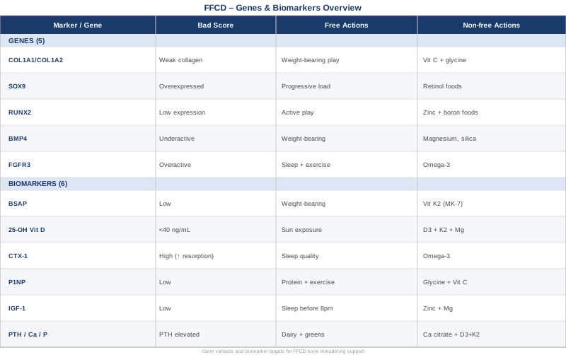
6 Biomarkers Worth Tracking in Focal Fibrocartilaginous Dysplasia
The biomarkers below were chosen because they reflect the core biological processes disrupted in FFCD: bone formation, bone resorption, mineralization sufficiency, and growth factor signaling. None of them are exotic or expensive. Most can be ordered by a general pediatrician. Together they give a working picture of whether the skeleton has the biological resources it needs to remodel the dysplastic lesion over time.
Because FFCD-specific biomarker research is essentially nonexistent — the condition is too rare to have generated dedicated cohort studies — the evidence base here draws from the broader pediatric bone disease literature, including rickets, hypophosphatasia, osteogenesis imperfecta, and general bone development science. The clinical logic for applying these markers to FFCD is strong even when direct FFCD studies are absent.
1. Bone-Specific Alkaline Phosphatase (BSAP)
Why it matters. Alkaline phosphatase exists in multiple tissue isoforms. The bone-specific isoform, produced by osteoblasts, reflects how actively new bone matrix is being laid down. In FFCD, the central problem is a failure of normal periosteal ossification at the medial tibial metaphysis. The fibrocartilaginous tissue that has taken the place of normal bone needs to be progressively replaced through osteoblast activity. If BSAP is low for age, bone formation is sluggish and the remodeling process is likely to be slow. If BSAP is elevated beyond the expected range for a growing child, it can indicate a compensatory response that is worth monitoring.
How to measure it. A standard alkaline phosphatase blood test costs $15–$40. BSAP fractionation (separating the bone isoform from liver alkaline phosphatase) adds $30–$80 and is worth requesting if total ALP is elevated or ambiguous. In children under two, total ALP normally runs higher than adult reference ranges, so using pediatric-specific reference intervals is essential. Labs at children's hospitals routinely have these.
If the score is low — the plan without supplements. Mechanical loading of bone is one of the most reliable stimulators of osteoblast activity. For an infant or toddler who is not yet walking, supervised tummy time, assisted standing with supported weight-bearing, and hydrotherapy in a warm pool all provide the mechanical stimuli that upregulate alkaline phosphatase. Weight-bearing encourages piezoelectric signaling in bone, which directly activates osteoblast precursors. Even brief daily sessions — 10 to 15 minutes twice daily — are supported by pediatric physical therapy literature as safe and effective for promoting bone formation in young children with developmental bone conditions. There are no side effects to weight-bearing within normal developmental bounds.
If the score is low — the plan with supplements or equipment. Vitamin K2 (MK-7 form) activates osteocalcin, the protein that draws calcium into bone matrix. In children, doses of 25–45 mcg daily are generally considered safe and relevant to bone formation. Studies on vitamin K2 and bone formation markers suggest measurable increases in BSAP within 8–12 weeks of supplementation in children with suboptimal values. Vibration platforms designed for bone stimulation (whole-body vibration at 25–50 Hz) have pediatric safety data in conditions like cerebral palsy and have been studied for their ability to activate osteoblasts. These should only be used under medical supervision in toddlers. Cycle: 5 days on, 2 days off. No supplementation should be started without confirming vitamin D and calcium sufficiency first.
2. 25-Hydroxyvitamin D
Why it matters. Vitamin D is not simply a bone vitamin — it is a steroid hormone precursor that regulates over 200 genes, including several directly involved in chondrocyte differentiation, osteoblast maturation, and the cartilage-to-bone transition. In the growth plate and at the periosteum, vitamin D receptor activation is required for the orderly mineralization of cartilage matrix. Deficiency does not just cause rickets; at subclinical levels it slows periosteal bone formation and can compromise the very remodeling process that needs to occur in FFCD.
How to measure it. A 25-OH vitamin D serum test costs $30–$60. Many labs include it in standard pediatric panels. The functional optimal range for children with active bone conditions is generally considered 40–60 ng/mL by clinicians like Peter Attia who specialize in metabolic health — notably higher than the 20 ng/mL threshold used in most standard references. Testing every 3–4 months during the active remodeling phase is reasonable.
If the score is low — the plan without supplements. Safe sun exposure remains the most physiologically complete source of vitamin D. For fair-skinned infants and toddlers, 10–15 minutes of midday sun on arms and legs (without sunscreen during this brief window) can produce meaningful skin-synthesized vitamin D. This is highly dependent on latitude, season, and skin pigmentation. Dark-skinned children require significantly longer exposure for equivalent synthesis. Outdoor play that includes adequate sun exposure should be structured as a daily habit, not an afterthought.
If the score is low — the plan with supplements or equipment. Vitamin D3 (cholecalciferol) is the form used in pediatric supplementation. Typical corrective doses range from 1,000–2,000 IU daily for infants and toddlers with confirmed deficiency, always paired with vitamin K2 to ensure calcium is directed to bone rather than soft tissue. Randomized studies on vitamin D in infants support this pairing. Magnesium glycinate (50–75 mg/day for toddlers, based on body weight) is essential because magnesium is required to convert vitamin D to its active form — a fact often overlooked in pediatric supplementation protocols. Recheck 25-OH D after 8 weeks. Side effects are minimal at these doses; toxicity risk begins above 4,000 IU/day sustained without monitoring.
3. CTX-1 (C-Terminal Telopeptide of Type 1 Collagen)
Why it matters. CTX-1 is released into the bloodstream when osteoclasts break down type I collagen, the dominant structural protein in bone. It is the most widely used marker of bone resorption. In FFCD, understanding the resorption side of the remodeling equation matters because effective remodeling requires coordinated formation and resorption — the fibrocartilaginous lesion needs to be resorbed as new bone matrix is deposited. Excessive resorption relative to formation indicates an imbalance that may slow resolution.
How to measure it. CTX-1 is measured in serum or urine. Serum testing costs $50–$90. Pediatric reference ranges vary significantly with age and growth velocity, so results must be interpreted with age-matched norms. The sample should ideally be taken fasting in the morning, as CTX-1 has a diurnal variation of up to 40% and is suppressed postprandially. Reference range studies for CTX-1 in children are available and should be used to contextualize results.
If the score is high — the plan without supplements. Weight-bearing exercise and mechanical load are the most reliable non-pharmacological signals that reduce osteoclast activity. Sleep quality has a direct effect on bone resorption: most osteoblast activity occurs during deep sleep, and poor sleep shifts the balance toward resorption. Ensuring a consistent, dark sleep environment for the affected child and maximizing total sleep time creates a favorable biochemical environment for bone formation over resorption. Reducing inflammatory dietary triggers — including high-sugar, ultra-processed foods, and excess omega-6 fats in the parent's breast milk if still nursing — can lower the systemic inflammatory tone that drives osteoclast activation.
If the score is high — the plan with supplements or equipment. Omega-3 fatty acids (DHA/EPA) have been shown in multiple human studies to reduce CTX-1 levels by dampening osteoclast-activating inflammatory signaling. For nursing mothers, 1–2 g EPA+DHA daily passes relevant levels to the infant through breast milk. For toddlers eating solids, fish-based foods or pediatric fish oil supplements at 250–500 mg DHA daily are appropriate. Cycling: continuous with food is generally safe; reassess CTX-1 after 12 weeks. No significant side effects at these doses.
4. P1NP (Procollagen Type 1 N-Terminal Propeptide)
Why it matters. P1NP is the most sensitive and specific marker of bone formation currently available. When osteoblasts synthesize type I collagen — the scaffold of new bone — they cleave propeptides that enter the bloodstream as P1NP. Unlike alkaline phosphatase, P1NP is not influenced by liver disease, making it a cleaner signal in children who might have elevated total ALP for other reasons. In the context of FFCD monitoring, P1NP alongside CTX-1 gives a ratio — the formation-to-resorption balance — that is more informative than either marker alone.
How to measure it. Serum P1NP costs $60–$110. It is measured by immunoassay and requires a reliable pediatric lab with age-specific reference intervals. Some children's hospitals include P1NP in their bone metabolism panels. Like CTX-1, morning fasted collection improves consistency. The International Osteoporosis Foundation guidelines on bone turnover markers consider P1NP the reference standard for formation monitoring.
If the score is low — the plan without supplements. Protein intake is one of the most underappreciated drivers of P1NP. Collagen is protein, and inadequate dietary protein directly limits how much type I collagen osteoblasts can synthesize. For a toddler being weaned, ensuring adequate protein from whole food sources — eggs, meat, fish, legumes — directly supports bone formation. Resistance exercise (in pediatric-appropriate forms: crawling against resistance, assisted stair climbing, play that involves pulling and pushing) stimulates collagen gene expression in osteoblasts.
If the score is low — the plan with supplements or equipment. Glycine and proline are the two amino acids that dominate the triple-helix structure of type I collagen. Bone broth, gelatin, or hydrolyzed collagen peptides (in age-appropriate food-based forms for toddlers) provide these amino acids directly. Vitamin C is required as a cofactor for collagen hydroxylation — the step that gives collagen its structural stability. Supplementing vitamin C at 50–100 mg daily in a toddler who eats limited fruit and vegetables is safe and supports P1NP-relevant biology. Recheck P1NP at 12 weeks. No meaningful cycling is needed for food-derived collagen sources.
5. IGF-1 (Insulin-Like Growth Factor 1)
Why it matters. IGF-1 is a growth hormone-dependent signal that is one of the primary anabolic drivers of long bone growth and bone formation. It stimulates osteoblast proliferation, collagen synthesis, and chondrocyte activity in growth plates. In a condition where the bone's periosteal environment is disrupted and new bone formation needs to be upregulated, IGF-1 is one of the systemic signals doing the work. Children with low IGF-1 for their age have measurably slower bone formation and longer recovery from bone pathology. IGF-1 testing in an infant or toddler with FFCD is a simple way to assess whether the anabolic bone-building signal is adequate.
How to measure it. Serum IGF-1 costs $70–$120. Pediatric reference ranges are tightly age- and sex-adjusted. The test is typically covered when growth concerns are documented. IGF-binding protein 3 (IGFBP-3) is often ordered alongside IGF-1 to assess the free-to-bound fraction, adding $50–$80. A pediatric endocrinologist should interpret the results in context.
If the score is low — the plan without supplements. Sleep is the primary driver of growth hormone secretion in young children, and growth hormone is what the liver converts to IGF-1. Maximizing slow-wave sleep — through a consistent bedtime before 8pm, a dark cool room, and no screens within an hour of sleep — directly optimizes IGF-1 in young children. This is one of the most powerful interventions available and requires no prescription or supplement. Protein adequacy and meal timing also matter: IGF-1 synthesis requires sufficient dietary protein, and growth hormone pulses are largest in a fasted state (overnight fast for toddlers is naturally achieved with a good bedtime).
If the score is low — the plan with supplements or equipment. Zinc and magnesium both serve as cofactors in growth hormone signaling and IGF-1 synthesis. In children with confirmed deficiency of either mineral, supplementation can meaningfully raise IGF-1 over 8–12 weeks. Studies on zinc supplementation in children with poor growth show IGF-1 increases of 15–30% in deficient individuals. For toddlers: zinc gluconate 5–10 mg/day (cycle with a copper supplement 1:10 ratio to avoid copper depletion) and magnesium glycinate 50–75 mg/day. Always test zinc status before supplementing, as excess zinc at high doses competes with copper. Reassess IGF-1 at 12 weeks.
6. PTH, Calcium, and Phosphorus Panel
Why it matters. Parathyroid hormone (PTH), calcium, and phosphorus operate as an interconnected regulatory triad that governs mineral delivery to bone. PTH is the main alarm signal: when serum calcium falls or when vitamin D is insufficient, PTH rises and instructs the kidney and bone to release calcium, often at the expense of bone density. Phosphorus is equally important because hydroxyapatite — the mineral crystal that gives bone its rigidity — requires both calcium and phosphorus in the correct ratio. In a child whose bone needs to actively mineralize and remodel a periosteal lesion, any imbalance in this triad represents a direct constraint on recovery.
How to measure it. A basic metabolic panel covering calcium and phosphorus costs $20–$40. Adding intact PTH costs $50–$90. This triad should ideally be measured alongside 25-OH vitamin D, since the interpretation is incomplete without it. Elevated PTH with low-normal calcium and low vitamin D is the most common pattern indicating that the body is cannibalizing bone to maintain serum calcium — the opposite of what is needed in FFCD.
If the scores are imbalanced — the plan without supplements. Dietary calcium from whole food sources — dairy, leafy greens, bone-in fish — provides the most bioavailable forms. High-phytate foods (unsoaked grains and legumes) bind calcium in the gut and reduce absorption. Reducing phytate load through soaking, sprouting, or fermentation of grains improves calcium availability without adding supplements. Adequate protein intake preserves the acid-base balance in blood, which has a direct effect on urinary calcium excretion — a high-protein, whole-food diet loses less calcium through the kidneys than a low-protein, high-carbohydrate diet.
If the scores are imbalanced — the plan with supplements or equipment. Calcium carbonate versus calcium citrate: in toddlers, calcium citrate is better absorbed and does not require stomach acid for dissolution. Doses of 200–400 mg elemental calcium daily are appropriate for toddlers not meeting needs from food. Phosphorus is rarely deficient in children eating whole foods, but if PTH is elevated and phosphorus is low, a phosphorus-containing electrolyte supplement under medical supervision is warranted. Vitamin D3 and K2 must be optimized before supplementing calcium to ensure proper routing to bone rather than arterial calcification. Meta-analyses on calcium and vitamin D co-supplementation in children consistently show improved bone mineral density only when both are adequately supplied. Reassess the full triad every 3 months.
With a clear biomarker picture in hand, the next logical layer of investigation is understanding whether an individual child's genetic background may explain why the bone-to-cartilage transition is disrupted in the first place — and what that means for the pace of recovery.
The Genetic Architecture Behind Focal Fibrocartilaginous Dysplasia
FFCD does not yet have a well-characterized Mendelian genetics profile. Most reported cases are sporadic, and no single causative gene has been identified in the way that, for example, FBN1 is identified in Marfan syndrome. What is understood, however, is that the biological processes FFCD disrupts — periosteal chondrogenesis, the fibrocartilage-to-bone transition, collagen matrix organization, and local growth factor signaling — are each regulated by genes whose common variants influence how robustly an individual child will navigate and resolve this kind of periosteal disruption. Genetic testing in this context is not about finding a cause of FFCD. It is about identifying biological tendencies that may explain a slower or faster recovery, and that point toward specific nutritional or environmental adjustments.
The limited literature on FFCD etiology suggests a periosteal vascular or mechanical insult triggers abnormal fibrocartilaginous repair. Whether genetic susceptibility modulates which children develop permanent lesions versus those who spontaneously resolve is an open and clinically important question.
Gene 1: COL1A1 and COL1A2 — Type I Collagen Architecture
What these genes do. COL1A1 and COL1A2 encode the alpha chains of type I collagen, the primary structural protein of bone, periosteum, and fibrocartilage. Type I collagen is not only the scaffold of normal bone — it is also the dominant protein in the fibrocartilaginous tissue that characterizes FFCD lesions. Variants in these genes affect collagen fiber thickness, cross-linking efficiency, and tensile strength.
Relevant variant. The COL1A1 Sp1 binding site polymorphism (rs1800012) has been associated in multiple studies with reduced collagen synthesis and lower bone mineral density. It is found in a meaningful proportion of the general population. Studies linking COL1A1 variants to bone outcomes are well replicated, particularly in populations with osteoporosis. The implication for FFCD is that a child carrying this variant may produce less competent periosteal collagen during the repair phase.
If the gene has an unfavorable variant — the plan without supplements. Mechanical loading is one of the primary signals that upregulates COL1A1 and COL1A2 gene expression in osteoblasts and periosteal cells. Daily weight-bearing activity — walking, crawling, pushing — physically stimulates collagen gene transcription through mechanotransduction pathways. Prioritizing this even before the child is fully weight-bearing (through assisted standing and hydrotherapy) activates collagen production independently of genetic background.
If the gene has an unfavorable variant — the plan with supplements or equipment. Vitamin C (ascorbate) is essential for the prolyl hydroxylase enzyme that stabilizes the collagen triple helix. Without adequate vitamin C, even genetically normal collagen production produces weaker fibers. In a child with a COL1A1 variant, ensuring consistent vitamin C from food (citrus, kiwi, bell pepper) or supplementation (50–100 mg daily for toddlers) is a direct compensatory strategy. Glycine-rich food sources (bone broth, gelatin desserts made with unflavored gelatin) supply the rate-limiting amino acid for collagen synthesis. No meaningful cycling is needed; these are food-level interventions. Side effects are minimal.
Gene 2: SOX9 — Master Regulator of Chondrogenesis
What this gene does. SOX9 is arguably the most important transcription factor in cartilage biology. It controls the differentiation of mesenchymal stem cells into chondrocytes, regulates type II collagen and aggrecan expression in cartilage, and acts as a brake on the transition from cartilage to bone. In FFCD, the periosteal tissue fails to complete the normal transition from fibrocartilage to lamellar bone. SOX9 activity that is inappropriately sustained or upregulated in the periosteal repair zone could be one mechanism that explains why fibrocartilaginous tissue persists rather than resolving to bone.
Relevant variants. Common non-coding variants near the SOX9 locus have been associated with altered chondrogenesis in developmental orthopedic conditions. While no specific SOX9 variant has been studied in FFCD, the pathway is mechanistically central. Expression of SOX9 is downregulated as chondrocytes hypertrophy and give way to osteoblasts — any factor that delays this downregulation delays the cartilage-to-bone transition.
If the gene is unfavorably expressed — the plan without supplements. Controlled mechanical stress accelerates chondrocyte hypertrophy and the SOX9-to-RUNX2 transcription factor switch in growth cartilage. This is why weight-bearing is not just mechanical support — it is a biological signal that times the cartilage-to-bone transition. Daily progressive weight-bearing within the child's developmental capacity directly works against persistent SOX9 overexpression in the periosteal repair tissue.
If the gene is unfavorably expressed — the plan with supplements or equipment. Retinoic acid (vitamin A as retinol, not beta-carotene) suppresses SOX9 and accelerates chondrocyte hypertrophy in cell and animal models. The evidence in humans is indirect, but vitamin A adequacy is a reasonable and safe target. Liver, egg yolks, and full-fat dairy provide preformed retinol. Avoid high-dose supplemental vitamin A (above the tolerable upper limit for age), as excess retinol is teratogenic and can be toxic. At food-level doses, this is a safe nutritional strategy. Recheck vitamin A status (as retinol) if there are concerns about excess, but deficiency is the more common issue in children on restricted diets.
Gene 3: RUNX2 — Osteoblast Commitment Switch
What this gene does. RUNX2 (Runt-related transcription factor 2) is the master transcription factor for osteoblast differentiation. Without functional RUNX2, bone does not form. Heterozygous loss-of-function mutations in RUNX2 cause cleidocranial dysplasia — a severe bone development disorder. But common population variants that reduce (not eliminate) RUNX2 expression are associated with subtly lower bone formation rates, slower fracture healing, and weaker periosteal ossification. In FFCD, where periosteal ossification needs to accelerate to replace fibrocartilaginous tissue, RUNX2 activity is a critical bottleneck.
Relevant variants. Several RUNX2 single nucleotide polymorphisms have been associated with bone mineral density in genome-wide association studies. Their effect sizes are modest individually but can be meaningful in combination. GWAS studies on RUNX2 and bone outcomes have replicated associations across multiple populations.
If the gene is unfavorably variant — the plan without supplements. Resistance and weight-bearing exercise is the most potent known upregulator of RUNX2 expression in bone progenitor cells. The Wnt signaling pathway — activated by mechanical load — directly induces RUNX2 transcription. Even in toddlers, structured active play that involves pulling, pushing, and weight-bearing stimulates the RUNX2 pathway in a way that dietary interventions alone cannot replicate. This is not hypothetical: pediatric physical therapy protocols for bone development routinely exploit this mechanism.
If the gene is unfavorably variant — the plan with supplements or equipment. Boron is a trace mineral that has been shown in cell and animal studies to upregulate RUNX2 expression and support osteoblast differentiation. Human evidence for boron in pediatric bone disorders is limited, but dietary boron from whole plant foods (raisins, almonds, avocado, dried apricots) is safe and relevant. Targeted boron supplementation in toddlers should wait for more human evidence; food-based intake is appropriate now. Zinc, as noted in the IGF-1 section, also supports RUNX2-mediated osteoblast differentiation and is a more evidence-supported concurrent target.
Gene 4: BMP4 — Bone Morphogenetic Protein Signaling
What this gene does. Bone morphogenetic proteins — BMP2, BMP4, BMP7 — are members of the TGF-beta superfamily and are among the most powerful inducers of osteoblast differentiation and bone matrix production known in biology. They are produced locally in bone and cartilage, act on nearby progenitor cells, and are critically involved in the replacement of cartilage by bone during development and repair. BMP4 in particular is expressed in the periosteum and regulates the differentiation of periosteal cells toward the osteoblast lineage.
Relevant variants. BMP4 promoter polymorphisms affecting expression levels have been reported in craniofacial and skeletal development contexts. While FFCD-specific BMP4 studies do not exist, the pathway is directly relevant: BMP signaling through SMAD1/5/8 is a primary driver of periosteal bone formation, the exact process that needs to be enhanced to resolve the FFCD lesion.
If the pathway is underactive — the plan without supplements. BMP signaling is potentiated by mechanical stimulation through integrin signaling cascades. The same weight-bearing activities that upregulate RUNX2 also potentiate BMP pathway activity in periosteal progenitor cells. Low-level vibration (in supervised clinical settings) has been shown in animal models to specifically increase BMP-2 and BMP-4 expression in periosteal cells — providing a rationale for the vibration platforms mentioned earlier.
If the pathway is underactive — the plan with supplements or equipment. Magnesium is a cofactor for BMP signaling transduction, and magnesium deficiency has been shown to reduce BMP pathway activity in bone cells. Ensuring magnesium adequacy (as described in the biomarker sections) is directly supportive of BMP signaling. Silica, provided as orthosilicic acid from food sources such as oats and green beans, has been shown in in vitro human osteoblast studies to stimulate collagen type I synthesis and BMP-2 expression. Food-level intake is safe; targeted silica supplementation has limited pediatric human data and should be approached cautiously.
Gene 5: FGFR3 — Fibroblast Growth Factor Receptor 3
What this gene does. FGFR3 encodes a receptor that normally limits chondrocyte proliferation and acts as a brake on long bone growth. Gain-of-function mutations in FGFR3 cause achondroplasia (the most common form of short-limbed dwarfism). Common variants that shift FGFR3 signaling have subtler effects on the pace of bone development, cartilage-to-bone transitions, and the shape of long bones including the tibia. An FGFR3 signaling environment that is excessively active suppresses chondrocyte differentiation toward hypertrophy — delaying the transition that needs to occur at the FFCD lesion site.
Relevant variants. The FGFR3 p.Ala391Glu variant has been associated with altered bone geometry. More broadly, FGFR3 pathway polymorphisms that subtly shift signaling balance have been reported in pediatric orthopedic developmental studies. Studies on FGFR3 variants and skeletal development have described modest but measurable effects on bone morphology.
If the gene is unfavorably variant — the plan without supplements. FGFR3 signaling is downregulated by C-type natriuretic peptide (CNP), which is stimulated by exercise and adequate sleep. Prioritizing physical activity and sleep quality in the affected child directly modulates the FGFR3 signaling environment. Reducing chronic low-grade inflammation also reduces FGFR3 pathway overactivation — anti-inflammatory dietary patterns (whole foods, low refined sugar, adequate omega-3) support this.
If the gene is unfavorably variant — the plan with supplements or equipment. Omega-3 fatty acids have been shown to reduce FGF-signaling-related inflammation in musculoskeletal tissue. The EPA/DHA supplementation strategy described for CTX-1 normalization is doubly relevant here. No direct FGFR3-targeting supplement exists that is appropriate for pediatric use without medical supervision. The most evidence-supported approach remains anti-inflammatory nutrition and sleep optimization, with genetic testing informing monitoring intensity rather than a dramatic supplementation change.
The genetic layer gives a deeper understanding of biological vulnerabilities. But synthesizing this into a practical daily framework requires integrating multiple signals at once — which is exactly what some of the most compelling recent work in bone biology attempts to do.
What Andrew Huberman's Work on Bone Biology Can Teach You
Andrew Huberman, a Stanford neuroscientist and host of the Huberman Lab podcast, has dedicated several episodes to bone biology, growth, and the integrative physiology of skeletal health. His approach is notable for synthesizing recent mechanistic science into actionable protocols, challenging the conventional "eat calcium, avoid fractures" model of bone care. While his work is primarily framed around adult bone health, the core mechanisms he describes are highly relevant to pediatric bone remodeling and FFCD recovery. The following are the ten most clinically impactful insights from his bone biology framework.
1. Bone Remodeling Is Continuous and Controlled by Load
Huberman emphasizes that bone is not inert tissue. It is in a constant state of remodeling driven by the mechanical forces placed on it. Osteocytes — the most abundant bone cell — sense strain and direct osteoblast and osteoclast activity accordingly. This means that the best stimulus for bone repair in FFCD is controlled, progressive loading — not rest.
2. Impact Forces Stimulate Bone More Than Sustained Load
His synthesis of the bone biomechanics literature highlights that brief, high-impact forces (jumping, stomping, sudden loading) are more osteogenic per unit time than sustained low-level load. For toddlers, this translates to jumping, bouncing, and active play. Even brief sessions of 10 minutes of high-impact movement have been shown to produce measurable bone formation responses in pediatric populations.
3. Sleep Is the Primary Recovery Window for Bone
Huberman repeatedly emphasizes that slow-wave sleep drives the largest growth hormone pulse of the day, and growth hormone is the primary systemic signal driving IGF-1 synthesis and bone formation. A child getting suboptimal sleep is physiologically constrained in their bone repair capacity, regardless of diet. This is not a wellness platitude — it is a mechanistic fact with direct implications for FFCD recovery.
4. Vitamin D Is a Hormone, Not a Supplement
His framing of vitamin D as a steroid hormone precursor — rather than a simple nutrient — shifts the way clinicians and parents should think about sufficiency targets. He advocates for 40–60 ng/mL as a functional minimum, substantially higher than the standard clinical definition of "sufficiency" (20 ng/mL). This matters enormously for pediatric bone conditions where active mineralization is occurring.
5. Calcium Without K2 Is Incomplete
Huberman draws on the work of cardiologists and nutritional scientists to explain that calcium supplementation without vitamin K2 does not reliably route calcium to bone. Vitamin K2 (MK-7 form) activates matrix Gla protein and osteocalcin, the two proteins responsible for directing calcium into bone matrix rather than arterial walls. This pairing principle is particularly important when supplementing calcium in growing children.
6. Collagen Is the Scaffold That Mineralization Follows
Before hydroxyapatite can form in bone, there must be an organized collagen scaffold for it to deposit on. Huberman discusses how vitamin C, glycine, and proline availability directly limit how quickly and how competently this scaffold is built. This mechanistically supports the P1NP-based intervention strategy described earlier — bone formation markers reflect both collagen scaffold quality and mineral deposition rate.
7. Magnesium Is Systematically Under-Supplemented
Huberman has highlighted in multiple episodes that magnesium deficiency is widespread even in individuals who think they are eating well. Magnesium is a cofactor for vitamin D activation, ATP production in osteoblasts, and BMP signaling transduction. Its deficiency silently limits bone formation even when all other inputs appear adequate. This is particularly relevant for children on limited diets.
8. Cortisol and Chronic Stress Suppress Bone Formation
Elevated cortisol — from poor sleep, psychological stress, or chronic illness — directly suppresses osteoblast activity and upregulates osteoclast activity. In pediatric terms, this means that a child who is experiencing recurrent illness, sleep disruption, or significant stress has a less favorable biochemical environment for resolving a bone lesion. Addressing the stress biology is not secondary to the bone biology — it is part of it.
9. Omega-3 Fats Change the Inflammatory Tone of Bone
His synthesis of the inflammation-bone axis draws on human studies showing that the EPA:arachidonic acid ratio in cell membranes directly influences how aggressively osteoclasts are activated. Higher omega-3 intake shifts this ratio in a direction that reduces bone resorption signaling. This aligns with the CTX-1 reduction data cited earlier and provides a second mechanism justifying omega-3 optimization in FFCD care.
10. The Bone-Microbiome Axis Is Emerging as Clinically Relevant
Among Huberman's more forward-looking discussions is the emerging evidence that gut microbiome composition influences systemic calcium absorption, vitamin K2 production, and immune signaling at the bone surface. Short-chain fatty acid-producing bacteria improve mineral absorption and reduce osteoclast-activating inflammatory cytokines. For children with FFCD, attending to gut health through fermented foods, diverse fiber intake, and avoiding unnecessary antibiotic courses is an upstream investment in bone health that is rarely discussed in the orthopedic context.
Complementary Approaches With Real Evidence
Given how early-stage the science of FFCD specifically remains, the following complementary approaches are selected for their evidence in bone healing, periosteal remodeling, and pediatric musculoskeletal conditions generally — not for FFCD directly, where clinical trials do not exist. The evidence quality is noted honestly for each.
Low-Level Laser Therapy / Photobiomodulation
Photobiomodulation (PBM) uses near-infrared and red light wavelengths to stimulate mitochondrial activity in cells through cytochrome c oxidase absorption. In bone and cartilage tissue, PBM has been shown in human randomized controlled trials to accelerate bone healing by increasing osteoblast proliferation, collagen synthesis, and local growth factor production including TGF-beta and IGF-1. A 2019 systematic review of PBM for bone regeneration found consistent evidence of accelerated healing in controlled human studies. The periosteal tissue type involved in FFCD (fibrocartilage undergoing ossification) is precisely the type of tissue shown to respond to PBM. Equipment cost ranges from professional clinical devices ($5,000–$20,000) to home-grade near-infrared panels ($300–$800). For toddlers, PBM should only be applied under medical supervision, with appropriate eye protection and distance management. Treatment protocol: typically 3 sessions per week, 3–5 minutes per site, at wavelengths of 810–850 nm, cycling 6 weeks on, 2 weeks off. Evidence is promising but FFCD-specific trials are absent; this should be considered an adjunct, not a primary treatment.
Massage Therapy
The fibrocartilaginous lesion in FFCD exerts a tethering force on the proximal tibia that contributes mechanically to varus bowing. The surrounding soft tissue — periosteum, fascia, and muscle — adapts to this altered loading pattern with changes in tone and tension. Therapeutic massage targeting the lower leg musculature can reduce this compensatory tension, improve local circulation, and support periosteal blood flow — which is essential for delivering the growth factors and minerals required for bone repair. Human studies on massage and pediatric musculoskeletal conditions show measurable improvements in tissue perfusion and pain markers, though direct bone healing evidence is limited. The practical protocol: gentle effleurage and petrissage of the calf and tibial soft tissues by a pediatric-trained massage therapist, 2–3 times per week, 20 minutes per session. Parents can learn simplified home techniques for daily application. No significant adverse effects at appropriate pediatric pressure levels.
Breathing-Based Therapies
Slow, controlled diaphragmatic breathing activates the parasympathetic nervous system and reduces the cortisol output that, as noted in the Huberman section, suppresses bone formation. In infants and toddlers, direct participation in breathing exercises is not possible, but the parent's stress state significantly influences the child's autonomic regulation through co-regulation. Guided breathing practices for parents and caregivers of children with chronic conditions have been shown in clinical research to reduce family stress biomarkers and improve the quality of the caregiving environment. Beyond this parent-facing application, age-appropriate soothing practices that promote deep, slow breathing in the child — including white noise, rocking, and infant massage — reduce cortisol and create a biochemically favorable bone repair environment. Studies on breathing and cortisol regulation in pediatric populations consistently show HRV improvement and cortisol reduction within 4–8 weeks of consistent practice. No adverse effects. This is low cost, universally available, and mechanistically grounded.
Conclusion
Focal fibrocartilaginous dysplasia is rare enough that most families and even most clinicians encounter it without a clear framework for what to monitor and what to do beyond imaging follow-up. The biomarkers covered here — BSAP, 25-OH vitamin D, CTX-1, P1NP, IGF-1, and the PTH-calcium-phosphorus triad — provide a practical, affordable way to assess whether the child's biology has the resources it needs to resolve the lesion. The genetic context — COL1A1/COL1A2, SOX9, RUNX2, BMP4, and FGFR3 — helps explain why some children's bone repair biology may need more targeted nutritional support than others.
The most actionable next step is straightforward: work with the child's pediatrician or a pediatric orthopedist to order the six biomarkers at the next scheduled visit. Results in hand, you can have a far more specific conversation about whether vitamin D, protein, collagen support, or sleep optimization should be prioritized. Better data does not replace clinical judgment — but it makes clinical judgment better. That is what a monitoring-first approach to FFCD offers: not a cure promise, but a clearer map.
Musculoskeletal Endocrine & Metabolic
Musculoskeletal: Bone Conditions
Autoimmune: Connective Tissue Conditions