This article contains AI generated content.
Foreign Body Synovitis: 4 Genes And 7 Biomarkers To Track
If you've been dealing with a joint that swells, aches, or just doesn't feel "clean" long after some forgotten scrape, splinter, thorn, or puncture wound, you already know the frustrating part: the story doesn't match the exam. You mention the old injury, and it gets waved off as irrelevant. You get told it's "probably just arthritis" or "wear and tear," and sent home with anti-inflammatories and a shrug. Meanwhile the joint keeps flaring, quietly, on its own schedule.
That mismatch happens because foreign body synovitis is a mechanical and immunological problem hiding inside a generic-sounding symptom. A sliver of plant material, wood, coral, or sea urchin spine can lodge in or near a joint, get walled off by the immune system, and sit there for weeks or months before triggering a slow-building, granulomatous synovitis — inflammation built around the retained material rather than around a virus or an autoimmune trigger. Generic joint-pain advice isn't built for this. It doesn't ask what your synovial fluid actually looks like, whether there's still material in there, or why your immune system is reacting this aggressively to something a splinter usually causes only mild, self-limited irritation.
A more useful approach starts with data instead of guesses: specific lab values, imaging findings, and fluid analysis that tell you what's actually happening inside the joint, plus an honest look at the genetic and immune factors that might explain why some people wall off a foreign body quietly while others develop chronic, destructive synovitis. None of this promises a shortcut around a splinter that needs to come out — but it does mean fewer wasted months and a much sharper conversation with whoever is treating you.
This article works through both angles. The main section walks through seven biomarkers worth tracking — what they reveal, how they're measured, what it costs, and what a sensible response looks like when a result comes back abnormal. A shorter section after that looks at the genetic side: the innate-immune genes that may shape how forcefully your body reacts to retained foreign material. After that, a research-based detour and a look at complementary approaches round out a picture that's meant to be practical, not theoretical.
Summary
Foreign body synovitis rarely announces itself clearly. It hides behind vague swelling, an old and half-forgotten injury, and lab results that look unremarkable at first glance — which is exactly why the right biomarkers matter. Ahead, you'll find out which seven measurements (from a $10 blood test to a specialized enzyme assay) actually distinguish a retained-splinter reaction from an infection, from gout, or from early rheumatoid arthritis — and what a sensible next step looks like for each one. You'll also see why certain immune-system genes, the same ones implicated in rare granulomatous diseases like Blau syndrome, may explain why some joints overreact to material that should barely cause a ripple. Beyond that, a science-heavy podcast breakdown and a review of complementary approaches with real supporting trials fill in the parts that blood work alone can't answer: how to calm a chronically inflamed joint while you sort out its root cause.
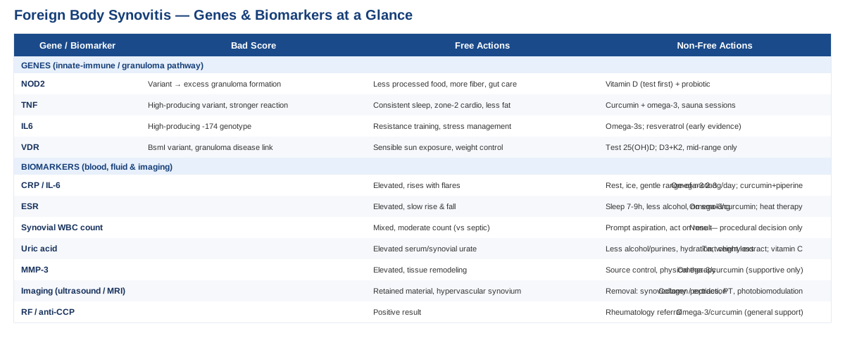
Key Biomarkers For Diagnosing And Tracking Foreign Body Synovitis
Foreign body synovitis is ultimately confirmed by finding retained material and granulomatous tissue with foreign body giant cells on histology, but you rarely get to that point without a trail of supporting lab and imaging data. The biomarkers below are the ones that matter most in practice: they help separate a foreign-body reaction from its two biggest mimics — septic arthritis and crystal arthropathy (gout or pseudogout) — while also tracking whether the joint is settling down or still actively inflamed.
C-reactive protein (CRP) and IL-6
CRP is produced by the liver in direct response to interleukin-6 (IL-6) signaling coming from the inflamed synovium. In a joint reacting to retained foreign material, CRP tends to rise with flares and fall as the reaction quiets down, making it one of the most useful trend-tracking markers available. IL-6 itself sits further upstream and is worth knowing about even though it's tested far less often, since it reflects synovial cytokine activity directly rather than the liver's downstream response to it, as described in this review of interleukin-6 biology in inflammatory joint disease.
How to measure it
Standard or high-sensitivity CRP is a routine blood draw, typically $10 to $30 out of pocket and covered by most insurance when ordered for joint symptoms. Serum IL-6 is the more advanced option — not part of a standard panel, usually $50 to $100, and mainly available through specialty or research-oriented labs. The affordable-first, advanced-if-needed approach favored by physicians like Peter Attia applies well here: start with hs-CRP, and only add IL-6 if the picture stays unclear after imaging and fluid analysis.If the score is bad, the plan without supplements
An elevated CRP in this context is a signal to look for a mechanical cause, not just to treat the number. That means prioritizing imaging to locate retained material and, if found, surgical or ultrasound-guided removal — the only treatment shown to be curative for plant-thorn and related foreign body synovitis, per this case series on plant thorn synovitis of the elbow. In the meantime, rest, ice, and gentle range-of-motion work reduce mechanical aggravation, and CRP should be rechecked every 4 to 6 weeks to confirm the trend is improving rather than plateauing.If the score is bad, the plan with supplements or equipment
Omega-3 fatty acids (roughly 2 to 3 grams combined EPA/DHA daily, taken continuously with food) have the most consistent evidence for lowering systemic inflammatory markers, though effects are modest and take 8 to 12 weeks to show up in blood work. Curcumin with piperine (500 to 1000 mg, once or twice daily) is a reasonable adjunct, best cycled 8 to 12 weeks on with a 2-week break to reassess need. Side effects to know: fish oil can increase bleeding risk at higher doses and interacts with blood thinners; curcumin can cause GI upset and should be used cautiously with gallstones or anticoagulant medication. None of this substitutes for addressing the retained material itself — it only supports the body while that gets sorted out.Erythrocyte sedimentation rate (ESR)
ESR reflects changes in plasma proteins like fibrinogen and rises more slowly than CRP, but it also falls more slowly — which makes it a good marker for the kind of low-grade, weeks-to-months smoldering inflammation typical of a forgotten foreign body sitting in a joint, rather than an acute infection.
How to measure it
A Westergren-method blood draw, typically $10 to $20, almost always covered by insurance and often ordered alongside CRP.If the score is bad, the plan without supplements
Same core logic as CRP: treat ESR as a signal to keep pursuing the anatomic source rather than the number itself. Consistent sleep (7 to 9 hours), reduced alcohol intake, and smoking cessation all measurably lower background ESR over 2 to 3 months and are worth addressing regardless of the joint issue.If the score is bad, the plan with supplements or equipment
The same omega-3 and curcumin approach described above applies, with the same dosing, cycling, and side-effect cautions. Heat therapy (a heating pad or infrared device, 15 to 20 minutes daily) can help with comfort during flares, though it has no measurable effect on the ESR value itself.Synovial fluid white blood cell count and differential
This is the single most informative test available, because it samples the joint environment directly rather than inferring from blood. Foreign body synovitis typically shows a mixed, moderately elevated cell count with lymphocytes, histiocytes, and multinucleated giant cells — quite different from the neutrophil-dominant, very high counts seen in septic arthritis. One study found that without recent antibiotic use, a synovial leukocyte cutoff around 33,000 cells/µL had 96% sensitivity and 95% specificity for diagnosing native joint septic arthritis — useful context for understanding where a foreign-body reaction usually falls short of that threshold. It's worth knowing this overlap isn't perfect: cell counts also poorly predict septic arthritis when crystal arthropathy is also present, which is exactly why the next two biomarkers matter as well.
How to measure it
Arthrocentesis (joint aspiration) performed by an orthopedist or rheumatologist, with the fluid sent for cell count and differential, Gram stain, culture, and crystal analysis. Cost typically runs $200 to $600 for the procedure plus lab fees, usually covered when medically indicated by joint swelling or effusion.If the score is bad, the plan without supplements
This isn't a value you improve with lifestyle changes — it's a diagnostic result that determines the next procedural step. A pattern consistent with foreign body reaction, combined with imaging showing retained material, points toward synovectomy or targeted removal. A pattern consistent with infection changes the plan entirely toward antibiotics and urgent drainage. There is no supplement pathway here; the "plan" is getting the aspiration done promptly and acting on what it shows.Serum and synovial uric acid
Gout is one of the most common mimics of foreign body synovitis, and the two can even coexist, since retained material can provoke local inflammation in a joint that's separately prone to urate crystal deposition. Hyperuricemia is present in roughly 95% of acute gout cases, but an elevated level alone doesn't confirm the diagnosis, and a normal one doesn't rule it out — polarized light microscopy of synovial fluid remains the gold standard, as detailed in this review on synovial fluid uric acid and gout diagnosis.
How to measure it
Serum uric acid is a simple blood test, $10 to $20. Synovial uric acid and crystal analysis are done from the same joint aspiration used for the white cell count above, so it typically doesn't add extra cost if fluid is already being collected.If the score is bad, the plan without supplements
Reducing alcohol (beer in particular), cutting back on high-purine and high-fructose foods (organ meats, shellfish, sugary drinks), consistent hydration, and gradual weight loss all lower serum urate meaningfully over 2 to 3 months.If the score is bad, the plan with supplements or equipment
Tart cherry extract (480 to 1000 mg daily) has modest supporting evidence for lowering urate and flare frequency; vitamin C (500 mg daily) has a small urate-lowering effect as well. Both can be used continuously, though vitamin C at high doses raises kidney stone risk in people already prone to them. If true gout is confirmed rather than just elevated uric acid, urate-lowering medication (allopurinol or febuxostat) is a medical decision to make with a physician — not a supplement, but worth naming so it isn't confused with one.Matrix metalloproteinase-3 (MMP-3)
MMP-3 is an enzyme secreted by activated synovial cells that breaks down cartilage and connective tissue components. It's been studied mainly in rheumatoid arthritis, where it correlates with disease activity and predicts structural joint damage, as shown in this comparison of serum MMP-3 against acute phase proteins in early RA. It hasn't been specifically validated in foreign body synovitis, but the underlying biology — synoviocytes revved up by chronic granulomatous inflammation — is directly relevant, which makes it a reasonable marker of ongoing tissue remodeling to track if the diagnosis remains unclear.
How to measure it
A specialized serum ELISA test, not part of routine panels, typically $50 to $150 and ordered through a rheumatology-oriented lab. It's not always covered by insurance outside of an RA workup, so it's worth asking about cost before ordering.If the score is bad, the plan without supplements
As with CRP, an elevated MMP-3 should redirect attention to source control — confirming and removing any retained material — combined with physical therapy to preserve range of motion while that's being sorted out. Recheck every 8 to 12 weeks alongside the treating clinician rather than in isolation.If the score is bad, the plan with supplements or equipment
No supplement has been shown to specifically lower MMP-3 in this condition, so the honest answer is that the same broad anti-inflammatory support described for CRP (omega-3s, curcumin) is supportive at best, not corrective. Treat this marker as a monitoring tool rather than a target.Imaging-based synovial markers (high-resolution ultrasound and MRI)
This is arguably the most important entry on this list, because it's the one that actually finds the foreign material driving everything else. High-resolution ultrasound can detect retained plant material smaller than half a millimeter and assess synovial thickening and blood flow (via power Doppler), as described in this case report on diagnosing plant-thorn synovitis by ultrasonography. MRI adds more detail on soft tissue involvement when ultrasound is inconclusive.
How to measure it
Ultrasound is the sensible first step: inexpensive ($150 to $400 cash-pay), repeatable, and radiation-free. MRI runs considerably higher, roughly $500 to $3000 depending on region, contrast use, and insurance coverage, and is generally reserved for cases where ultrasound doesn't clarify the picture.If the score is bad, the plan without supplements
If imaging identifies retained material or a markedly thickened, hypervascular synovium, removal — via synovectomy or ultrasound-guided extraction — is the treatment with actual curative evidence behind it. This is a procedural fix, not something a supplement protocol replaces.If the score is bad, the plan with supplements or equipment
Post-procedure, collagen peptides (10 to 15 g daily) may support connective tissue recovery, and a structured physical therapy program with resistance bands helps restore joint function. Photobiomodulation devices (covered in more detail later in this article) can also support recovery, but as an adjunct to removal, not a substitute for it.Rheumatoid factor (RF) and anti-CCP antibodies
Chronic foreign body synovitis can look a lot like early rheumatoid arthritis on exam — persistent swelling, morning stiffness, reduced range of motion — which makes this pair of tests an important branch point. RF and anti-CCP positivity point toward an autoimmune process requiring a completely different treatment approach (rheumatology referral, disease-modifying medication) rather than orthopedic source removal.
How to measure it
A standard blood draw, often bundled into an "arthritis panel," costing $30 to $80 and typically covered when ordered to work up unexplained joint swelling.If the score is bad, the plan without supplements
A positive result should prompt a rheumatology referral rather than continued focus on foreign body removal alone — the two conditions can coexist, but the autoimmune component needs its own management plan.If the score is bad, the plan with supplements or equipment
This result reroutes care rather than something to correct directly, but general anti-inflammatory support (the omega-3 and curcumin approach described earlier) remains reasonable regardless of which path the workup takes.Taken together, these seven biomarkers form a practical decision tree: CRP, ESR, and MMP-3 track how active the inflammation is; synovial fluid analysis and imaging locate and characterize the cause; and uric acid plus RF/anti-CCP rule out the two most common mimics. None of them replace a clinician's judgment, but showing up to an appointment already knowing which of these have been checked — and which haven't — tends to shorten the path to an actual answer considerably.
What Your Genes May Have To Do With It
Foreign body synovitis itself hasn't been the subject of dedicated genetic studies — it's simply too rare and too often misdiagnosed as something else to attract large research cohorts. But the underlying biology, an innate immune system deciding how aggressively to wall off and attack retained foreign material, overlaps directly with genes studied in related granulomatous and inflammatory joint conditions. Geneticists like Ali Torkamani, whose work on polygenic risk has pushed precision medicine toward looking at inherited variants rather than just family history, and Gary Brecka, who popularized functional genetic panels covering inflammation and detoxification pathways, both point to the same idea: your baseline genetic wiring shapes how your immune system responds to a trigger, even when the trigger itself (a splinter) is banal. The honest caveat up front: most of what follows comes from studying rare monogenic granulomatous diseases and common inflammatory arthritis, not foreign body synovitis directly, so treat it as a plausible framework rather than settled fact.
NOD2
NOD2 is an intracellular receptor that recognizes bacterial cell wall fragments and activates the NF-κB inflammatory pathway. Rare NOD2 mutations cause Blau syndrome, a genetic disease defined by granulomatous arthritis, uveitis, and skin rash — essentially an inherited tendency to form excessive granulomas, described in detail in this review of Blau syndrome as the prototypic autoinflammatory granulomatous disease. Common NOD2 variants (distinct from the rare Blau-causing mutations) are also linked to Crohn's disease, suggesting a broader role in how the body handles chronic granuloma formation, including plausibly around retained foreign material in a joint.
If the gene is bad, the plan without supplements
No lifestyle change reverses a gene variant, but NOD2 senses gut bacterial signals, so supporting gut barrier health is a reasonable downstream lever: a lower intake of ultra-processed food, adequate fiber, and identifying and addressing any personal food intolerances.If the gene is bad, the plan with supplements or equipment
Vitamin D repletion (test first, then supplement to a mid-normal range, typically 1000 to 2000 IU daily, rechecking levels every 3 months) and a targeted probiotic (strains studied in Crohn's such as Saccharomyces boulardii) are the most defensible additions. Avoid megadosing vitamin D without monitoring — hypercalcemia is a real risk, especially relevant given vitamin D's separate role in granuloma biology discussed below.TNF (tumor necrosis factor-alpha)
TNF-alpha is a central cytokine in granuloma formation, and baseline production levels are substantially genetically determined, largely through promoter polymorphisms in the TNF gene region on chromosome 6, as summarized in this review of TNF-alpha gene polymorphisms and expression. Higher-producing variants have been linked to a range of inflammatory and autoimmune conditions and plausibly to a more aggressive local reaction around retained foreign material.
If the gene is bad, the plan without supplements
Sleep deprivation reliably raises TNF-alpha, so consistent sleep is one of the higher-leverage, zero-cost interventions available. Regular zone-2 cardiovascular exercise and reducing visceral fat (adipose tissue is itself a source of TNF-alpha) both lower baseline levels over 2 to 3 months.If the gene is bad, the plan with supplements or equipment
Curcumin and omega-3s (dosing as described earlier, cycled 8 to 12 weeks on with breaks) are reasonable adjuncts. Sauna use (3 to 4 sessions weekly, 15 to 20 minutes) has some evidence for transiently reducing inflammatory cytokines, though it's contraindicated in certain cardiovascular conditions and should be cleared with a physician first.IL6
The IL6 promoter polymorphism at position -174 (rs1800795) affects how much IL-6 synovial fibroblasts produce, with the high-producing genotype linked to greater disease activity in inflammatory arthritis, as detailed in this review of IL-6 in rheumatoid arthritis. Since IL-6 drives CRP production directly, a high-producing variant may partly explain why some people run persistently elevated CRP during a foreign body reaction while others barely register one.
If the gene is bad, the plan without supplements
Resistance training lowers tonic IL-6 levels over time, as does consistent stress management — cortisol dysregulation from chronic stress independently raises IL-6. Reducing glycemic spikes (via meal composition and timing) is another lever worth prioritizing.If the gene is bad, the plan with supplements or equipment
Omega-3s at the dosing described earlier can be used continuously. Resveratrol has early, less consistent evidence for lowering IL-6 — worth flagging clearly as preliminary — and if used, is reasonable cycled 8 weeks on and 4 weeks off, watching for GI upset and avoiding it alongside blood thinners.VDR (vitamin D receptor)
VDR polymorphisms, including the well-studied BsmI variant, are associated with sarcoidosis, another granulomatous condition, as shown in this study of vitamin D receptor gene polymorphism in sarcoidosis. The nuance here matters: granuloma macrophages can locally synthesize active vitamin D, which itself promotes giant cell formation — meaning more vitamin D isn't automatically better in a granulomatous context, and this is genuinely different from how vitamin D is usually framed for general immune health.
If the gene is bad, the plan without supplements
Sensible sun exposure and weight management (vitamin D is fat-soluble and gets sequestered in excess adipose tissue) are the non-supplement levers available.If the gene is bad, the plan with supplements or equipment
Test blood 25(OH)D before supplementing, and if replacing, pair vitamin D3 with K2 and target a mid-normal range rather than the higher end often recommended for general wellness — this is one case where more is not clearly better given the granuloma biology involved. Recheck every 3 to 6 months, and don't supplement without monitoring if a granulomatous condition is confirmed, given the hypercalcemia risk.Genetic information like this is best used as context, not diagnosis — it explains predisposition, not destiny, and none of these variants change the basic fact that a retained foreign body generally needs to come out. With both the biomarker and genetic pictures in view, it's worth stepping back to the bigger immune-system picture, which is where the next section picks up.
10 Immune-System Lessons From The Huberman Lab Deep Dive On Inflammation
The Huberman Lab podcast's episodes on immune function, including its conversation with pulmonologist and immunologist Dr. Roger Seheult, dig into how innate immunity — the same system responsible for walling off and reacting to foreign material in a joint — actually resolves inflammation, and where common assumptions break down. Here are ten of the more useful, occasionally counterintuitive takeaways relevant to a chronically inflamed joint.
1. Inflammation resolution is an active process, not just an absence of inflammation
The body doesn't simply "run out" of inflammatory signal over time. Specialized pro-resolving mediators, largely derived from omega-3 fatty acids, actively shut down the inflammatory response. This is part of why omega-3 intake shows up repeatedly as a supportive intervention throughout this article — it's not just "anti-inflammatory," it's substrate for the resolution machinery itself.2. Sleep is when a large share of immune recalibration happens
Even one night of shortened sleep measurably shifts inflammatory cytokine levels, including TNF-alpha. For a joint already fighting a chronic low-grade reaction, inconsistent sleep is one of the few daily variables working directly against resolution.3. Chronic stress reroutes immune resources
Sustained cortisol elevation doesn't just feel bad — it shifts immune cell behavior in ways that can prolong low-grade inflammatory states rather than resolve them, reinforcing why stress management isn't a soft add-on but a mechanistic lever.4. Deliberate heat exposure has a measurable, if modest, immune effect
Sauna use raises core body temperature enough to trigger heat shock proteins and transient shifts in circulating immune cells. It's not a treatment for a retained foreign body, but it's a reasonable adjunct for general inflammatory tone.5. Cold exposure needs more nuance than "cold is always anti-inflammatory"
Aggressive cold exposure immediately after tissue injury or intense exercise can blunt some of the adaptive signaling the body needs to remodel tissue. For an actively inflamed joint recovering from a procedure, this is a reason to be more cautious with ice than the reflexive "ice everything" approach.6. Gut health and systemic inflammation are tightly linked
A meaningful share of immune signaling originates from gut-resident immune tissue, which is part of why NOD2 — a gene that senses gut bacterial fragments — shows up in both inflammatory bowel disease and granulomatous joint conditions.7. Vitamin D is an immune modulator, not just a bone nutrient
Vitamin D receptors are present on nearly every immune cell type, and vitamin D status shapes both how aggressively the innate immune system responds and how well it eventually stands down — which lines up directly with the VDR discussion above.8. Alcohol has an outsized effect on innate immune function
Even moderate alcohol intake measurably disrupts innate immune cell function for 24 hours or more, which is relevant both for general inflammation and specifically for uric acid and gout risk discussed earlier.9. Exercise intensity matters more than exercise volume for inflammatory tone
Moderate, consistent aerobic exercise (zone 2) lowers baseline inflammatory markers over time, while very high-intensity training without adequate recovery can transiently raise them — a useful nuance for anyone managing a flaring joint who's tempted to "push through it."10. The immune system needs a clear signal to stand down
Chronic, low-level irritants — including retained foreign material — can keep the innate immune system in a persistent, low-grade "on" state indefinitely, because there's no clear resolution signal until the irritant itself is gone. This is arguably the single most important takeaway for foreign body synovitis specifically: no amount of general immune optimization fully substitutes for removing the thing the immune system is reacting to.These lessons reinforce a theme that runs through both the biomarker and genetic sections: supporting the immune system generally is worthwhile, but it works alongside — not instead of — addressing the physical cause. That same principle applies to the complementary approaches covered next, which are genuinely useful for symptom management but shouldn't be mistaken for a substitute for source control.
Complementary Approaches Worth Considering
None of the modalities below have been studied specifically in foreign body synovitis — the condition is too rare for dedicated trials — but each has meaningful human evidence in closely related inflammatory joint conditions like knee osteoarthritis and rheumatoid arthritis, where synovial inflammation and joint pain overlap substantially with what's happening in a foreign-body-affected joint.
Tai chi
Tai chi combines slow, controlled movement with weight shifting and breath control, and is one of the better-studied movement-based therapies for joint conditions involving synovial inflammation and pain. For a joint recovering from foreign body synovitis, particularly after removal of the material, it offers a low-impact way to rebuild proprioception and range of motion without the mechanical stress of higher-impact exercise.
A commonly studied protocol involves twice-weekly, 12-week programs using modified Yang-style forms, which showed meaningful improvements in pain and physical function compared to control groups, as summarized in this review on the effectiveness of tai chi for knee osteoarthritis. The review also notes that overall study quality in this area remains inconsistent, so results should be read as promising rather than definitive.
In practice, this means starting tai chi only once acute swelling has settled and, ideally, after any retained material has been addressed — using it as a rehabilitation and maintenance tool rather than a treatment for active inflammation. Two sessions a week, 20 to 30 minutes each, is a reasonable and sustainable starting cadence, with no notable side effects beyond normal exercise soreness.
Massage therapy
Massage therapy targets the surrounding soft tissue and musculature rather than the joint itself, which can help with the compensatory stiffness and guarding that develop around a chronically inflamed joint. It won't touch the underlying granulomatous reaction, but it addresses a real and often-overlooked source of secondary discomfort.
A randomized dose-finding trial in knee osteoarthritis found that 60-minute weekly Swedish massage sessions over 8 weeks produced significantly greater improvement in pain and function scores than usual care, though the benefit faded by 24 weeks without continued sessions, according to this randomized dose-finding trial of massage therapy for knee osteoarthritis.
This suggests massage works best as an ongoing maintenance practice rather than a one-time fix — weekly sessions during an active flare, tapering to biweekly or monthly once the joint stabilizes, avoiding direct deep pressure over a visibly swollen or acutely inflamed joint itself.
Photobiomodulation (low-level laser therapy)
Photobiomodulation uses specific wavelengths of low-intensity light, generally in the 780 to 900 nm range, thought to act at the mitochondrial level to modestly reduce local inflammation and pain. It's a plausible adjunct for a joint recovering from either the foreign body reaction itself or a removal procedure, given its localized, non-invasive application directly over the affected area.
A double-blind, placebo-controlled trial in knee osteoarthritis using a 790 nm laser at 4 J per treatment point across nine areas of the knee found significant reductions in pain and improvements in WOMAC scores compared to sham and no-treatment groups, as reported in this randomized controlled trial of low-level laser therapy for knee osteoarthritis. Evidence in rheumatoid arthritis has been more mixed, with some meta-analyses finding benefits for stiffness and grip strength but not pain specifically, so expectations should stay modest.
A reasonable approach is two to three sessions weekly for 4 to 6 weeks, ideally using a device or in-office protocol matching the wavelength and dosing used in trials, then reassessing. Side effects are minimal, though it should be avoided directly over any area with an open wound or recent surgical incision until healed.
Mindfulness meditation / MBSR
Mindfulness-Based Stress Reduction won't change what's happening inside the joint, but chronic, unresolved joint pain has a well-documented psychological toll, and MBSR is one of the more rigorously studied tools for managing that burden without medication.
A randomized trial in rheumatoid arthritis patients found MBSR improved psychological distress and well-being at 6 months, though it's worth being direct about the limits here: the intervention did not measurably change disease activity itself, as shown in this randomized trial of mindfulness-based stress reduction in rheumatoid arthritis. In other words, it helps with coping, not with the underlying inflammatory process.
Used realistically, an 8-week structured MBSR course (the standard format used in most trials) is a reasonable investment for anyone finding the uncertainty and duration of a foreign body synovitis workup genuinely stressful — but it should be framed to yourself honestly as support, not treatment.
Bringing It Together
Foreign body synovitis rewards precision more than most joint conditions do, because the fix — when material is actually present — is genuinely curative rather than palliative. The biomarkers covered here exist to get you to that clarity faster: CRP, ESR, and MMP-3 to track how active things are; synovial fluid analysis and imaging to find and characterize the cause; uric acid and RF/anti-CCP to rule out the two most common lookalikes. The genetic layer helps explain why some people mount a bigger reaction to the same kind of injury, and the immune-system lessons and complementary approaches fill in the space around that — supporting a joint through inflammation and recovery without ever pretending to replace removing the thing that started it all.
If there's one practical next step, it's this: if a joint has been acting up since some injury you'd otherwise dismiss, ask specifically for a joint aspiration and high-resolution ultrasound before accepting a generic arthritis diagnosis. Bring this list of biomarkers to that appointment, track the trend over weeks rather than a single reading, and loop in a rheumatologist if RF or anti-CCP come back positive. Better information here doesn't just mean more data — it means a shorter, more targeted path to actually resolving what's going on.
Autoimmune: Inflammatory Conditions