This article contains AI generated content.
· UpdatedFreeman-Sheldon Syndrome: 5 Genes and 7 Biomarkers to Track
If your family has just received a Freeman-Sheldon syndrome diagnosis, or you're a clinician trying to build a coherent monitoring plan for a patient with this rare condition, you've probably noticed that most of what's written online falls into one of two buckets: dense genetics papers that don't translate into a plan, or generic "rare disease" pages that don't go deep enough to be useful day to day. Neither tells you what to actually watch for at the next appointment, or why.
Freeman-Sheldon syndrome isn't a lifestyle-driven condition with dozens of tunable risk factors. It's caused by a small number of well-characterized genes that disrupt how fetal muscle fibers contract and relax, and its downstream effects — on the airway, the lungs, the spine, the joints, the eyes, the mouth — are the things that actually need tracking over a lifetime. Generic advice tends to either overstate what genetics alone can tell you, or understate how much can be done with careful, structured monitoring once you know what to look for.
This article takes the more precise route. It walks through the specific biomarkers and clinical measurements worth tracking over time, what each one reveals, how it's measured, and what realistic management looks like when a value drifts in the wrong direction. It then goes a layer deeper into the genes themselves — what MYH3 and its close relatives actually do inside a developing muscle fiber, and why "fixing" a structural gene is a different problem from optimizing a metabolic one.
None of this promises a cure, because Freeman-Sheldon syndrome doesn't have one. But a clearer map of what to measure, when, and why tends to produce calmer, better-informed decisions than either panic or vague reassurance — and that's a genuinely useful place to start.
Summary
Freeman-Sheldon syndrome is caused, in roughly 90% of cases, by mutations in a single gene — MYH3 — that encodes a form of myosin used only in developing fetal muscle. A handful of related genes (TNNT3, TNNI2, TPM2, MYH8) explain most of the remaining cases and the closely overlapping distal arthrogryposis spectrum. None of these are genes you can "optimize" with diet or supplements — they're structural sarcomere genes, fixed from conception, and the real leverage comes from knowing which one is involved and monitoring its downstream effects closely.
That's where the second half of this article does the heavy lifting: seven measurable markers — from creatine kinase and pulmonary function to spinal curvature, joint range of motion, and mouth opening — that give a family and their care team an early warning system for the complications that actually cause harm in Freeman-Sheldon syndrome, along with realistic, cost-aware plans for each one when a number moves in the wrong direction. There's also a look at what a biomechanics book with no connection to rare disease can still teach about long-term joint care, and a review of which complementary therapies have real human evidence behind them for this population — and which don't.
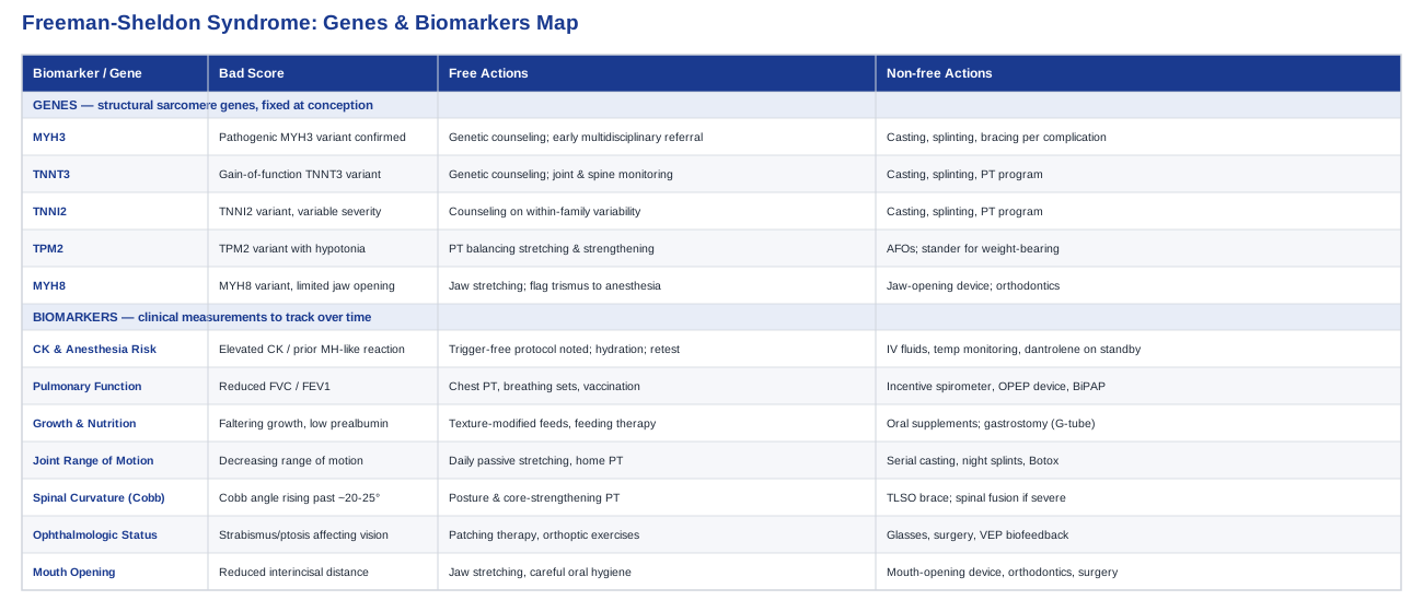
The Biomarkers Worth Tracking in Freeman-Sheldon Syndrome
Because Freeman-Sheldon syndrome is a structural, congenital condition rather than a metabolic one, "biomarkers" here means something slightly broader than a lipid panel — it includes blood tests, functional measurements, and imaging findings that, tracked over months and years, tell you whether the condition is stable or drifting toward a complication that needs intervention. Multidisciplinary reviews of the syndrome consistently point to the same handful of systems as the ones that actually determine quality of life: the airway and lungs, the spine and joints, growth and feeding, and the eyes and mouth Freeman-Sheldon Syndrome — comprehensive review. The seven markers below cover those systems.
1. Creatine kinase and anesthesia-risk markers
Creatine kinase (CK) is a simple blood marker of muscle breakdown, and in Freeman-Sheldon syndrome it matters less as a routine wellness check and more as a safety signal before any procedure requiring general anesthesia. Case reports describe elevated CK and malignant-hyperthermia-like reactions — muscle rigidity, rising temperature — in patients undergoing anesthesia with triggering agents, which is why anesthesiologists caring for this population typically default to trigger-free protocols regardless of the CK result Anesthetic considerations in Freeman-Sheldon syndrome Pediatric anesthetic management case report.
How to measure it
A basic blood draw, typically $15–40 out of pocket if not covered, drawn before any elective surgery and any time unexplained muscle pain, weakness, or dark urine appears.If the score is bad, the plan without supplements
Document the elevated CK and any prior reaction in the medical record so every future anesthesia team defaults to a trigger-free (non-succinylcholine, non-volatile-agent) protocol; hydrate well in the 24 hours before any planned procedure; avoid unaccustomed intense exertion in the days before a scheduled test, since exercise alone can transiently raise CK; retest in 2–4 weeks to see whether the elevation was transient or persistent.If the score is bad, the plan with supplements or equipment
There's no supplement that lowers a genetically-influenced baseline CK, and none should be used to try. The actual safety net is procedural: intravenous fluids during surgery, continuous temperature monitoring, and dantrolene kept available in the operating room in case of a true hypermetabolic crisis. Dantrolene is reserved for an active crisis, not used prophylactically, because it carries its own side effects (muscle weakness, liver enzyme changes) that aren't justified without an emergency.2. Pulmonary function (FVC/FEV1)
Restrictive lung disease — smaller lung volumes from a combination of scoliosis and weakened respiratory muscles — has been documented in Freeman-Sheldon syndrome and is a leading cause of recurrent pneumonia if unaddressed Pulmonary function evaluation in Freeman-Sheldon syndrome.
How to measure it
Spirometry ($50–300 depending on setting) or a full pulmonary function test with lung volumes ($200–500 at a pulmonology lab), typically every 6–12 months, more often if scoliosis is progressing.If the score is bad, the plan without supplements
Assisted coughing techniques and chest physiotherapy, deep-breathing sets two to three times daily, upright positioning during the day, prompt antibiotic treatment for respiratory infections, and up-to-date flu and pneumococcal vaccination — one case series noted no further pneumonia episodes once structured respiratory care was in place.If the score is bad, the plan with supplements or equipment
Daily incentive spirometer practice (roughly ten breaths, three times a day) has randomized-trial support for reducing pulmonary complications around surgery Preoperative incentive spirometry RCT; an oscillating positive-expiratory-pressure device can help if secretions are hard to clear; nighttime non-invasive ventilation (BiPAP) may be added if a sleep study shows hypoventilation. Side effects to watch: lightheadedness from over-vigorous spirometer use, and mask-related skin breakdown with BiPAP, which needs padding and regular fit checks.3. Growth and nutrition markers
Microstomia and swallowing difficulty make failure to thrive a real risk, and growth trajectory is one of the most sensitive early indicators that feeding support needs to be escalated.
How to measure it
Growth charts at every pediatric visit (no added cost), prealbumin or albumin blood tests ($20–80), and a videofluoroscopic swallow study ($300–800) if aspiration is suspected.If the score is bad, the plan without supplements
Texture-modified meals, smaller and more frequent feeds, careful positioning during eating, and oral-motor feeding therapy with a speech-language pathologist once or twice a week.If the score is bad, the plan with supplements or equipment
Calorie-dense food fortification and pediatric oral nutrition supplements under a dietitian's guidance; if oral intake still can't meet needs, a gastrostomy (G-tube), typically fed as four to six boluses a day or overnight continuous feeds. Watch for reflux or vomiting from overfeeding and keep the tube site cleaned daily to avoid infection.4. Joint range of motion and contracture tracking
Camptodactyly, ulnar deviation, and clubfoot are hallmark features, and how they change over time — not just their presence — drives the timing of bracing, casting, or surgery.
How to measure it
Goniometer measurement by a physical therapist ($75–200 per visit) or a smartphone goniometer app for home tracking between visits, ideally every three months during active growth.If the score is bad, the plan without supplements
Daily passive stretching (twice a day, five to ten repetitions, holding each 15–30 seconds) and active-assisted range-of-motion exercises taught by a physical therapist for a family home program. Stay within a pain-free range — aggressive stretching of an already-contracted joint can cause micro-tearing and set progress back.If the score is bad, the plan with supplements or equipment
Serial casting, with the cast changed weekly over several weeks, or dynamic night splints worn 8–12 hours to gradually lengthen shortened tissue. Botulinum toxin injections are sometimes used alongside casting to reduce muscle resistance, typically re-dosed every three to six months, with transient weakness or bruising as the main side effects. Reassess range of motion at every casting or splinting cycle to confirm it's still working.5. Spinal curvature (Cobb angle)
Scoliosis is common in this syndrome and is a direct contributor to the restrictive lung pattern discussed above, which makes it one of the more consequential numbers to track.
How to measure it
A standing spine X-ray ($100–500) or low-dose EOS imaging ($300–600), every 6–12 months during growth spurts and less frequently once skeletally mature.If the score is bad, the plan without supplements
For curves under roughly 20–25 degrees, posture training, core-strengthening physical therapy, and continued monitoring are usually sufficient.If the score is bad, the plan with supplements or equipment
For moderate curves (roughly 20–40 degrees) in a still-growing child, a thoracolumbosacral orthosis (TLSO) brace worn 16–23 hours a day is standard. Progressive curves beyond about 45–50 degrees, or curves compromising breathing, are typically referred for spinal fusion. Bracing side effects include skin irritation and pressure points, so refit checks every three to four months as the child grows are essential.6. Ophthalmologic markers
Deep-set eyes, ptosis, and strabismus are frequent features, and untreated misalignment in early childhood carries a real risk of amblyopia on top of the structural eye differences.
How to measure it
A pediatric ophthalmology exam ($100–300) using a prism cover test for strabismus angle, margin-reflex distance for ptosis, and standard visual acuity testing, recommended every 6–12 months starting in infancy.If the score is bad, the plan without supplements
Patching therapy for amblyopia — commonly two hours a day, based on NIH-supported treatment trials — along with orthoptic exercises.If the score is bad, the plan with supplements or equipment
Corrective glasses or prism lenses, strabismus surgery for significant misalignment, and ptosis repair if the eyelid is obstructing vision. For residual amblyopia after the usual treatment window, visual-evoked-potential biofeedback training has shown measurable gains in small clinical studies VEP biofeedback vision training in amblyopia, and biofeedback-based fixation training has also improved eye stability after strabismus surgery Biofeedback fixation training after strabismus surgery. Patching overuse can paradoxically cause amblyopia in the stronger eye, so it should follow a clinician-set schedule.7. Mouth opening and dental-orthodontic markers
The whistling-face microstomia that gives this syndrome its distinctive appearance also complicates feeding, dental care, and airway access during anesthesia, so tracking mouth opening is more than cosmetic.
How to measure it
Interincisal distance measured with a simple ruler at dental visits, a panoramic dental X-ray ($75–150), and orthodontic consultation ($100–300), roughly every six months.If the score is bad, the plan without supplements
Gentle jaw-stretching exercises, a small-headed soft toothbrush and meticulous home hygiene given the limited opening, and more frequent dental checks, since decay is harder to treat once it develops.If the score is bad, the plan with supplements or equipment
Dynamic mouth-opening stretching devices used briefly several times a day, orthodontic appliances tailored to the malocclusion pattern, as described in a published case report of comprehensive orthodontic management in this syndrome Orthodontic therapy in Freeman-Sheldon syndrome, and surgical commissurotomy to widen the mouth corners in more limiting cases. Over-aggressive jaw stretching can cause temporomandibular joint soreness, so gains should be gradual.Once these seven markers are being tracked consistently, it becomes much easier to have a focused, specific conversation with each specialist — which is really the point of measuring anything in a condition like this. It also sets up the next question naturally: what's actually driving these patterns at the gene level, and does knowing the specific gene involved change anything about the plan?
The Genes Behind Freeman-Sheldon Syndrome
Unlike common polygenic health conditions, where dozens of small-effect gene variants combine with lifestyle to shape risk, Freeman-Sheldon syndrome is driven almost entirely by rare, high-impact mutations in a small set of genes that build the contractile machinery of muscle. These are structural genes — active mainly before birth — not metabolic pathways that respond to diet, exercise, or supplementation. Genetics researchers such as Ali Torkamani, whose work focuses on how rare high-penetrance variants differ from the common variants used in polygenic risk scoring, and clinicians like Gary Brecka, who popularized actionable gene-and-biomarker panels for common conditions, both draw a sharp line between the two categories — and Freeman-Sheldon syndrome sits firmly in the rare, structural camp. That distinction matters practically: there is no "protocol" that compensates for a mutated myosin gene the way a B-vitamin regimen might support a slow MTHFR variant. What follows is what each gene does, and what a genuinely useful response looks like.
MYH3 — the primary gene
MYH3 encodes embryonic myosin heavy chain, the motor protein that drives muscle contraction specifically during fetal development, before adult myosin isoforms take over. Mutations in MYH3 are found in roughly 90% of Freeman-Sheldon cases and about 40% of the closely related Sheldon-Hall syndrome, making it by far the most common known cause across the distal arthrogryposis spectrum MYH3 mutations cause Freeman-Sheldon and Sheldon-Hall syndrome. The leading model is that these mutations prolong muscle contraction and impair relaxation in the developing fetus, so joints form and fuse in a contracted position before birth rather than moving freely through their normal range MYH3 gene overview, NCBI. Inheritance is autosomal dominant, and most cases arise from a new (de novo) mutation rather than being inherited from a parent, though some families do show inheritance across generations with variable severity.
If the gene is bad, the plan without supplements
Genetic confirmation of MYH3 doesn't change what needs to happen medically, but it does remove ambiguity: it confirms the diagnosis, ends further diagnostic testing for unrelated conditions, and opens the door to genetic counseling for future pregnancies, since recurrence risk and prenatal testing options differ depending on whether the variant is de novo or inherited. From there, the practical response is the same multidisciplinary referral pattern covered in the biomarkers section above — orthopedics, pulmonology, feeding therapy, ophthalmology — started as early as possible.If the score is bad, the plan with supplements or equipment
No supplement or device corrects a myosin structural defect. The equipment-based interventions that actually help are the ones already detailed above: serial casting and splinting for contractures, bracing for scoliosis, and non-invasive ventilation if breathing is affected. Framing these as "compensating for MYH3" rather than generic advice at least makes clear why they're being recommended and how often to expect them to be reassessed — typically every 3–6 months during growth.TNNT3 and TNNI2 — the troponin genes
TNNT3 and TNNI2 encode troponin T and troponin I, two of the three proteins in the troponin complex that regulates when fast-twitch muscle fibers contract in response to calcium. Unlike MYH3's mechanism, mutations here tend to be gain-of-function — they increase the ATPase activity of the contractile apparatus, effectively making developing muscle fibers generate more tension than they should, which pulls joints into the same kind of fixed contractures seen with MYH3 Spectrum of mutations causing distal arthrogryposis types 1 and 2B. These two genes, together with TPM2 and MYH3, account for disease-causing variants distributed nearly evenly across a large multi-family study of the Sheldon-Hall and Freeman-Sheldon spectrum Sheldon-Hall syndrome genetics.
If the gene is bad, the plan without supplements
Because TNNT3/TNNI2-related presentations sit on the same clinical spectrum as MYH3-related Freeman-Sheldon syndrome, the monitoring plan doesn't diverge much — the same joint, spine, and respiratory tracking applies. What does change is genetic counseling detail, since some TNNI2 families show marked variability in severity even within the same family, which is worth discussing explicitly so expectations aren't set by the most severe relative's presentation.If the score is bad, the plan with supplements or equipment
Same toolkit as above — casting, splinting, PT — since the downstream muscle tension problem looks similar regardless of which contractile gene is involved. There's no troponin-targeted supplement or device in current use; the "fix" is mechanical and rehabilitative, not biochemical.TPM2 — the tropomyosin gene
TPM2 encodes a tropomyosin isoform that sits alongside troponin on the thin filament and helps regulate the calcium sensitivity of muscle contraction. Mutations here overlap not only with the distal arthrogryposis spectrum but also with a group of congenital myopathies, meaning some TPM2-positive individuals have a component of underlying muscle weakness or hypotonia in addition to contractures — a detail worth flagging to a physical therapist, since a weak muscle needs a different strengthening approach than a purely contracted one.
If the gene is bad, the plan without supplements
Ask specifically whether the clinical picture includes hypotonia or fatigability, not just contractures — this changes physical therapy goals from pure stretching toward a balance of stretching and graded strengthening.If the score is bad, the plan with supplements or equipment
Standard contracture equipment (splints, casting) still applies, with the addition of supportive equipment for weakness if present — ankle-foot orthoses for foot drop, or a stander for a child who needs assisted weight-bearing time to support bone and joint development.MYH8 — the trismus-linked gene
MYH8 encodes perinatal myosin heavy chain, active in the same developmental window as MYH3 but with a phenotype that leans more heavily toward limited jaw opening (trismus) alongside hand and foot contractures. This is directly relevant to the mouth-opening biomarker covered earlier, since MYH8-related presentations are a reminder that microstomia and restricted jaw movement can have a distinct genetic driver from the classic MYH3 picture, even though the clinical management — jaw stretching, orthodontic support, careful airway planning for anesthesia — ends up looking very similar.
If the gene is bad, the plan without supplements
Flag trismus specifically to the anesthesia team ahead of any procedure, since a limited jaw opening changes airway management planning independent of the malignant-hyperthermia-type precautions already discussed. Start jaw-stretching exercises early, since range of motion here tends to be easier to maintain than to recover.If the score is bad, the plan with supplements or equipment
Dynamic jaw-opening devices and orthodontic management, as covered in the mouth-opening biomarker section, are the primary tools. There is no supplement that improves jaw range of motion in this context.Knowing which of these five genes is involved won't change the broad shape of the care plan much, but it sharpens the details — what to ask the anesthesiologist, what to expect from physical therapy, and what to discuss at a genetic counseling appointment. From there, it's worth looking at how families actually sustain a decades-long stretching and mobility routine, since adherence, not knowledge, is usually the harder problem.
What Move Your DNA Gets Right About Lifelong Joint Care
Katy Bowman's book Move Your DNA: Restore Your Health Through Natural Movement has nothing to do with rare genetic syndromes — it's a biomechanics-focused critique of how modern, chair-bound life restricts the range of motion the human body evolved to use. But its central argument, that tissue adapts to the loads and positions it actually experiences rather than the ones you intend for it, is directly useful for any family managing a lifelong contracture-prone condition, because it reframes daily positioning and micro-movement as part of the treatment, not just the formal PT session. It should sit alongside, never in place of, an orthopedic and physical therapy plan, but the mental model is worth borrowing.
1. Sedentism is a movement deficiency, not a moral failing
Bowman argues that a body deprived of varied movement inputs develops adaptations to stillness the same way it would adapt to any other repeated input — a useful reframe for thinking about why consistent daily stretching, not occasional intensive sessions, is what actually shapes joint range over years.2. Alignment is a full-time input, not a gym-time input
How a joint is positioned during ordinary daily activities — sitting, standing, resting — shapes its long-term shape far more than a 20-minute exercise block, which supports the case for consistent positioning habits between formal PT sessions.3. Tissue follows Wolff's law, not intention
Bone and connective tissue remodel according to the mechanical loads actually placed on them, a well-established principle in orthopedic biomechanics that underlies why splinting and bracing protocols specify exact hours of wear rather than "as much as convenient."4. Footwear shapes the joints above it
Bowman's argument that rigid, narrow footwear restricts natural foot and ankle mechanics is a useful prompt to discuss footwear choice with an orthopedist for a child managing clubfoot correction, even though final footwear decisions should follow the surgical and bracing plan, not a general wellness book.5. Micro-movement adds up more than isolated stretching
Small positional shifts accumulated across a day — changing sitting position, floor-based play instead of chair-based sitting — are framed as meaningfully additive to formal stretching sessions, not a replacement for them.6. Environment beats willpower
Bowman's practical suggestion to change the physical environment (floor seating, lower furniture) rather than relying on remembering to stretch translates well into building a home environment that makes the prescribed home exercise program easier to actually complete consistently.7. Range of motion is genuinely "use it or lose it"
This is the one place the book's core thesis maps almost directly onto contracture management: joints that aren't regularly moved through their available range tend to lose more of it, which is the entire rationale behind daily passive stretching protocols.8. Variety protects joints better than repetition
Varying the positions and angles used during a stretching or play routine, rather than repeating the identical sequence, is presented as protective against overuse patterns — a reasonable principle to bring up with a physical therapist designing a home program.9. Rest positions are still movement inputs
How a child sits, sleeps, or is positioned during quiet time counts as a movement input in Bowman's framework, which is a useful lens for thinking about night splinting and daytime seating together as one continuous positioning strategy rather than separate interventions.10. Frequent small doses beat rare intense ones
The book's general recommendation to distribute movement in small frequent doses throughout the day, rather than concentrating it, aligns with the standard rehabilitation advice already given for stretching contracted joints — short sessions several times a day rather than one long session.None of this replaces a physical therapist's specific prescription, and Bowman's book was written for a general audience with typical joints, not congenitally contracted ones — but the underlying biomechanical logic is sound and worth having in mind between clinic visits. That same idea, that small, well-chosen daily practices can meaningfully support a medical plan without replacing it, carries over directly into the complementary approaches below.
Complementary Approaches Worth Considering
None of the following modalities treat Freeman-Sheldon syndrome itself, and none should replace orthopedic, pulmonary, or surgical management. What they can do is support specific, well-defined pieces of the care plan — pain, anxiety around frequent procedures, residual amblyopia, and joint mobility — where there is genuine human evidence, even if that evidence isn't always specific to this exact syndrome.
Massage therapy
Massage is a manual therapy that can reduce muscle tension and support the general mobility work around a stiff or contracted joint, which makes it a plausible adjunct for the joint-focused care that dominates Freeman-Sheldon management. It's not a substitute for stretching or casting, but as a way to make a child more comfortable before or after a PT session, it has a reasonable rationale.
The best-documented protocol comes from work outside this specific syndrome: a systematic review and meta-analysis of massage therapy for shoulder range of motion found the evidence base genuinely mixed, with some trials showing modest gains and others showing none, and noted a real shortage of well-designed trials overall Effectiveness of massage therapy on shoulder range of motion, systematic review. That mixed picture should set expectations honestly — this is a supportive comfort measure, not a proven range-of-motion treatment.
In practice, a short massage session before a stretching routine, delivered by a caregiver trained by the physical therapist or by a licensed pediatric massage therapist, is a low-risk way to make daily mobility work more tolerable. It should never be used on an acutely swollen or recently casted limb without clearance from the treating orthopedist.
Biofeedback
Biofeedback gives real-time visual or auditory feedback tied to a physiological signal, and in the ophthalmology space it has been used specifically to retrain visual fixation and processing after standard amblyopia treatment has plateaued — directly relevant given how common strabismus and ptosis are in this syndrome.
A study using visual-evoked-potential biofeedback in children aged 8–17 with residual amblyopia after the typical treatment window found measurable improvements over a ten-week, twenty-session training program VEP biofeedback vision training in amblyopia, and separate biofeedback-based fixation training after strabismus surgery improved eye stability in amblyopic eyes Biofeedback fixation training after strabismus surgery.
This is worth raising specifically with a pediatric ophthalmologist if standard patching hasn't fully resolved amblyopia after the usual treatment window closes, rather than pursued independently — it's typically delivered in a specialized vision therapy or ophthalmology clinic, not at home.
Progressive muscle relaxation and guided imagery
Children with Freeman-Sheldon syndrome often face a higher-than-average number of medical procedures and surgeries over childhood, and progressive muscle relaxation paired with guided imagery has decent pediatric evidence as a non-drug way to manage the anxiety and chronic discomfort that comes with that.
A randomized controlled trial of children aged 5–18 with recurrent pain found that those taught guided imagery combined with progressive muscle relaxation had significantly greater reductions in pain days and missed activity days than those taught breathing exercises alone, both at one and two months Guided imagery for recurrent pain in children, randomized trial.
A therapist or child psychologist experienced in pediatric procedural anxiety can teach this in a handful of sessions, and it costs nothing to practice at home afterward. There are no meaningful side effects beyond the time investment, which makes it one of the lower-risk additions on this list.
Music therapy
Given how many procedures and surgeries this condition can involve — feeding gastrostomy, mouth-corner surgery, orthopedic releases, spinal surgery — reducing preoperative anxiety without added medication has real practical value, and music is one of the better-studied tools for exactly that.
A Cochrane review of music interventions for preoperative anxiety, covering 26 trials, found a genuine benefit, with one large study showing music listening was as effective as the sedative midazolam at reducing preoperative anxiety and equally effective at calming physiological stress responses Music interventions for preoperative anxiety, Cochrane review.
Playing a child's preferred music in the pre-op holding area, or arranging a session with a certified music therapist ahead of a scheduled surgery, is a simple, no-risk addition many hospitals already support — it's worth asking the surgical team directly whether this can be arranged.
Breathing-based therapies
Given how central restrictive lung disease is to long-term outcomes in this syndrome, structured breathing practice has a clear rationale beyond general wellness — it's one of the few interventions with a direct line to the pulmonary function biomarker discussed earlier.
The clearest evidence comes from surgical populations rather than this syndrome specifically: a randomized controlled trial of preoperative incentive spirometry before coronary artery bypass surgery found it reduced postoperative pulmonary complications and shortened hospital stays Preoperative incentive spirometry, randomized controlled trial, a mechanism that extrapolates reasonably well to any patient with reduced lung volumes facing surgery.
In practice, this means asking a pulmonologist or physical therapist for a daily incentive spirometer routine — commonly ten breaths, three times a day — both as an ongoing habit and intensified in the weeks before any planned surgery, with the main caution being mild lightheadedness if done too vigorously in one sitting.
Conclusion
Freeman-Sheldon syndrome is driven by a small, well-understood set of genes — MYH3 most often, with TNNT3, TNNI2, TPM2, and MYH8 accounting for most of the rest — that disrupt fetal muscle contraction in a way no supplement or lifestyle change can reverse. The genuinely useful lever isn't trying to modify the gene; it's building a consistent monitoring routine around the seven markers that actually predict complications: creatine kinase and anesthesia risk, pulmonary function, growth and nutrition, joint range of motion, spinal curvature, ophthalmologic status, and mouth opening. Each has a clear measurement method, a realistic cost, and a specific plan for when it drifts in the wrong direction — supplement-free and equipment-based options alike.
The complementary approaches and movement principles covered here are exactly that — complementary. They support comfort, adherence, and specific well-defined problems like procedural anxiety or residual amblyopia, but they sit around the edges of a plan built on orthopedics, pulmonology, genetics, and physical therapy, not in place of it. If there's one next step worth taking after reading this, it's a practical one: pick two or three of the seven biomarkers that haven't been checked recently, and bring this list to the next specialist appointment as a starting point for the conversation.