This article contains AI generated content.
· UpdatedGeniculate Artery Aneurysm — 5 Genes and 6 Biomarkers to Track
Introduction
A geniculate artery aneurysm is one of those diagnoses that tends to arrive without much context. The geniculate arteries — a network of small vessels supplying the knee joint — are not frequently discussed in standard cardiovascular care, and most clinicians encounter these aneurysms rarely enough that the guidance offered often defaults to watchful waiting. That posture is sometimes appropriate, but it leaves patients without the tools to understand what is actually happening inside the vessel wall, or whether their broader vascular biology is creating conditions for the problem to worsen.
What makes peripheral arterial aneurysms in small vessels different from their aortic counterparts is partly their cause. Where aortic aneurysms are dominated by atherosclerosis and hypertension, geniculate artery aneurysms are more often linked to trauma, connective tissue fragility, or inflammatory remodeling of the arterial wall. That distinction matters, because it changes which markers are worth tracking. Broad cardiovascular screening panels — the ones ordered by default — tend to miss the more specific signals that govern arterial wall integrity, extracellular matrix degradation, and small-vessel thrombotic risk.
The biology behind any aneurysm, regardless of location, involves a balance between structural proteins holding the vessel wall together and enzymes gradually dismantling it. That balance is measurable. It is also influenced by genetics, lifestyle, and modifiable metabolic factors. For patients living with a geniculate artery aneurysm, knowing where you stand on that spectrum provides something genuinely useful: a basis for decisions beyond surveillance alone.
This article explores both sides of that biology. The first angle is blood-based biomarkers — markers that reflect what is happening in your vascular system right now, including inflammation, matrix degradation, clotting activity, and arterial atherogenicity. The second angle is genetics, which addresses the upstream question of why your arterial walls may be vulnerable in the first place. A third section draws from Peter Attia's framework for vascular longevity, and a final section covers complementary approaches with clinical evidence for supporting vascular health. Taken together, these four angles do not replace medical management, but they substantially raise the quality of information available to the people making decisions about this condition.
Summary
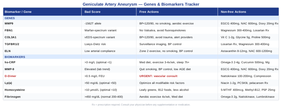
This article covers six targeted blood biomarkers for monitoring geniculate artery aneurysm activity and risk — including inflammation, matrix breakdown, clotting dynamics, and pro-atherogenic particles — with specific cost ranges, optimal values, and practical intervention plans for each. It also profiles five genetic variants linked to arterial wall fragility, from MMP9 polymorphisms to connective tissue disorder genes like FBN1 and COL3A1, with concrete plans for compensating for high-risk profiles. Beyond that, you will find Peter Attia's ten most impactful vascular insights from Outlive, plus three evidence-based complementary modalities for supporting vascular tone and blood pressure. If you have been told to simply "monitor" this aneurysm, this article gives you a framework for doing so with precision.
6 Biomarkers to Track If You Have a Geniculate Artery Aneurysm
Most of what is known about aneurysm biomarkers comes from research on abdominal aortic and intracranial aneurysms, simply because those are common enough to study at scale. Geniculate artery aneurysms are rare — most of the clinical literature consists of case reports. That said, the fundamental biology of arterial wall remodeling, inflammation, and thrombosis is shared across vessel locations. The six biomarkers below are the most directly relevant to that biology, and each has a meaningful evidence base in peripheral vascular disease, even when geniculate-specific studies are limited.
Biomarker 1: High-Sensitivity C-Reactive Protein (hs-CRP)
Why it matters for arterial aneurysms: CRP is synthesized by the liver in response to interleukin-6 signaling, which is itself produced when the arterial wall is inflamed. In aneurysmal tissue, this inflammatory cascade directly activates matrix metalloproteinases — the enzymes that degrade collagen and elastin in the vessel wall. Elevated hs-CRP is consistently associated with both peripheral arterial disease progression and aneurysm expansion rates in aortic studies. Ridker et al.'s landmark JUPITER trial (2008) demonstrated that even modest elevations in hs-CRP predict major vascular events independently of LDL cholesterol — a finding with clear implications for anyone managing a vascular lesion.
How to measure it: A standard blood draw, available through virtually any lab or as part of a cardiovascular panel. Cost ranges from $15 to $40 without insurance, and it is frequently covered when cardiovascular risk is a documented concern. Ensure the order specifies high-sensitivity CRP — the standard CRP test is insufficiently sensitive for vascular monitoring.
Optimal range: below 1 mg/L. Between 1 and 3 mg/L reflects moderate vascular inflammation; above 3 mg/L is clinically significant and warrants investigation. Values above 10 mg/L typically indicate acute infection rather than chronic vascular risk, and should be interpreted accordingly.
If the score is bad, the plan without supplements
Chronic hs-CRP elevation almost always has a lifestyle root cause. The most evidence-supported interventions are dietary: a Mediterranean-pattern diet reduces hs-CRP by 20–30% within 12 weeks in most studies. This means replacing refined carbohydrates with vegetables, legumes, and whole grains; eliminating industrial seed oils; and prioritizing oily fish at least twice weekly. Sleep is equally important — habitual short sleep (under six hours) elevates IL-6 and CRP consistently. Aerobic exercise three to four times per week, even brisk walking, significantly reduces systemic inflammation over eight to twelve weeks. Addressing untreated gum disease, sleep apnea, or chronic low-grade infections also matters more than most people realize.
If the score is bad, the plan with supplements or equipment
Omega-3 fatty acids (EPA+DHA): 2–4g per day of combined EPA and DHA from a high-quality fish oil or algae oil. Start at 2g and increase after four weeks. Run for 12 weeks, reassess with blood test. Cycle: 12 weeks on, 4 weeks off, or use continuously at 2g if monitored. Side effects include a fishy aftertaste and, at higher doses, mild blood thinning — important to discuss with your physician if you are on antiplatelet or anticoagulant therapy.
Curcumin with piperine: 500–1000mg/day of a bioavailable curcumin formulation (look for BCM-95 or Meriva liposomal forms). Piperine increases bioavailability by 20-fold. Run continuously, reassess at 8 weeks. Avoid at high doses if on blood thinners.
Magnesium glycinate: 300–400mg/day. Magnesium deficiency is extremely common and independently elevates hs-CRP. This is a foundational supplement with minimal downside when used at physiologic doses.
Biomarker 2: Matrix Metalloproteinase-9 (MMP-9)
Why it matters: MMP-9 is the enzyme most directly responsible for breaking down elastin and type IV collagen in the arterial extracellular matrix. It is produced by macrophages infiltrating the aneurysm wall in response to oxidative stress and inflammatory signaling. Research in abdominal aortic aneurysm has shown that MMP-9 is dramatically elevated in aneurysm tissue compared to normal aorta, and plasma MMP-9 correlates with aneurysm growth rate. For peripheral aneurysms, the same logic applies: higher MMP-9 activity accelerates structural weakening of the arterial wall. Smoking is one of the most potent known activators of MMP-9 expression.
How to measure it: A serum ELISA assay available through specialty and functional medicine labs. Not a standard clinical order at most hospitals — you will likely need to request it explicitly or access it through a functional medicine physician. Cost ranges from $100 to $300 depending on the lab. Quest Diagnostics and LabCorp both offer it as a sendout test. Because normal ranges are not yet standardized across labs, trend over time matters more than a single absolute value.
If the score is bad, the plan without supplements
Smoking cessation is the single most impactful intervention — smoking increases MMP-9 gene expression through NF-κB activation and can elevate levels several-fold. Sustained aerobic exercise (not intense acute bouts) reduces chronic MMP-9 elevation by improving endothelial function and reducing macrophage activation. Blood pressure control matters directly here: hypertensive shear stress is a mechanical trigger for MMP-9 release from vascular smooth muscle cells. Dietary AGEs (advanced glycation end-products from high-heat cooking, processed meats) also upregulate MMP-9 and should be reduced.
If the score is bad, the plan with supplements or equipment
EGCG (epigallocatechin gallate, green tea extract): 400–800mg/day of standardized green tea extract. EGCG inhibits MMP-9 at the transcription level through effects on AP-1 and NF-κB. Use 8 weeks on, 4 weeks off. Side effects: avoid on an empty stomach (nausea); avoid at high doses if you have liver sensitivity.
N-Acetylcysteine (NAC): 600mg twice daily. NAC replenishes glutathione, the primary intracellular antioxidant, which reduces the oxidative signaling that drives MMP-9 expression. Continuous use or 12-week cycles. Generally well-tolerated; avoid high doses alongside nitroglycerin medications.
Sub-antimicrobial doxycycline (20mg twice daily): This is a physician-supervised intervention that has been specifically studied for MMP inhibition in aortic aneurysm (published trials through Johns Hopkins). At sub-antimicrobial doses, doxycycline inhibits MMP activity without significant antibiotic effects. This requires a prescription and ongoing physician monitoring. Not suitable for long-term use without regular reassessment.
Biomarker 3: D-Dimer
Why it matters: D-dimer is a fibrin degradation product, meaning it is released whenever the body is actively forming and dissolving blood clots. In geniculate artery aneurysms, one of the most significant complications is intraluminal thrombus — a clot that forms within the aneurysm sac and can embolize downstream to the tibial vessels, causing acute limb ischemia. An elevated D-dimer suggests that this process is active. It is a nonspecific marker (it rises with any clotting activity, including DVT, PE, or recent surgery), but in the context of a known aneurysm, a persistently elevated D-dimer should prompt urgent vascular re-evaluation.
How to measure it: A standard blood test available at any clinical lab, typically as part of a coagulation panel. Cost ranges from $20 to $60. Most labs report results in mg/L FEU (fibrinogen equivalent units); the standard cutoff is 0.5 mg/L FEU for clinical concern.
If the score is bad, the plan without supplements
A significantly elevated D-dimer in the context of a known aneurysm is primarily a clinical emergency signal — this is not a biomarker to attempt to optimize with lifestyle changes before speaking to your vascular surgeon. If your D-dimer is persistently and significantly elevated without another clear explanation, a vascular consultation should happen promptly. Preventively, avoiding prolonged immobility (sitting for more than two hours without movement), maintaining adequate hydration, and regular low-impact movement reduce background clotting activation.
If the score is bad, the plan with supplements or equipment
Anticoagulation decisions (heparin, warfarin, DOACs) belong entirely to your physician based on your specific anatomy and bleeding risk — do not self-prescribe. As a supportive measure, nattokinase (100–200mg/day) is a fibrinolytic enzyme from fermented soybeans that has modest evidence for reducing clotting tendency — however, it interacts with anticoagulant medications and should only be used with physician knowledge. Compression therapy (medical-grade compression stockings or pneumatic compression devices) reduces venous stasis and is a low-risk mechanical intervention for preventing clot formation in the lower extremity.
Biomarker 4: Lipoprotein(a) — Lp(a)
Why it matters: Lp(a) is a modified LDL particle with an additional apolipoprotein(a) attached. It is both pro-atherogenic — accumulating in arterial walls and accelerating plaque formation — and pro-thrombotic, because apolipoprotein(a) structurally resembles plasminogen and competes with it, impairing the body's ability to dissolve clots. For anyone with a peripheral arterial aneurysm, this dual mechanism matters: Lp(a) both accelerates the underlying arterial disease and compounds the thrombotic risk within the aneurysm. Peter Attia considers Lp(a) one of the highest-leverage single biomarkers in all of cardiovascular medicine.
A landmark GWAS study published in the New England Journal of Medicine (Clarke et al., 2009) demonstrated that genetic variants in the LPA gene that raise Lp(a) are causally associated with cardiovascular events, establishing Lp(a) as a true risk factor rather than a passive marker.
How to measure it: A single blood test that must be specifically ordered — it is not included in standard lipid panels. Cost ranges from $20 to $100 depending on the lab. It only needs to be measured once or twice in a lifetime because Lp(a) is over 90% genetically determined and highly stable over time.
Optimal: below 50 mg/dL (or below 125 nmol/L in mass-based units). Above 180 mg/dL (450 nmol/L) represents substantially elevated cardiovascular and thrombotic risk.
If the score is bad, the plan without supplements
There is no dietary or exercise intervention that significantly lowers Lp(a) — it is largely genetically fixed. However, an elevated Lp(a) result changes the risk calculus: it means other modifiable risk factors (blood pressure, hs-CRP, metabolic health) should be optimized more aggressively to compensate. Specifically: achieving optimal LDL-C and ApoB levels, maintaining blood pressure below 120/80, eliminating smoking, and following an anti-inflammatory diet all matter more if your Lp(a) is elevated. Think of Lp(a) as a fixed background risk — your job is to drive all modifiable risks as low as possible around it.
If the score is bad, the plan with supplements or equipment
Extended-release niacin: 1–2g/day can reduce Lp(a) by 20–30% but carries significant side effects (flushing, hepatotoxicity at high doses, glucose dysregulation). Should only be used under physician supervision. Flush-free niacin (inositol hexaniacinate) does not appear to have the same effect on Lp(a).
PCSK9 inhibitors (evolocumab, alirocumab — prescription medications): reduce Lp(a) by approximately 20–30% in addition to their primary LDL-lowering effect. If your cardiologist has not discussed PCSK9 inhibitors in the context of elevated Lp(a), this is worth raising directly.
RNA-targeted therapies (pelacarsen, olpasiran): Currently in Phase 3 clinical trials. These medications specifically target Lp(a) production in the liver and can reduce levels by 80–90%. If you have significantly elevated Lp(a), ask your cardiologist about eligibility for ongoing trials or emerging prescriptions as these reach approval.
Biomarker 5: Homocysteine
Why it matters: Homocysteine is a sulfur-containing amino acid produced during methionine metabolism. When it accumulates above normal levels, it becomes directly toxic to endothelial cells through multiple mechanisms: it increases reactive oxygen species production, activates MMP pathways, promotes smooth muscle proliferation, and enhances platelet aggregation and clot formation. In peripheral arterial disease, elevated homocysteine is independently associated with accelerated progression and worse outcomes. For patients with geniculate artery aneurysms, elevated homocysteine amplifies both the structural degradation of the arterial wall and the thrombotic risk within the aneurysm.
The important clinical note is that homocysteine is highly modifiable — it is one of the few cardiovascular biomarkers where supplementation reliably produces clinically meaningful changes.
How to measure it: Fasting blood draw available at any standard lab. Cost ranges from $20 to $60. Ideally measured fasting (4–6 hours minimum) to avoid transient post-meal fluctuations.
Optimal: below 10 μmol/L. Between 10 and 15 is mildly elevated; between 15 and 30 is moderately elevated; above 30 is severely elevated and usually indicates significant metabolic or renal pathology requiring investigation.
If the score is bad, the plan without supplements
Increase dietary folate from food sources: dark leafy greens (spinach, romaine, arugula), legumes, and asparagus are all high in natural folate. Reduce alcohol consumption — alcohol directly impairs folate absorption. Increase B12-rich foods (meat, eggs, fish, dairy). If kidney function is impaired, treating the underlying renal cause is essential, as the kidneys are the primary route for homocysteine clearance. Check thyroid function — hypothyroidism is a common and often overlooked contributor to elevated homocysteine.
If the score is bad, the plan with supplements or equipment
5-Methyltetrahydrofolate (5-MTHF): The active form of folate, bypassing the MTHFR enzyme that many people cannot activate efficiently. Dose: 400–1000mcg/day. This is preferable to folic acid for anyone with MTHFR variants. Continuous use; reassess homocysteine at 8 weeks.
Methylcobalamin (B12): 500–1000mcg/day sublingual or oral. Use the methylated form rather than cyanocobalamin for better cellular uptake. Continuous use.
Pyridoxal-5-phosphate (active B6): 25–50mg/day. The active form of B6 is involved in a separate homocysteine-clearing pathway (transsulfuration). Important to include when homocysteine is significantly elevated. Avoid doses above 100mg/day long-term (risk of peripheral neuropathy at high prolonged doses).
These three B vitamins together (the "methylation trio") typically normalize homocysteine within 8–12 weeks in most cases. Recheck at 12 weeks and adjust.
Biomarker 6: Fibrinogen
Why it matters: Fibrinogen is the precursor protein to fibrin — the structural scaffold of blood clots. Elevated fibrinogen both increases blood viscosity (forcing the heart to work harder and increasing shear stress on vessel walls) and directly promotes thrombus formation within the aneurysm lumen. It also behaves as an acute-phase reactant, rising with systemic inflammation, making it a useful cross-check for hs-CRP. In peripheral vascular disease, elevated fibrinogen is associated with increased risk of arterial occlusion — a particular concern when an aneurysm is present.
How to measure it: Standard blood test included in many coagulation panels. Cost ranges from $15 to $50. Reference ranges vary slightly by lab but are typically 200–400 mg/dL for normal; above 450 mg/dL in the context of vascular disease warrants attention.
If the score is bad, the plan without supplements
Sustained aerobic exercise is the most consistent lifestyle reducer of fibrinogen — multiple studies demonstrate significant reductions with 30–45 minutes of moderate-intensity exercise four or more days per week over 12 weeks. Smoking cessation dramatically lowers fibrinogen. Mediterranean diet adherence, independent of weight loss, reduces fibrinogen. Treating underlying chronic infection or inflammation (including periodontitis) is also an important step, since fibrinogen elevation often reflects an inflammatory trigger that needs to be identified.
If the score is bad, the plan with supplements or equipment
Omega-3 fatty acids (EPA+DHA): 2–4g/day reduces fibrinogen as well as hs-CRP. Coordinate with physician if on anticoagulants.
Nattokinase: 100–200mg/day (2000–4000 FU) is a systemic proteolytic enzyme with specific fibrinolytic activity. Modest evidence in humans. Use only under physician supervision if on anticoagulants. Cycle: 8 weeks on, 4 weeks off.
Lumbrokinase: A more potent fibrinolytic enzyme derived from earthworms, used in traditional Chinese medicine and increasingly studied in Western vascular medicine. Doses vary by formulation; consult a functional medicine physician familiar with systemic enzymes. This requires physician oversight, particularly for anyone already on blood-thinning medications.
Moving from what the blood reveals to what the genome may already have written, genetics can explain why some individuals develop peripheral arterial aneurysms without obvious traumatic or atherogenic causes — and what to do about a high-risk genetic profile.
What Your Genetics May Reveal About Vascular Wall Fragility
When a geniculate artery aneurysm develops in a relatively young person, in the absence of clear trauma or severe atherosclerosis, genetics deserve serious consideration. The arterial wall is a complex structure held together by collagen, elastin, and smooth muscle cells — all encoded and regulated by genes that vary between individuals. Variants in these genes can silently reduce arterial tensile strength for decades before a focal weakness becomes visible on imaging.
It is worth noting that for rare peripheral aneurysms specifically, there is limited direct genetic research. Most of what is known comes from hereditary arterial aneurysm syndromes (Marfan, Ehlers-Danlos, Loeys-Dietz) and from population-level GWAS studies on aortic aneurysm. The principles extrapolate meaningfully to small vessel aneurysms, but individual genetic variants should always be interpreted by a clinical geneticist or cardiologist with expertise in connective tissue disorders.
Gene 1: MMP9 (Matrix Metalloproteinase 9)
What it affects: The MMP9 gene encodes the primary elastin- and collagen-degrading enzyme discussed in the biomarker section. A well-studied functional polymorphism in the promoter region — the -1562 C/T variant — is associated with significantly increased MMP-9 transcription. Carriers of the T allele may have elevated baseline MMP-9 enzymatic activity in arterial tissue even when serum levels appear normal. Research in abdominal aortic aneurysm has linked this variant to increased aneurysm risk and expansion rate.
Testing for MMP9 variants is available through consumer genomic platforms (23andMe data can be analyzed through Promethease) or through clinical panels ordered by a physician.
If the gene is bad, the plan without supplements
The -1562T allele means your arterial wall is structurally more vulnerable to inflammatory activation than average. The primary response is to keep the downstream triggers of MMP-9 activation controlled: strict blood pressure management (targeting below 120/80), no smoking under any circumstances, regular moderate aerobic exercise to maintain endothelial protective mechanisms, and an anti-inflammatory diet. Fluoroquinolone antibiotics (ciprofloxacin, levofloxacin) are known to upregulate MMP expression and degrade connective tissue — avoid them unless there is no alternative, and inform every prescribing physician of your vascular history.
If the score is bad, the plan with supplements or equipment
EGCG (green tea extract): 400–800mg/day standardized to ≥40% EGCG. Runs through 8-week cycles with a 4-week break. Side effects: mild gastric irritation on empty stomach; avoid very high doses with liver conditions.
NAC (N-acetylcysteine): 600mg twice daily, 12-week cycles. Supports glutathione synthesis and attenuates oxidative NF-κB signaling that drives MMP9 expression.
Sub-antimicrobial doxycycline (physician prescription required): 20mg twice daily. This specific dosing protocol directly inhibits MMP enzymatic activity without antibiotic side effects. Requires physician monitoring.
Gene 2: FBN1 (Fibrillin-1)
What it affects: FBN1 encodes fibrillin-1, the major structural protein of elastic microfibrils found throughout the arterial wall. Full loss-of-function mutations in FBN1 cause Marfan syndrome — characterized by aortic root aneurysm, lens dislocation, and skeletal abnormalities. But milder, partial-loss variants exist on a spectrum and may subtly reduce arterial wall elasticity without meeting diagnostic criteria for Marfan syndrome. In patients with peripheral aneurysms and no clear traumatic cause, FBN1 sequencing is clinically warranted.
FBN1 also regulates TGF-β signaling within the extracellular matrix — its dysfunction not only weakens structural scaffolding but also dysregulates growth factor signaling that controls smooth muscle cell behavior.
If the gene is bad, the plan without supplements
Imaging surveillance frequency should be discussed with your vascular surgeon — FBN1 variants change the risk calculus toward more frequent imaging. Avoid activities that produce sharp spikes in blood pressure through Valsalva (heavy powerlifting with breath-holding, straining). Swimming, cycling, and moderate resistance training at controlled intensity are preferred. Critically: avoid fluoroquinolone antibiotics — there is now significant clinical evidence that this class of antibiotics damages connective tissue and has caused aneurysm rupture in FBN1 mutation carriers. Carry documentation of your variant when seeking medical care.
If the score is bad, the plan with supplements or equipment
Magnesium glycinate: 300–400mg/day. Magnesium has been studied as a supplement for TGF-β pathway modulation in Marfan syndrome patients. Animal studies showed attenuation of aortic dilation; human data is limited but safety is high. Continuous use.
Losartan (prescription ARB medication): Has been studied specifically in Marfan syndrome for its TGF-β-blocking properties via angiotensin II inhibition. Not standard of care for mild FBN1 variants, but worth a conversation with a cardiologist familiar with connective tissue disorders, especially if blood pressure is in the high-normal range.
Avoid: excess vitamin A supplementation (can impair collagen synthesis); very high-dose curcumin may dysregulate TGF-β signaling in connective tissue contexts — use moderate doses only.
Gene 3: COL3A1 (Collagen Type III Alpha 1)
What it affects: COL3A1 encodes type III collagen, the primary structural protein of the walls of arteries and hollow organs. Mutations causing vascular Ehlers-Danlos syndrome (vEDS) dramatically reduce arterial tensile strength — the vessels literally lack the structural integrity to contain normal blood pressure. vEDS carries a risk of spontaneous arterial rupture and aneurysm. Even subclinical partial-loss variants, not meeting diagnostic criteria, may increase susceptibility to arterial injury under mechanical stress — including the kind of repetitive microtrauma experienced by the geniculate arteries of the knee during daily ambulation.
Any patient with a geniculate artery aneurysm at an unusually young age, or with a family history of early arterial rupture or aneurysm, should be evaluated for COL3A1 variants.
If the gene is bad, the plan without supplements
Blood pressure control is the most critical lifestyle intervention — every millimeter of mercury above optimal represents additional wall stress on structurally compromised collagen. Target strictly below 120/80. Avoid contact sports, heavy manual labor, and any activity with significant risk of blunt knee trauma. Alert all medical providers to your variant before any vascular procedures — arterial access sites in vEDS patients carry significantly elevated complication risk.
If the score is bad, the plan with supplements or equipment
Vitamin C (ascorbic acid): 1–2g/day in divided doses. Vitamin C is a required cofactor for prolyl hydroxylase and lysyl hydroxylase — the enzymes that cross-link collagen chains into stable triple-helix structures. Deficiency directly impairs whatever collagen synthesis capacity remains. Liposomal formulations at 500mg twice daily improve absorption. Continuous use.
Glycine: 5–10g/day, the dominant amino acid in collagen (making up ~33% of its residues). Emerging evidence suggests glycine supplementation supports endogenous collagen production. Well-tolerated; mix in water or add to food. Continuous use.
Proline: 500mg/day alongside glycine and vitamin C — the combination supports collagen synthesis more than any single agent alone. These are supportive nutritional interventions, not treatments for COL3A1 mutations.
Gene 4: TGFBR1 and TGFBR2 (TGF-β Receptor Genes)
What they affect: Mutations in TGF-β receptor genes 1 and 2 cause Loeys-Dietz syndrome — a hereditary arterial aneurysm disorder characterized by arterial tortuosity and aneurysm formation throughout the vasculature. Unlike Marfan syndrome, Loeys-Dietz aneurysms can occur in arteries far from the aortic root, including peripheral vessels. TGF-β signaling governs smooth muscle cell differentiation and extracellular matrix homeostasis; its disruption leads to a paradoxical upregulation of downstream TGF-β activity within the arterial wall, driving maladaptive remodeling.
Testing: included in standard hereditary aortopathy panels (often covering FBN1, COL3A1, TGFBR1, TGFBR2, SMAD3, ACTA2). Clinical genetic testing is appropriate when the clinical picture is suggestive.
If the gene is bad, the plan without supplements
Regular vascular imaging surveillance is essential — the scope and frequency should be determined by a geneticist or specialist in hereditary aortopathies. Blood pressure management is critical and should target the lower end of normal. Physical activity modifications similar to those for FBN1 variants apply. A referral to a center with expertise in hereditary arterial aneurysm syndromes (these exist at major academic medical centers) is strongly recommended for any confirmed TGFBR variant.
If the score is bad, the plan with supplements or equipment
Losartan (prescription): Has been studied in both Marfan and Loeys-Dietz syndromes for its ability to attenuate TGF-β signaling. Clinical trials have shown slower aortic dilation in some patient populations. This is a physician decision, but one worth specifically raising if you carry a TGFBR variant.
Magnesium glycinate: 300–400mg/day as a low-risk supportive measure with plausible TGF-β modulating properties.
Avoid curcumin at doses above 500mg/day — TGF-β pathway effects are complex and context-dependent in connective tissue disorders; the evidence is insufficient to justify high-dose use when TGF-β dysregulation is already the core pathology.
Gene 5: ELN (Elastin)
What it affects: The ELN gene encodes elastin, the protein responsible for the elastic recoil of arterial walls — the ability of vessels to stretch under systolic pressure and return to resting diameter during diastole. Loss of ELN function reduces compliance of the arterial wall, increasing pulse wave velocity (arterial stiffness), raising local mechanical stress at vessel bifurcations and curves, and creating conditions for focal dilation. Complete ELN deletion causes supravalvular aortic stenosis, but milder variants reduce arterial compliance in a dose-dependent manner. Because the geniculate arteries operate under mechanical stress from knee movement and loading, reduced elastin content may predispose to focal dilation at these sites.
If the gene is bad, the plan without supplements
Reducing arterial stiffness through lifestyle is meaningfully possible: regular low-impact aerobic exercise (walking, cycling, swimming) consistently reduces pulse wave velocity in human trials, independent of genetics. Blood pressure reduction is directly protective — stiff, non-compliant arteries tolerate hypertension less well than healthy ones. Adequate sleep and stress management reduce sympathetic tone, which otherwise maintains elevated resting vascular resistance. Smoking destroys elastin directly through neutrophil elastase activation — this is the most damaging single modifiable factor for any elastin-compromised individual.
If the score is bad, the plan with supplements or equipment
No supplement directly restores degraded elastin — elastin is synthesized primarily in early development and has extremely low turnover in adults. The goal is preventing further degradation.
Astaxanthin: 8–12mg/day. A powerful carotenoid antioxidant that protects extracellular matrix proteins from reactive oxygen species-mediated degradation, including elastin. Continuous use; well-tolerated, safe.
NAC: 600–1200mg/day reduces oxidative elastase activation. 12-week cycles.
Arterial stiffness monitoring device (SphygmoCor or similar): Pulse wave velocity measurement devices are now available in clinical settings and at some specialty labs ($100–300/test). Tracking arterial stiffness over time provides a functional read-out of vascular wall compliance — more sensitive to change than imaging alone.
Ten Things Peter Attia's Outlive Teaches About Vascular Longevity That Most Doctors Don't Mention
Peter Attia's 2023 book Outlive: The Science and Art of Longevity synthesizes decades of cardiovascular and metabolic research into a coherent, aggressive framework for preventing vascular disease — one that goes considerably further than standard clinical guidelines. For anyone managing a peripheral arterial aneurysm, the vascular chapters are among the most practically useful things available in mainstream health literature.
1. ApoB Is What Actually Matters — Not LDL-C
Attia argues that ApoB (apolipoprotein B) is the single most important lipid marker to know and optimize. Every atherogenic lipoprotein particle — LDL, IDL, VLDL, Lp(a) — carries exactly one ApoB molecule. ApoB therefore counts the number of particles that can penetrate and accumulate in arterial walls. Two people with identical LDL-C can have very different ApoB levels and very different arterial disease risk. Standard lipid panels routinely miss this. Ask for ApoB specifically.
2. Lp(a) Should Be Measured in Everyone at Least Once
Attia is emphatic that Lp(a) is severely underdiagnosed. Because it is genetically fixed and over 90% heritable, measuring it once tells you something permanent about your baseline vascular risk. About 20% of people have levels high enough to materially change their risk management strategy — yet most have never been tested. This is a meaningful clinical gap.
3. Blood Pressure Should Be Treated More Aggressively Than Guidelines Suggest
The SPRINT trial data, which Attia discusses in detail, showed that treating blood pressure to a target below 120 mmHg systolic significantly reduced cardiovascular events compared to the standard 140 target. For vascular patients, particularly those with arterial aneurysms where wall stress is a direct concern, this more aggressive target is well-supported. Home blood pressure monitors should be used regularly, not just at clinic visits.
4. Arterial Stiffness Is a Separate and Underappreciated Risk Factor
Attia highlights pulse wave velocity — the speed at which a pressure wave travels through the arterial tree — as one of the most predictive measures of vascular aging and risk. It is independent of blood pressure and cholesterol. Exercise, and specifically zone 2 aerobic training, is the most evidence-backed intervention for reducing arterial stiffness over time.
5. Zone 2 Training Is the Single Most Vascularly Protective Form of Exercise
Zone 2 — sustained aerobic effort at a pace where you can maintain a conversation but feel moderately exerted — improves mitochondrial density, reduces systemic inflammation, improves insulin sensitivity, and reduces arterial stiffness. Attia recommends three to four hours per week minimum. For people with known vascular disease, the emphasis should be on consistency and avoiding the blood-pressure spikes associated with high-intensity exertion.
6. Metabolic Health Is Upstream of Vascular Disease
Insulin resistance and hyperinsulinemia precede arterial disease by years and drive it through multiple mechanisms: increased inflammation, oxidative stress, endothelial dysfunction, and dyslipidemia. Fasting insulin and HOMA-IR are the most useful markers to track, and neither is included in routine panels. A fasting insulin above 8 μIU/mL already suggests developing insulin resistance even when fasting glucose is normal.
7. Sleep Is a Non-Negotiable Cardiovascular Intervention
Attia treats sleep as a first-line intervention with the same rigor as pharmacology. Short sleep (under 6 hours) reliably elevates cortisol, raises blood pressure, increases hs-CRP, and disrupts endothelial repair. The mechanism is direct and dose-dependent. Using a sleep tracker, optimizing sleep environment (temperature, darkness, timing), and treating sleep apnea are priorities that rival any supplement in cardiovascular impact.
8. Stress-Induced Hemodynamic Spikes Are Arterially Damaging
Acute psychological stress reliably elevates blood pressure by 20–40 mmHg through sympathetic activation. In someone with a compromised arterial wall, these repeated spikes increase mechanical stress and may accelerate wall remodeling. Stress management is not soft advice — it is a hemodynamic intervention. Attia supports mindfulness, structured relaxation, and therapy as legitimate clinical tools.
9. VO2 Max Is the Strongest Predictor of Longevity in the Data
Attia cites observational data showing that VO2 max is among the strongest predictors of all-cause and cardiovascular mortality ever identified. Moving from the bottom quartile to above median on VO2 max roughly halves mortality risk. Improving VO2 max through progressive aerobic training is therefore not about athletic performance — it is about changing the long-term probability of vascular events.
10. Continuous Glucose Monitoring Reveals Hidden Metabolic Stress
Attia recommends periodic use of continuous glucose monitoring (CGM) even in non-diabetic individuals, because post-meal glucose spikes — which are invisible on standard fasting labs — drive endothelial glycation, oxidative stress, and inflammation. Many people who appear metabolically normal on a fasting panel have significant post-meal dysregulation that accelerates vascular aging. A two-week CGM trial costs approximately $50–80 and can reveal dietary patterns causing repeated spike cycles.
Complementary Approaches With Evidence for Vascular Support
The three approaches below have meaningful human clinical evidence for reducing cardiovascular risk factors — blood pressure, vascular tone, and systemic stress response — that are directly relevant to aneurysm management. None should replace medical care, and evidence is strongest for these risk factors rather than geniculate artery aneurysm specifically.
Mindfulness-Based Stress Reduction (MBSR)
MBSR is an 8-week structured program developed by Jon Kabat-Zinn combining body scanning, sitting meditation, and mindful movement. Its cardiovascular relevance stems from its documented ability to reduce sympathetic nervous system activation and cortisol, with secondary effects on blood pressure and systemic inflammation. For a person with a peripheral arterial aneurysm, chronic psychological stress represents a direct source of repeated hemodynamic stress on the vessel wall — each sympathetic activation spike raises blood pressure acutely and sustains vascular wall tension.
A meta-analysis of 13 randomized controlled trials published in the Journal of Hypertension found that mindfulness-based interventions significantly reduced both systolic and diastolic blood pressure compared to control conditions, with effects comparable to some antihypertensive medications in mildly hypertensive populations. MBSR also consistently reduces hs-CRP in multiple controlled trials, providing an additional mechanistic link to aneurysm wall biology.
Practically: the gold-standard format is an in-person 8-week MBSR course (offered through hospitals, mindfulness centers, and online platforms). A home practice of 20–30 minutes daily of body-scan or breath-focused meditation achieves meaningful physiologic benefit within 8 weeks. The key is daily consistency — irregular, occasional meditation does not produce the same autonomic recalibration as a sustained daily practice. Track resting heart rate variability (via a wearable device) as an objective proxy for the nervous system changes MBSR is producing.
Breathing-Based Therapies
Slow, controlled breathing — typically at a rate of 5–6 breaths per minute — activates the baroreceptor reflex and increases parasympathetic tone. This direct effect on the autonomic nervous system reduces resting blood pressure, lowers heart rate, and decreases vascular resistance. Device-guided slow breathing (using a biofeedback device that paces your breathing) has been specifically approved by the FDA as an adjunct blood pressure intervention.
The evidence base is substantial: a meta-analysis of device-guided breathing studies (including the RESPeRATE device trials) published in the journal Hypertension demonstrated average systolic reductions of 8–15 mmHg with 15-minute daily sessions over 8 weeks. For someone with a vascular aneurysm where blood pressure control is a first-line protective measure, this is a meaningful, zero-risk adjunct. The mechanism operates independently of lifestyle modifications and medication, making it genuinely additive.
Practically: 15 minutes of device-guided slow breathing daily, or unguided 4-7-8 breathing (inhale 4 seconds, hold 7, exhale 8) for three sessions of 8 cycles per day. The RESPeRATE device costs approximately $100–150. Alternatively, free paced breathing apps achieve similar physiologic effects. Begin after meals or in the evening — blood pressure tends to peak in the morning and mid-afternoon, making these ideal intervention windows. No side effects; suitable for almost all patients.
Biofeedback
Biofeedback provides real-time physiologic data — heart rate, skin conductance, blood pressure, heart rate variability — allowing individuals to consciously influence these parameters through mental and behavioral techniques. For blood pressure management in vascular patients, heart rate variability (HRV) biofeedback is the most evidence-supported modality. By training patients to achieve coherent HRV patterns (through paced breathing and focused attention), biofeedback produces durable reductions in sympathetic tone and resting blood pressure.
A systematic review of biofeedback interventions for hypertension found consistent reductions in systolic blood pressure ranging from 4 to 12 mmHg across multiple randomized controlled trials — effects maintained at follow-up in most studies. For someone managing a vascular aneurysm, these reductions translate directly into reduced pulsatile wall stress on the aneurysm sac. Biofeedback is particularly useful for patients with white-coat hypertension or stress-reactive blood pressure — common patterns that standard medication dosing often fails to address.
Practically: HRV biofeedback is accessible through consumer devices (HeartMath Inner Balance, Polar H10 paired with HRV training apps, Elite HRV). Professional sessions with a biofeedback therapist ($80–150/session) provide training in techniques that can then be self-administered. Four to six sessions with a therapist to learn the technique, followed by daily 10–20 minute self-practice, produces most of the documented benefit. Track your HRV trend — a consistently rising HRV over weeks is objective evidence that your autonomic system is responding to the training.
Conclusion
A geniculate artery aneurysm occupies uncertain ground in clinical medicine — too rare for specific guidelines, too serious to dismiss. The most useful response to that uncertainty is not passive monitoring but informed, targeted vigilance. Tracking six specific biomarkers gives you a real-time picture of the inflammatory load, matrix degradation activity, and thrombotic risk that determine whether this aneurysm remains stable or progresses. Understanding your genetic profile — particularly variants in MMP9, FBN1, COL3A1, TGFBR1/2, and ELN — explains why the risk exists in the first place and points toward specific compensatory strategies.
None of this replaces the judgment of a vascular surgeon or a clinical geneticist. But it does give you better raw material for those conversations — and, in many cases, it raises questions that routine care would never think to ask. The next smart step is to request an Lp(a) test and a homocysteine panel at your next blood draw if you have not already, and to ask your physician whether a connective tissue disorder genetic panel is appropriate given your history. Better information consistently produces better decisions.
Musculoskeletal: Joint Conditions
Cardiovascular: Blood Vessel Conditions Vascular Conditions
Autoimmune: Inflammatory Conditions Connective Tissue Conditions