This article contains AI generated content.
· UpdatedGenu Recurvatum — 6 Genes And 7 Biomarkers To Track
Introduction
If your knees hyperextend — if they lock back past straight whenever you stand or bear weight — you probably know the feeling of being told to simply strengthen your quadriceps and wear a brace when things get bad. That advice is not wrong. But for many people with persistent genu recurvatum, it is incomplete in a way that matters. The exercises help, temporarily. The brace manages symptoms. And the underlying instability stays exactly where it was.
What rarely gets discussed is why some people develop significant genu recurvatum in the first place, while others with similar training histories and activity levels never do. The answer, in many cases, lives at the level of connective tissue biology — in the quality of the ligaments themselves, in the enzymatic environment that builds and maintains collagen, and in genetic patterns that make some people's joints structurally less able to resist hyperextension regardless of how much they train.
Generic rehabilitation protocols treat the mechanical problem. They rarely address the biological one. That gap is where this article sits. Understanding whether your connective tissue is being quietly degraded by a chronic inflammatory state, a micronutrient deficit that is impairing collagen crosslinking, or a gene variant that reduces ligament tensile strength changes what you should prioritize — and what might actually move the needle.
Better information does not guarantee a quick fix, but it consistently leads to better decisions. This article offers two complementary entry points. The first — and most immediately actionable — identifies seven biomarkers you can measure with standard or semi-standard blood tests to assess your connective tissue environment right now, with specific plans for addressing each one. The second examines six genes with established links to joint hypermobility and ligament laxity, with targeted strategies for each genetic pattern. Beyond these two tracks, you will find a summary of the movement rehabilitation framework that has quietly transformed knee recovery for thousands of people, followed by three complementary modalities with meaningful clinical evidence for joint stability.
Summary
If you have been managing genu recurvatum with exercises and bracing alone and progress has stalled, this article makes the case that the missing piece is often biological rather than mechanical. It covers seven measurable blood markers — including vitamin D, homocysteine, copper, ferritin, and collagen breakdown markers — that directly regulate ligament and tendon integrity, along with concrete action plans for improving each one. It then examines six connective tissue genes — COL5A1, COL1A1, TNXB, FBN1, MMP3, and ACAN — that are consistently linked to joint hypermobility and knee hyperextension, with gene-specific protocols including options both with and without supplementation. A dedicated section covers the Ben Patrick "Knees Over Toes" framework and the specific science behind why graduated connective tissue loading works. Three evidence-backed complementary modalities round out the picture. If your knee keeps hyperextending despite consistent effort, what follows may explain why — and what to do about it differently.
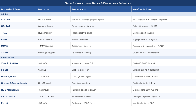
7 Biomarkers to Track for Genu Recurvatum
The connective tissue of your knee ligaments and joint capsule is not static. It is in a constant state of synthesis and degradation, driven by enzymes, cofactors, inflammatory signals, and hormonal cues — all of which are measurable in a blood sample. When that environment is poor, ligaments become mechanically inferior even in people who train consistently. Seven biomarkers stand out as the most clinically useful for understanding and improving connective tissue quality in the context of genu recurvatum.
Biomarker 1: Vitamin D (25-OH)
Why it matters. Vitamin D receptors are expressed in skeletal muscle, ligament tissue, and joint capsule cells. Adequate levels are necessary for neuromuscular control, proprioceptive feedback quality, and the muscle tone that protects against passive hyperextension. Studies consistently link vitamin D deficiency to generalized muscle weakness and increased joint laxity. For people with genu recurvatum, quadriceps and hamstring inhibition at end range is often a core problem — and low vitamin D is a direct contributor. The optimal functional range for musculoskeletal health is generally considered 40–60 ng/mL (100–150 nmol/L), meaningfully higher than the laboratory "normal" floor of 20 ng/mL.
How to measure it. A 25-OH vitamin D blood test, available as part of most routine panels or as a standalone test. Cost: $30–60 in the US; often covered by insurance with a relevant clinical indication. Results come back within a few days. Retest after 12 weeks of any intervention.
If the score is low — plan without supplements. Midday sun exposure of 15–25 minutes on arms and legs (without sunscreen), three to five times per week, depending on skin type and latitude. Fatty fish two to three times per week (salmon, sardines, mackerel). Egg yolks daily. These measures can raise levels by 5–10 ng/mL over 8–12 weeks in mild deficiency.
If the score is low — plan with supplements or equipment. Vitamin D3 (not D2) at 2,000–5,000 IU daily, taken with a fat-containing meal for absorption. Pair with vitamin K2 (MK-7 form) at 100–200 mcg/day to direct calcium appropriately. At therapeutic doses (4,000–5,000 IU), retest at 12 weeks and adjust. Side effects at these doses are rare but include hypercalcemia with very prolonged excessive dosing — hence the retesting. Do not supplement above 5,000 IU without physician supervision. Light therapy lamps do not produce vitamin D; they are not a substitute for sun or oral supplementation.
Biomarker 2: hs-CRP (High-Sensitivity C-Reactive Protein)
Why it matters. Chronic low-grade inflammation — even at levels that cause no obvious symptoms — activates matrix metalloproteinases (MMPs), a family of enzymes that degrade collagen, elastin, and other extracellular matrix proteins in ligaments and joint capsules. An hs-CRP above 1 mg/L suggests an inflammatory environment that is actively working against connective tissue integrity. This is particularly relevant for people who notice that their knee stability worsens after poor sleep, stress, or dietary periods that are high in refined carbohydrates — all known drivers of hs-CRP elevation.
How to measure it. Standard blood test, often included in cardiovascular risk panels. Cost: $10–30. Optimal target: below 0.5 mg/L. Anything above 3 mg/L warrants clinical investigation.
If the score is elevated — plan without supplements. Eliminate refined seed oils (linoleic acid drives arachidonic acid cascade), reduce refined sugars and ultra-processed foods, prioritize sleep (7–9 hours — sleep deprivation raises CRP measurably within days), and increase cold-water fatty fish intake. These changes can reduce hs-CRP by 30–50% within 4–8 weeks in lifestyle-driven elevations.
If the score is elevated — plan with supplements or equipment. High-dose omega-3 fatty acids (EPA+DHA combined, 2–4 g/day) are the most evidence-supported anti-inflammatory supplement. Take with a meal. Cycling is not strictly required but a 12-week assessment is practical. Curcumin with piperine (500–1000 mg curcumin, 5–10 mg piperine daily) adds meaningful MMP-suppressing activity; cycle 8 weeks on, 2–3 weeks off to avoid adaptation and reduce any theoretical risk of excessive inhibition of wound-healing MMP activity. Side effects of high-dose omega-3 include mild blood thinning; caution if on anticoagulants.
Biomarker 3: Homocysteine
Why it matters. Homocysteine is not often associated with joint health in mainstream clinical practice, but the mechanism is direct and well-established. Elevated homocysteine inhibits lysyl oxidase, the copper-dependent enzyme responsible for forming the crosslinks between collagen and elastin fibers. Without proper crosslinks, ligament fibers are structurally analogous to a rope where the strands are braided but not fused — significantly weaker under tensile load. Homocysteine above 10 µmol/L is associated with reduced connective tissue mechanical integrity; optimal for joint health is generally considered below 7 µmol/L.
How to measure it. Standard blood test, sometimes included in cardiovascular risk panels. Cost: $30–50. Fasting is recommended. Retest 8–12 weeks after intervention.
If the score is elevated — plan without supplements. Increase methyl-donor foods: leafy dark greens (folate), eggs (choline, B12), organ meats (B12, folate). Reduce alcohol, which depletes B vitamins. Address gut health if B12 absorption is a concern (intrinsic factor, H. pylori).
If the score is elevated — plan with supplements or equipment. The most effective and evidence-supported protocol is methylfolate (5-MTHF, 400–1000 mcg/day) + methylcobalamin B12 (1000 mcg/day) + P5P (pyridoxal-5-phosphate, the active form of B6, 25–50 mg/day). This triple combination directly drives the remethylation pathway that clears homocysteine. Critically: if you know you carry an MTHFR C677T or A1298C polymorphism, folinic acid or methylfolate is essential — folic acid supplementation is not the same and can actually worsen outcomes in MTHFR variants by competing for receptor sites. Retest at 8 weeks. Side effects are rare at these doses; very high B6 (>200 mg/day) over prolonged periods causes peripheral neuropathy, but P5P at 25–50 mg is well within safe range.
Biomarker 4: Copper and Ceruloplasmin
Why it matters. Copper is the essential cofactor for lysyl oxidase, the same enzyme mentioned in the homocysteine section. Without adequate copper, lysyl oxidase activity drops — regardless of whether homocysteine is elevated — and collagen crosslinking is impaired at the enzymatic level. This is not a theoretical concern: frank copper deficiency produces a connective tissue phenotype that mimics Ehlers-Danlos syndrome, with joint laxity and skin fragility as prominent features. Ceruloplasmin is the primary copper-carrying protein and a more stable marker of copper status than serum copper alone. Optimal serum copper: 80–120 µg/dL; ceruloplasmin: 20–55 mg/dL.
How to measure it. Serum copper and ceruloplasmin panel. Cost: $30–60. Note that inflammation artificially elevates ceruloplasmin (it is an acute phase reactant), so measure alongside hs-CRP for context.
If the score is low — plan without supplements. Beef liver is by far the most bioavailable source of copper — 85g (3 oz) provides roughly 12 mg. Consuming it once per week is adequate for most people. Oysters, dark chocolate (70%+), and cashews are additional sources. Reducing zinc supplementation also helps, as high-dose zinc directly competes with copper absorption.
If the score is low — plan with supplements or equipment. Copper bisglycinate 1–2 mg/day is the preferred supplemental form (high bioavailability, fewer GI effects). This is a case where precision matters: the therapeutic window for copper is narrow. Target the low end of normal and retest every 3–6 months. If you are also supplementing zinc, maintain a zinc-to-copper ratio no higher than 10:1 (e.g., 20 mg zinc paired with 2 mg copper). Excess copper is toxic and accumulates — do not self-prescribe above 2 mg without lab confirmation of deficiency. No strict cycling is needed at low physiological doses.
Biomarker 5: RBC Magnesium
Why it matters. Standard serum magnesium is largely useless for assessing functional magnesium status — the body maintains serum levels at the expense of intracellular stores, so serum can appear normal while tissue deficiency is significant. RBC (red blood cell) magnesium reflects intracellular concentrations and is the clinically meaningful measurement. Magnesium is a cofactor for over 300 enzymatic reactions, including ATP production, neuromuscular transmission, and protein synthesis. For genu recurvatum specifically, low intracellular magnesium impairs the neuromuscular signaling that governs dynamic knee stabilization — the millisecond-by-millisecond muscle activation that prevents passive hyperextension during gait and athletic activity. Optimal RBC magnesium: 5.2–6.5 mg/dL.
How to measure it. RBC magnesium (not serum magnesium) — request specifically. This is a slightly specialty test, not always available in standard panels. Cost: $40–80. Some functional medicine labs include it routinely.
If the score is low — plan without supplements. Pumpkin seeds (highest food source per gram), spinach, black beans, dark chocolate, and mineral water (look for Mg²⁺ content above 50 mg/L). Cooking vegetables in hard water adds small but meaningful amounts. Reduce alcohol and excess caffeine, which both increase renal magnesium wasting.
If the score is low — plan with supplements or equipment. Magnesium glycinate (200–400 mg elemental magnesium/day) is the preferred form — high absorption, minimal laxative effect. Take in the evening; magnesium supports sleep quality as a secondary benefit. Magnesium L-threonate (2000 mg of the compound, delivering ~144 mg elemental Mg) is better researched for neurological applications and may have additional relevance for neuromuscular function. Avoid magnesium oxide — poor absorption, primarily laxative effect. Side effects at 300–400 mg: mild loosening of stools in some people. No cycling required for maintenance dosing. Retest RBC Mg at 12 weeks.
Biomarker 6: CTX-I and P1NP (Collagen Turnover Markers)
Why it matters. CTX-I (C-terminal telopeptide of type I collagen) is a marker of collagen breakdown — it rises when collagen is being degraded faster than it is rebuilt. P1NP (procollagen type I N-terminal propeptide) reflects collagen synthesis. Looking at both together gives you a "balance score" for connective tissue remodeling. In genu recurvatum, a high CTX paired with low P1NP suggests a catabolic connective tissue state: ligaments are being broken down faster than they are being rebuilt — a situation no amount of strengthening exercise can fully compensate for. These markers are well-established in bone medicine (they guide osteoporosis treatment) but are underused in joint laxity assessment.
How to measure it. CTX-I (fasting morning sample — CTX is highest in the early morning and suppressed by food), P1NP can be drawn at any time. Often requires a specialist order; not standard in general panels. Cost: $50–120 each. Reference ranges vary by age and sex; compare against age-matched norms.
If the score is catabolic (high CTX, low P1NP) — plan without supplements. Load-bearing exercise is the primary driver of P1NP — even moderate progressive loading increases collagen synthesis markers within days. Adequate dietary protein is essential (at least 1.6 g/kg bodyweight/day). Sleep quality matters more here than most other interventions: growth hormone — which peaks during deep sleep — directly stimulates collagen synthesis and P1NP elevation. Reducing cortisol load (stress, sleep deprivation) is also relevant, as glucocorticoids suppress collagen formation.
If the score is catabolic — plan with supplements or equipment. Hydrolyzed collagen peptides (10–15 g/day), taken 30–60 minutes before joint-loading exercise, have been shown in randomized trials to measurably increase joint collagen synthesis. This timing is critical — the amino acid pulse coincides with exercise-driven blood flow to tendons and ligaments. Vitamin C (500–1000 mg) taken alongside collagen peptides is required as a cofactor for prolyl hydroxylase. Vitamin K2 (MK-4 or MK-7, 100–200 mcg/day) supports bone-side P1NP. Cycling is not required for these; they function as cofactors rather than pharmacological agents. Side effects are minimal.
Biomarker 7: Ferritin
Why it matters. Ferritin is most commonly discussed in the context of anemia, but its relevance to connective tissue is underappreciated. Iron is a required cofactor for two enzymes: prolyl-4-hydroxylase and lysyl hydroxylase — both of which hydroxylate specific amino acid residues on procollagen chains in the endoplasmic reticulum. Without adequate hydroxylation, collagen triple helices cannot form correctly, and the resulting fibers have reduced tensile strength. Ferritin below 50 ng/mL — which is common, particularly in premenopausal women — is sufficient to impair this process even when serum iron appears adequate. The functional optimal for connective tissue is generally considered 70–100 ng/mL, not merely above the laboratory floor of 12 ng/mL.
How to measure it. Ferritin is part of most routine blood panels. Cost: $20–40. Measure alongside transferrin saturation and TIBC for full iron status context. Retest 8–12 weeks after intervention.
If the score is low — plan without supplements. Heme iron (red meat, organ meat, dark meat poultry) is 2–3 times more bioavailable than non-heme iron and is not suppressed by phytates or calcium. Eating red meat 3–4 times per week alongside vitamin C-rich foods meaningfully increases ferritin in iron-insufficient individuals within 8–12 weeks. Avoid coffee and tea within 1 hour of iron-rich meals.
If the score is low — plan with supplements or equipment. Iron bisglycinate 25–50 mg every other day (not daily — alternate-day dosing increases absorption by allowing hepcidin to reset, a strategy validated in recent research). Take on an empty stomach with 500 mg vitamin C. Avoid co-administration with calcium supplements, antacids, or zinc. Side effects: constipation is common with ferrous sulfate; iron bisglycinate is significantly better tolerated. Critical safety note: do not supplement iron without confirming deficiency through blood testing. Excess iron is a pro-oxidant and associated with cardiovascular and liver risk. Retest at 8–12 weeks. Hemochromatosis should be excluded if ferritin is being tested for the first time.
The Genetic Side of Knee Hyperextension: 6 Key Genes
Understanding biomarkers tells you about the current state of your connective tissue environment. Genetics tells you something different: whether your baseline connective tissue architecture is structurally more vulnerable to hyperextension regardless of external factors. These are not separate questions — genetic variants often directly impair the same enzymatic and structural pathways reflected in your biomarkers, meaning genetics can help you understand why certain biomarkers keep drifting out of range despite your best efforts.
Gene 1: COL5A1 — The Collagen V Blueprint
COL5A1 encodes the alpha-1 chain of type V collagen, which plays a regulatory role in collagen fibril diameter. Collagen V is not the main structural component of ligaments — that is collagen I — but it controls how collagen I fibers are organized. Variants in COL5A1, particularly in the 3' untranslated region (the rs12722 polymorphism has been studied extensively in the context of ACL and ligament laxity), are associated with abnormally large, disorganized collagen fibrils that are mechanically weaker than tightly packed smaller fibrils. This gene is also the classic Ehlers-Danlos syndrome type I/II locus — relevant even for people who don't have full EDS, as subclinical variants produce subclinical but real ligament vulnerability.
If the gene variant is present — plan without supplements. The structural deficit cannot be corrected, but it can be compensated mechanically and neurologically. Proprioceptive training (unstable surfaces, single-leg balance progressions, wobble boards) builds neuromuscular compensation for ligament slack. Eccentric loading protocols — particularly for hamstrings and gastrocnemius, which act as secondary stabilizers against hyperextension — are more effective than concentric-focused exercise for strengthening the tissue that protects lax ligaments. Avoid extreme end-range loading of the knee in hyperextension; use wedge insoles or proprioceptive taping to prevent passive recurvatum during daily activities and early rehabilitation.
If the gene variant is present — plan with supplements or equipment. Vitamin C (500–1000 mg/day, split dose), glycine (5–10 g/day as glycine powder or from bone broth), and proline (1–2 g/day) provide the rate-limiting amino acids for collagen synthesis. Hydrolyzed collagen peptides (10–15 g pre-exercise as above) provide both the amino acids and small peptide signals that stimulate fibroblast activity. Copper bisglycinate (1–2 mg) supports LOX-mediated crosslinking of whatever collagen is synthesized. This is a lifelong maintenance approach rather than a short-term protocol.
Gene 2: COL1A1 — The Primary Collagen Structural Gene
COL1A1 encodes the primary structural chain of type I collagen — the most abundant protein in ligaments, tendons, and bone. The Sp1 polymorphism (rs1800012, a G→T change in intron 1) alters a transcription factor binding site and reduces the proportion of stronger α1 chains relative to α2 chains, producing a collagen triple helix with altered mechanical properties. The T allele is associated with osteoporosis, ligament rupture susceptibility, and in population studies, reduced joint mechanical stability. This is not a dramatic rare mutation — the T allele frequency is around 20% in European populations, making it relatively common.
If the gene variant is present — plan without supplements. Progressive resistance training acts as a direct stimulus to fibroblasts and osteoblasts to upregulate collagen I gene expression — meaning you can partially compensate for transcriptional efficiency deficits with mechanical loading. Prioritize compound loading patterns (squats, step-ups, deadlifts) with attention to not allowing the knee to passively hyperextend under load. Calcium-adequate diet (1000–1200 mg dietary calcium/day from food) supports the bone-side expression of this gene.
If the gene variant is present — plan with supplements or equipment. In addition to the collagen synthesis protocol described under COL5A1, consider orthosilicic acid (5–10 mg/day) from bamboo extract or stabilized silicic acid supplements — silicon is a cofactor for collagen biosynthesis and has evidence for increasing bone collagen density in limited trials. Vitamin D3 (targeting 50–60 ng/mL) is particularly important here, as VDR pathways interact with COL1A1 promoter regions. Cycle: orthosilicic acid can be taken continuously at low doses; reassess bone density and connective tissue function every 12 months.
Gene 3: TNXB — The Tenascin-X Stability Gene
TNXB encodes tenascin-X, an extracellular matrix glycoprotein that regulates collagen fibril spacing and mechanical transmission through the ECM. Unlike the collagen genes, TNXB haploinsufficiency (having only one functional copy) produces a clinically recognized syndrome: a form of Ehlers-Danlos syndrome with generalized joint hypermobility, skin hyperextensibility, and proprioceptive deficits — with the knee often among the most affected joints. Homozygous TNXB loss causes a more severe phenotype. This gene is uniquely important because its deficiency can be diagnosed biochemically (tenascin-X levels in serum can be measured at specialty labs) rather than only genetically.
If the gene variant is present — plan without supplements. Proprioceptive rehabilitation is arguably more critical here than for any other gene on this list, because TNXB deficiency impairs the sensory feedback from ligament mechanoreceptors — meaning the brain receives degraded position information from the knee joint. Vibration training platforms and joint position sense training (eyes-closed single leg balance, perturbation training) are specifically designed to address this. Physical therapy specializing in hypermobility (rather than standard sports rehabilitation) is important, as many standard protocols are too aggressive for TNXB-affected joints.
If the gene variant is present — plan with supplements or equipment. Similar connective tissue support protocol as COL5A1 and COL1A1. Additionally, if serum tenascin-X testing is available, it can guide monitoring. Compression garments and proprioceptive bracing during high-demand activities are not a supplement but are among the most evidence-supported tools for TNXB hypermobility. Any individual with suspected TNXB haploinsufficiency should be referred to a medical geneticist — this is one case where genetic diagnosis has direct management implications, including cardiac and vascular screening, that go well beyond the knee.
Gene 4: FBN1 — The Fibrillin-1 and Elastic Fiber Gene
FBN1 encodes fibrillin-1, a large glycoprotein that forms the scaffolding of elastic microfibrils in all connective tissues. It is the primary gene for Marfan syndrome, but heterozygous variants that don't produce the full Marfan phenotype are more common and can produce isolated joint hypermobility, including genu recurvatum, without the classical aortic root dilatation. FBN1 variants also alter TGF-β sequestration — excess free TGF-β drives further connective tissue remodeling and inflammation. This dual mechanism (structural weakness plus inflammatory signaling) makes FBN1 variants particularly impactful on joint stability.
If the gene variant is present — plan without supplements. Cardiovascular assessment is a priority — even variants not producing classical Marfan syndrome warrant aortic imaging at baseline. For the knee specifically, aquatic exercise and cycling are preferred over high-impact loading, as they load connective tissue without ballistic joint stress. Absolute avoidance of deep knee hyperextension during any physical activity is recommended.
If the gene variant is present — plan with supplements or equipment. Magnesium has evidence for modulating TGF-β signaling (the mechanism by which FBN1 variants drive secondary tissue damage). Standard magnesium glycinate at 300–400 mg/day is reasonable. High-dose omega-3 (2–4 g EPA+DHA) also modulates TGF-β. Note: losartan, an angiotensin II receptor blocker, has shown benefit for FBN1-driven aortic root dilatation in clinical trials and works partly through TGF-β inhibition — but this is a prescription medication requiring physician oversight and is not appropriate for self-directed supplementation. Any FBN1 variant finding should be disclosed to a physician.
Gene 5: MMP3 — The Collagen Breakdown Regulator
MMP3 (matrix metalloproteinase 3, also called stromelysin-1) is one of the most potent ECM-degrading enzymes in joint tissue. It degrades collagen types I, II, III, IV, V, and IX, fibronectin, laminin, and aggrecan. The 5A/6A promoter polymorphism (rs3025058) directly controls how much MMP3 is transcribed: individuals homozygous for the 5A allele produce roughly twice as much MMP3 as 6A/6A individuals. In any inflammatory context — even subclinical — 5A/5A individuals break down joint connective tissue significantly faster. This polymorphism is particularly relevant when hs-CRP or CTX-I is elevated in the same individual: it explains why inflammation causes disproportionate connective tissue damage in some people.
If the gene variant is present (5A carrier) — plan without supplements. Anti-inflammatory lifestyle measures (sleep quality, dietary pattern, stress load) have a disproportionately larger payoff in 5A carriers because the enzyme amplification means any inflammatory signal causes greater downstream connective tissue damage. Specifically: avoid training through joint pain or swelling, which signals tissue damage and MMP3 upregulation; allow adequate recovery time between loading sessions.
If the gene variant is present — plan with supplements or equipment. Curcumin with piperine (500–1000 mg curcumin + 5–10 mg piperine daily) has demonstrated MMP3 inhibitory activity in multiple cell and animal studies, with supporting human data in inflammatory joint conditions. Protocol: 8 weeks on, 2–3 weeks off. Resveratrol (200–500 mg/day from trans-resveratrol) and EGCG from green tea extract (400–800 mg/day) add complementary MMP-suppressing activity through different pathways. Avoid combining all three continuously — rotate or stack selectively. Side effects of curcumin include occasional GI upset; piperine increases bioavailability of many compounds (medications included), so disclose to a physician if taking prescription drugs.
Gene 6: ACAN — The Cartilage Matrix Gene
ACAN encodes aggrecan, the most abundant proteoglycan in articular cartilage. Aggrecan's primary function is to trap water in the cartilage matrix under compressive load, providing the shock-absorbing and load-distributing properties that protect joint surfaces. ACAN variants, including tandem repeat polymorphisms in the region encoding the chondroitin sulfate attachment domain, are associated with early-onset short stature, accelerated skeletal maturation, and joint hypermobility syndromes with disproportionate cartilage vulnerability. In the context of genu recurvatum, ACAN variants matter because hyperextended knees place abnormal compressive stress on areas of cartilage not designed for that load vector — and structurally inferior aggrecan makes that damage accumulate faster.
If the gene variant is present — plan without supplements. Impact management is the primary principle: avoid repetitive high-impact activities that load the knee in hyperextension (running on hard surfaces, high-volume jumping). Replace with low-impact loading: swimming, cycling, elliptical. Progressive joint loading that stays within normal knee range (not into hyperextension) builds the periarticular muscle mass that offloads cartilage stress.
If the gene variant is present — plan with supplements or equipment. Glucosamine sulfate (1500 mg/day) and chondroitin sulfate (1200 mg/day) provide the building blocks for GAG chains on proteoglycans including aggrecan. Evidence for these supplements is mixed overall but is strongest for individuals with genetic cartilage vulnerability who are under consistent joint load. Commitment of at least 3–6 months is needed before meaningful effect. Well-tolerated; no significant cycling required. Bone broth (high in glycosaminoglycans and collagen peptides) is a useful dietary analog.
What "Knees Over Toes" Revealed About Connective Tissue Loading
Ben Patrick — known online as the "Knees Over Toes Guy" and founder of the ATG (Athletic Truth Group) system — built a rehabilitation framework that, at its core, challenges one of the most persistent myths in orthopedic rehabilitation: that hyperextending or structurally vulnerable knees should be protected by limiting range of motion and avoiding end-range loading. His approach, documented in Knee Ability Zero and refined across hundreds of thousands of rehabilitation cases, is built on a different premise — that connective tissue adapts to the ranges of motion and loads it is progressively exposed to, and that the absence of loading at end-range produces exactly the structural weakness it was meant to prevent.
This is not fringe science. The biological mechanism is well established: fibroblasts in tendons and ligaments respond to mechanical loading by upregulating collagen synthesis and remodeling collagen fiber alignment. Immobilization and reduced loading, conversely, produce thinner, weaker, and more disorganized collagen fibers. The therapeutic implication for genu recurvatum — a condition where the ligaments and posterior capsule are consistently under-stimulated at end-range — is that graduated loading in controlled hyperextension-adjacent ranges may be necessary for connective tissue to develop adequate resistance to passive hyperextension.
The 10 Most Impactful Ideas From This Framework
1. Connective tissue adapts 3–10 times more slowly than muscle. Tendons and ligaments have a fraction of the blood supply of muscle. Collagen synthesis and remodeling in these tissues operates on a timeline of weeks to months, not days. Rehabilitation programs that feel "too easy" in terms of muscle fatigue may be exactly the right stimulus for connective tissue — the load is appropriate even if it doesn't feel challenging.
2. The posterior chain is the first line of defense against hyperextension. Hamstrings, gastrocnemius, and soleus act as dynamic stabilizers that prevent the knee from reaching passive hyperextension during stance. Strengthening these through full range of motion — particularly Nordic hamstring curls and tibialis raises — directly reduces hyperextension tendency.
3. Tibialis anterior weakness is a hidden driver. Ben Patrick's observation that tibialis anterior weakness (the shin muscle) leads to compensatory knee hyperextension during gait is underappreciated in classical physical therapy. Tibialis raises (walking and loaded) are a specific intervention.
4. The "ATG split squat" addresses connective tissue in the exact range where genu recurvatum occurs. Progressively loading the knee through anterior knee travel — starting unloaded and building over months — stimulates the posterior capsule, PCL, and surrounding connective tissue to remodel toward greater stiffness.
5. Sled pulling backward is specifically decompressive for the knee. Backward sled dragging loads the quadriceps eccentrically and the hamstrings concentrically without compression — a useful early-stage intervention when the knee is reactive or inflamed.
6. Elevation of foot on a wedge during early-phase loading reduces hyperextension tendency during exercise. A heel raise shifts the load slightly anteriorly and reduces the tendency to lock the knee into passive extension at the bottom of movement patterns. As strength improves, the wedge is progressively removed.
7. Zero-impact loading (like reverse sled) can be done daily — connective tissue responds to volume and frequency. Unlike muscle training which needs 48-hour recovery, low-load daily tendon and ligament stimulation may be beneficial, as the window for collagen synthesis stimulation is extended in connective tissue compared to muscle protein synthesis.
8. The L-sit and knees-over-toes patterns develop active range of motion, which transfers to passive joint stability. Active end-range strength closes the gap between where your muscles can actively stabilize and where your ligaments must take over passively — shrinking the "passive zone" where hyperextension occurs.
9. Progress is tracked by movement ability, not pain levels alone. Many people with genu recurvatum have chronically low-grade discomfort that fluctuates. Using functional movement milestones (unassisted step-up depth, Nordic hamstring completion, single-leg squat depth) provides more reliable progress tracking.
10. Collagen synthesis is enhanced when supplementation coincides with mechanical loading. The combination of loaded movement plus pre-exercise collagen peptides + vitamin C — as validated in a 2017 randomized trial by Shaw et al. — produces meaningfully greater connective tissue collagen synthesis than either loading or supplementation alone.
Complementary Approaches With Real Evidence
Moving beyond the biochemical and genetic framework, three complementary modalities have genuine human evidence relevant to joint stability, connective tissue rehabilitation, and neuromuscular control in conditions involving joint laxity.
Yoga for Joint Proprioception and Controlled Loading
Yoga is not a passive stretching practice when properly applied to hypermobility conditions — it is a system of sustained isometric and eccentric loading across joint ranges that can directly address the proprioceptive deficits and muscle activation failures that drive genu recurvatum. The caveat is important: standard yoga can worsen hypermobility if poses are practiced passively into joint end-range. Hypermobility-specific yoga protocols — focusing on muscular engagement rather than depth of stretch — are the appropriate model.
A 2013 systematic review published in the International Journal of Yoga Therapy examined yoga interventions in musculoskeletal conditions and found consistent evidence for improved proprioception, neuromuscular coordination, and reduced joint pain. More specifically relevant, protocols using yoga-based balance training have demonstrated improvements in knee joint position sense comparable to physiotherapy-based proprioception training in controlled trials.
For practical application in genu recurvatum: micro-bend the standing knee (slight flexion, never locked) in all standing poses, engage hamstrings actively, and avoid poses that passively lock the knee in extension (warrior I and III require special attention). Yin yoga and deep passive stretching styles are generally counterproductive for this condition. Two to three sessions per week of 30–45 minutes is a reasonable starting frequency, with progression guided by how well neuromuscular control holds under fatigue.
Biofeedback for Neuromuscular Re-Education
Genu recurvatum is fundamentally a motor control problem as much as a structural one — the timing and magnitude of quadriceps and hamstring co-activation during the stance phase of gait determines whether the knee passively hyperextends. Biofeedback — using electromyographic (EMG) surface sensors placed over the relevant muscles — gives patients real-time visual or auditory feedback about their muscle activation patterns, allowing conscious re-education of timing that would otherwise be inaccessible to voluntary control.
A 1997 randomized controlled trial by Krebs et al. in a stroke population with genu recurvatum found that EMG biofeedback-assisted gait training produced significantly greater reduction in hyperextension angle compared to conventional physical therapy alone — an important finding given that motor control deficits in stroke-related genu recurvatum are mechanistically similar (though not identical) to the neuromuscular dysfunction seen in ligamentous hypermobility. Multiple subsequent studies have supported biofeedback's superiority over verbal cueing alone for gait pattern retraining.
Practically: biofeedback for genu recurvatum typically involves 6–12 sessions with a physical therapist trained in EMG-guided rehabilitation, targeting the hamstrings and quadriceps for appropriate pre-activation during the loading response of gait. Home biofeedback units (portable EMG devices) are now available at $100–400 and allow self-directed practice between sessions. Combine with mirror feedback (watching your knee position in a wall mirror during walking) as a lower-cost complement.
Low-Level Laser Therapy and Photobiomodulation for Connective Tissue
Photobiomodulation (PBM) — delivered via low-level lasers or LED arrays in the red (630–700 nm) and near-infrared (800–1100 nm) ranges — has accumulated a substantial body of evidence for its effects on connective tissue repair. The primary mechanism is mitochondrial: red and near-infrared light is absorbed by cytochrome c oxidase, increasing ATP production in fibroblasts and accelerating the cellular machinery of collagen synthesis, MMP regulation, and anti-inflammatory signaling.
For joint and connective tissue applications, a 2015 systematic review by de Oliveira et al. in the Photomedicine and Laser Surgery journal found significant evidence for PBM in accelerating tendon and ligament healing, including upregulation of collagen I and III synthesis and improved tensile strength of repaired tissue in controlled models. Human RCTs in knee osteoarthritis and tendinopathy — conditions sharing connective tissue degradation as a mechanism — have demonstrated pain reduction and functional improvement with PBM.
For practical application: clinical-grade PBM devices (class IV lasers or high-power LED panels) can be accessed through physiotherapy clinics specializing in musculoskeletal rehabilitation. Sessions typically last 5–15 minutes over the posterior knee and medial/lateral ligament structures. Frequency: 3 times per week for 4–6 weeks as an initial course, tapering to weekly maintenance. Consumer-grade near-infrared panels ($150–500) can extend home treatment between clinic visits but lack the power density of clinical devices. The evidence base is most robust for wavelengths of 810–850 nm and 980 nm in near-infrared. Known contraindications: active cancer, pregnancy, directly over thyroid tissue.
Conclusion
Genu recurvatum is not a simple mechanical problem with a single-track solution. For a meaningful proportion of people who struggle with it despite doing the standard exercises, the issue lives in connective tissue biology — in the micronutrient environment that builds and maintains ligaments, in inflammatory processes that degrade them faster than they can be rebuilt, and in genetic predispositions that make those processes more difficult to control. None of this makes recovery impossible; it makes it more specific.
The most practical next step depends on where you are. If you have not tested your vitamin D, ferritin, homocysteine, or hs-CRP recently, those four markers are affordable, widely available, and likely to show something actionable. If you have access to genetic testing (23andMe data can be analyzed through third-party tools for most of the variants discussed here), reviewing the genes covered in this article can help you understand which of the connective tissue pathways deserves the most attention in your case. And if your rehabilitation has stalled, both the graduated loading principles from the ATG framework and the neuromuscular re-education approach via biofeedback offer evidence-based paths forward that go beyond standard physical therapy protocols.
Better biology-informed decisions, taken consistently over months, compound. The evidence base points clearly in that direction.
Musculoskeletal: Joint Conditions Tendon & Ligament Conditions Sports Injuries
Autoimmune: Inflammatory Conditions Connective Tissue Conditions