This article contains AI generated content.
· UpdatedGenu Valgum Genes And Biomarkers — 6 Genes And 7 Biomarkers To Track
Introduction
If your knees angle inward when you stand — what many call knock knees — you have probably heard the standard advice: do squats, try orthotics, or in some cases, simply accept the way your legs are built. That advice is not wrong exactly, but it skips over something important. Most people with genu valgum have no idea whether their condition is rooted in a nutritional deficiency, a connective tissue vulnerability from their genetics, a metabolic imbalance driving weight gain and excess joint load, or chronic inflammation silently degrading their cartilage. Without that knowledge, corrective strategies are largely guesswork.
Generic recommendations treat genu valgum as a purely mechanical problem. But the internal biological environment — the state of your bones, your inflammatory signals, your hormonal balance — determines whether your joints are quietly deteriorating or holding stable. Two people with visually similar knee alignment can have dramatically different prognoses based on what is happening beneath the surface. This is where biomarkers and genetic information become genuinely useful.
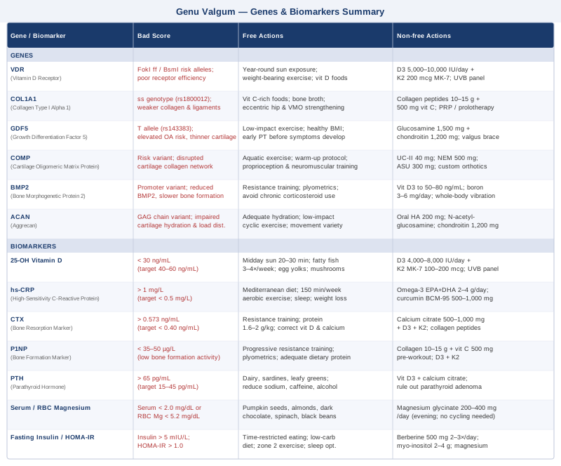
This article maps two layers of that biology. The first is your blood: seven specific biomarkers reveal whether your bones are in a healthy remodeling state, whether your inflammatory load is degrading joint tissue faster than it can be repaired, or whether a metabolic issue is adding unseen stress to your knee alignment. The second layer is your genetics: six gene variants affect how your body builds and maintains bone, cartilage, and connective tissue — and knowing your genotype allows you to target nutrition and supplementation far more precisely.
No article can promise a reversal, particularly for structural deformity in adults. But better information leads to better decisions, consistently. Understanding which biomarkers are out of range tells you which levers are actually worth pulling. Understanding your genetic predispositions helps you anticipate vulnerabilities before they become clinical problems. That is a genuinely different starting point than trial and error.
7 Biomarkers That Reveal What Is Really Happening in Your Knee Joints
Biomarkers do not diagnose genu valgum — imaging and physical examination do. What biomarkers do is map the biological environment that either supports joint integrity or undermines it. The seven below are the most actionable for this condition: each addresses a mechanism directly relevant to bone quality, cartilage health, or systemic load, and each can be measured affordably through a standard blood draw.
1. 25-OH Vitamin D
Why it matters
Vitamin D deficiency is one of the most well-documented causes of acquired genu valgum. Rickets — the classic presentation of severe deficiency in childhood — is characterized by bowing and angular deformity of the long bones, including the knock-knee pattern. In adults, persistent insufficiency does not cause the same dramatic deformity, but it contributes to bone softening, impaired bone mineral density, reduced muscle strength around the knee, and altered bone remodeling balance. Even moderate insufficiency in the 20–30 ng/mL range keeps the skeleton in a mildly demineralized state that is more susceptible to the mechanical stress of misalignment.
Peter Attia recommends maintaining 25-OH vitamin D in the 40–60 ng/mL range. Bone specialists frequently push for 50–80 ng/mL when optimizing for musculoskeletal function specifically.
How to Measure It
A serum 25-hydroxyvitamin D test is available at virtually any lab and typically costs $20–$60 out of pocket. Most primary care physicians will order it without question. Test fasted in the morning, at least twice a year if you live north of 35° latitude or spend limited time outdoors.
If the Score Is Bad: Plan Without Supplements
If your level is below 30 ng/mL, the first priority is consistent midday sun exposure — 20–30 minutes with arms and legs exposed can raise levels significantly over 4–8 weeks in lighter-skinned individuals. Add fatty fish (salmon, sardines, mackerel) 3–4 times per week, pasture-raised egg yolks, and fortified dairy. These dietary measures alone rarely restore severely low levels to optimal range, but they provide meaningful baseline support.
If the Score Is Bad: Plan With Supplements or Equipment
For levels below 30 ng/mL, most integrative practitioners recommend D3 (cholecalciferol) at 4,000–8,000 IU/day, always paired with vitamin K2 (100–200 mcg MK-7 form) to direct calcium toward bones rather than soft tissue. Retest after 12 weeks. For severe deficiency, short-term loading doses (50,000 IU weekly for 8 weeks) are sometimes used under medical supervision, followed by maintenance. A narrow-band UVB light panel can substitute for sun during winter months.
No cycling needed; maintain year-round and adjust dose based on bloodwork. Toxicity is rare below 10,000 IU/day but possible with prolonged use without monitoring — retest every 3–6 months to find your personal maintenance level.
2. hs-CRP (High-Sensitivity C-Reactive Protein)
Why it matters
hs-CRP is the most accessible blood marker of systemic low-grade inflammation. Genu valgum creates abnormal mechanical stress on the knee joint, which over time generates a local inflammatory response. When systemic inflammation is also elevated, cartilage degradation accelerates, ligament integrity weakens, and the regenerative capacity of joint tissue is impaired. In this context, elevated hs-CRP signals that corrective exercises and orthotics are fighting against a hostile internal environment — producing less durable results.
Thomas Dayspring and Peter Attia both use hs-CRP below 0.5 mg/L as a target for a genuinely healthy inflammatory state. Levels between 1–3 mg/L indicate moderate systemic inflammation warranting investigation. Above 3 mg/L points to significant inflammatory load that should be treated as a primary issue.
How to Measure It
hs-CRP is available on standard metabolic panels or as a standalone test, typically $15–$45 out of pocket. Always measure fasting in the morning. One critical caveat: hs-CRP spikes sharply with acute infections or recent injury. A single elevated reading during illness has no diagnostic value; use a baseline measurement when you are well and have not had strenuous exercise in the prior 24 hours.
If the Score Is Bad: Plan Without Supplements
Consistent moderate aerobic exercise (150 minutes per week at conversational intensity) lowers hs-CRP reliably over 12–16 weeks based on multiple clinical trials. Improving sleep quality and addressing chronic psychological stress have direct effects on inflammatory markers. A Mediterranean-style dietary pattern — emphasizing fatty fish, extra-virgin olive oil, vegetables, legumes, and limiting ultra-processed foods and refined carbohydrates — is the dietary intervention with the strongest and most consistent evidence. Weight loss of even 5–10% body weight meaningfully reduces CRP, which matters directly for genu valgum because excess weight amplifies mechanical knee load.
If the Score Is Bad: Plan With Supplements or Equipment
Omega-3 fatty acids (EPA+DHA, 2–4 g/day) from a high-quality, molecularly distilled fish oil have robust evidence for lowering hs-CRP in the 1–3 mg/L range. Curcumin (as BCM-95 or phospholipid complex for bioavailability) at 500–1,000 mg/day adds modest anti-inflammatory effects. Both can be used continuously without cycling. High-dose fish oil may mildly extend bleeding time — note this before any surgical procedure. Combine supplements with lifestyle interventions for maximal effect; supplements alone rarely normalize elevated hs-CRP.
3. CTX (C-Terminal Telopeptide of Type I Collagen)
Why it matters
CTX directly measures the rate of bone resorption — how fast osteoclasts are breaking down existing bone. In the context of genu valgum, elevated bone resorption progressively weakens the structural framework supporting knee alignment. The tibia and femur need a healthy mineralized matrix to resist and adapt to mechanical load. If resorption chronically outpaces formation, the angular forces acting on the knee joint work against increasingly compromised material. This dynamic matters particularly in adults over 40, in postmenopausal women, and in anyone with low vitamin D or inadequate dietary calcium.
Allan Sniderman and metabolic bone researchers increasingly advocate tracking CTX alongside P1NP (the bone formation marker, below) to evaluate the full remodeling picture rather than either marker in isolation.
How to Measure It
CTX requires a fasting blood draw in the morning — the marker shows significant circadian variation, peaking overnight and declining through the day. Eating suppresses CTX substantially; a non-fasted sample is essentially uninterpretable. Cost ranges from $50–$120 at specialty or functional medicine labs. Most conventional primary care offices do not routinely order it, but any endocrinologist or metabolic bone specialist can do so. Adult reference range: below 0.573 ng/mL; many functional medicine practitioners prefer below 0.40 ng/mL for bone health optimization.
If the Score Is Bad: Plan Without Supplements
Progressive resistance training — particularly exercises with compressive and impact loading — is the most reliable non-pharmacological strategy for reducing CTX over 8–12 weeks. Adequate dietary protein (1.6–2.0 g per kg of body weight) provides the substrate for bone matrix synthesis. Correcting vitamin D and calcium intake addresses the most common nutritional driver of elevated resorption. Sleep quality and cortisol management also matter: chronic stress elevates cortisol, which directly stimulates bone resorption through multiple pathways.
If the Score Is Bad: Plan With Supplements or Equipment
Calcium citrate (500–1,000 mg/day in divided doses with meals) combined with vitamin D3 and K2 addresses the most common nutritional drivers. Hydrolyzed collagen peptides (10–15 g/day) combined with resistance training have shown increases in bone formation markers in some controlled trials, with indirect benefits for the resorption/formation balance. For high-risk osteoporosis cases with severely elevated CTX, bisphosphonate medications are a medical option requiring physician supervision — not a first-line lifestyle intervention.
4. P1NP (Procollagen Type 1 N-Terminal Propeptide)
Why it matters
P1NP is the mirror of CTX: it measures the rate of new bone formation rather than resorption. Together, the two markers tell you whether your skeleton is in net gain or net loss. For genu valgum specifically, understanding bone turnover balance helps determine whether the structural framework is strengthening over time — which is the desired direction for anyone doing corrective exercises and optimization work. High P1NP with low CTX means active bone building. Low P1NP with elevated CTX is a concerning pattern indicating net bone loss. High values for both suggest high-turnover states that warrant medical evaluation.
How to Measure It
P1NP is measured from a standard blood draw, fasting preferred. Cost: $75–$150 at specialty labs including LabCorp and Quest. Reference ranges vary by age and sex; adults generally aim for P1NP above 35–50 µg/L. Values rising after starting a resistance training program are a positive signal of active bone formation — not a concern.
If the Score Is Bad: Plan Without Supplements
Resistance training is the most potent stimulus for P1NP elevation in healthy adults — controlled studies consistently show that progressive loading raises bone formation markers within 8–12 weeks. Brief plyometric loading (jumping rope, box jumps, low-impact variants) further amplifies the osteogenic response through mechanical strain on bone. Adequate dietary protein is non-negotiable: collagen synthesis for bone matrix depends on amino acid availability. Correcting vitamin D to the 40–80 ng/mL range enhances osteoblast activity downstream.
If the Score Is Bad: Plan With Supplements or Equipment
Hydrolyzed collagen peptides (10–15 g/day) taken with 500 mg of vitamin C approximately 30–60 minutes before resistance training have been shown in randomized controlled trials — including work by Keith Baar's research group at UC Davis — to increase collagen synthesis markers in tendon, cartilage, and bone tissue. Vitamin D3 and K2 (as described above) support osteoblast function directly. In cases of confirmed osteoporosis with persistently low P1NP, anabolic bone medications like teriparatide require endocrinologist guidance and represent a medical rather than lifestyle-level intervention.
5. PTH (Parathyroid Hormone)
Why it matters
PTH is released by the parathyroid glands when blood calcium drops — and when calcium or vitamin D is chronically insufficient, PTH rises persistently. Chronically elevated PTH is one of the strongest drivers of bone resorption: it mobilizes calcium from bone into circulation, progressively demineralizing the skeleton. In genu valgum, secondary hyperparathyroidism driven by nutritional deficiency creates a sustained catabolic bone state that compounds the mechanical stress of misalignment. In children who develop knock knees through nutritional rickets, elevated PTH is a consistent clinical finding alongside low vitamin D and low calcium. In adults, it reflects an ongoing imbalance that can continue silently for years.
How to Measure It
PTH is measured via a standard blood draw using the intact PTH (iPTH) assay. Cost: $30–$75 out of pocket. Conventional normal range: 15–65 pg/mL. Many functional medicine practitioners target the lower half of this range (15–45 pg/mL) for bone health optimization. Always interpret PTH alongside calcium, vitamin D, and phosphorus — PTH out of context is difficult to act on.
If the Score Is Bad: Plan Without Supplements
Correct the underlying calcium and vitamin D insufficiency through diet first: dairy products, fortified plant milks, sardines with bones, almonds, and leafy greens provide meaningful calcium. Reduce factors that increase urinary calcium loss: excessive animal protein, high sodium intake, and caffeine all contribute. Adequate magnesium (see below) is also required for normal PTH regulation. These changes alone often normalize mild secondary hyperparathyroidism within 8–12 weeks.
If the Score Is Bad: Plan With Supplements or Equipment
Vitamin D3 (4,000–8,000 IU/day) combined with calcium citrate (if dietary intake is inadequate) normalizes PTH driven by nutritional deficiency within 8–12 weeks of consistent use. Retest PTH alongside 25-OH vitamin D and serum calcium after 12 weeks. If PTH remains elevated despite normalized vitamin D and adequate calcium intake, evaluation for primary hyperparathyroidism (parathyroid adenoma) is warranted — this requires medical imaging and potentially surgical intervention, and supplementation alone will not resolve it.
6. Serum Magnesium (or RBC Magnesium)
Why it matters
Magnesium is a cofactor in over 300 enzymatic reactions, including the activation of vitamin D itself. The conversion of 25-hydroxyvitamin D to its active form (1,25-dihydroxyvitamin D) depends on magnesium-dependent enzymes — which means that supplementing vitamin D without addressing magnesium deficiency yields limited results. Beyond vitamin D activation, magnesium is essential for bone mineral matrix formation, normal neuromuscular function, and the muscle contractility that governs gait mechanics and joint load distribution. Chronically low magnesium — highly prevalent in Western diets — quietly undermines nearly every intervention targeting bone and joint health.
How to Measure It
Standard serum magnesium is available on most basic metabolic panels at $20–$40. However, serum magnesium is a poor proxy for intracellular stores — less than 1% of the body's magnesium is in serum, and the kidneys defend serum levels tightly. RBC magnesium (measuring intracellular magnesium inside red blood cells) is a more accurate reflection of tissue status and costs $50–$90 at specialty labs. Optimal serum range: 2.0–2.4 mg/dL. Optimal RBC magnesium range: 5.2–6.9 mg/dL. Many individuals with values in the low-normal serum range still show meaningfully improved function with increased magnesium intake.
If the Score Is Bad: Plan Without Supplements
The typical Western diet provides only 200–250 mg/day against a recommended intake of 400–420 mg (men) or 310–320 mg (women). Prioritize: pumpkin seeds, almonds, cashews, dark chocolate (70%+ cacao), black beans, spinach, Swiss chard, and avocado. Reduce alcohol consumption, which increases urinary magnesium excretion, and limit ultra-processed foods, which contain almost no magnesium. These dietary shifts alone can move RBC magnesium into optimal range over 6–8 weeks in mild cases.
If the Score Is Bad: Plan With Supplements or Equipment
Magnesium glycinate (200–400 mg elemental magnesium/day) is the best-tolerated and well-absorbed form. Magnesium malate and magnesium threonate are good alternatives with slightly different tissue affinities. Avoid magnesium oxide (poor absorption) and use magnesium citrate cautiously at higher doses due to its laxative effect. No cycling required; take daily, preferably in the evening — magnesium has mild relaxing effects that support sleep quality, creating additional downstream benefit for joint repair. Loose stools are a reliable signal to reduce dose. At standard doses, side effects are rare.
7. Fasting Insulin and HOMA-IR
Why it matters
Insulin resistance disrupts musculoskeletal health through several interconnected mechanisms. Chronic hyperinsulinemia drives systemic inflammation (elevating hs-CRP), promotes adipose accumulation (increasing mechanical knee load), and impairs bone quality through advanced glycation end products (AGEs) — proteins cross-linked by excess blood sugar — that make bone matrix more brittle and less flexible. For genu valgum specifically, every kilogram of excess body weight amplifies the angular forces acting on the knee joint. Reducing metabolic dysfunction is therefore both a direct and indirect intervention for knee health.
HOMA-IR (Homeostatic Model Assessment of Insulin Resistance) combines fasting insulin and fasting glucose into a single score. Peter Attia targets HOMA-IR below 1.0 for metabolic health; values above 2.0 begin to signal meaningful insulin resistance. The formula: (fasting insulin in mIU/L × fasting glucose in mmol/L) ÷ 22.5.
How to Measure It
Fasting insulin is a blood test available at most labs for $30–$60 out of pocket. It is not typically ordered on routine panels — you will likely need to request it specifically. Most conventional labs flag concern only above 20–25 mIU/L, which is an extremely permissive threshold for metabolic optimization. Peter Attia's preferred fasting insulin target is below 5 mIU/L. Calculate HOMA-IR yourself once you have both fasting insulin and fasting glucose values.
If the Score Is Bad: Plan Without Supplements
Time-restricted eating (16:8 or 14:10 intermittent fasting), combined with a dietary pattern lower in refined carbohydrates and ultra-processed foods, is the most powerful dietary intervention for reducing fasting insulin within 6–12 weeks. Progressive resistance training (2–3 sessions per week) and zone 2 aerobic exercise (150 minutes per week at conversational intensity) significantly improve insulin sensitivity through distinct and complementary mechanisms. Sleep optimization — including addressing undiagnosed sleep apnea — has a direct and frequently underestimated effect on insulin sensitivity; even moderate sleep restriction raises fasting insulin substantially.
If the Score Is Bad: Plan With Supplements or Equipment
Berberine (500 mg, 2–3 times/day with meals) has been shown in multiple randomized trials to lower fasting insulin and HOMA-IR with effect sizes comparable to metformin; cycle 8–12 weeks on, 4 weeks off to prevent tolerance. Myo-inositol (2–4 g/day) supports insulin receptor signaling. Magnesium supplementation (discussed above) independently improves insulin sensitivity in deficient individuals. Chromium picolinate (200–400 mcg/day) shows modest effects in some trials. For HOMA-IR persistently above 3.0 despite lifestyle optimization, metformin or GLP-1 receptor agonists are evidence-based medical options — discuss with a physician rather than self-managing.
The Genetic Layer: 6 Variants That Shape Your Bone and Joint Biology
Biomarkers tell you where you stand today. Genetics tell you which biological processes your body tends to run at a disadvantage — so you can compensate before problems emerge. The six variants below have the strongest relevance to the biological mechanisms underlying genu valgum: bone density, collagen quality, cartilage development, and vitamin D responsiveness.
VDR — Vitamin D Receptor Gene
The VDR gene encodes the nuclear receptor that allows cells throughout the body to respond to active vitamin D. Several well-studied polymorphisms — FokI (rs2228570), BsmI (rs1544410), ApaI (rs7975232), and TaqI (rs731236) — affect receptor efficiency and downstream biological signaling. Individuals with the FokI ff genotype have a less efficient receptor protein, meaning the same circulating vitamin D level produces less biological effect on bone mineralization, muscle function, and immune modulation. Multiple studies have linked BsmI and TaqI polymorphisms to lower bone mineral density in both postmenopausal women and growing children, making these among the most clinically relevant variants for skeletal conditions.
For genu valgum, the connection is direct: even with blood vitamin D in the so-called normal range, individuals with less efficient VDR variants may experience insufficient biological activity at the bone cell level — supporting bone softening and impaired remodeling.
If the gene is bad: plan without supplements — prioritize consistent year-round sun exposure to drive natural vitamin D synthesis at higher levels; add vitamin D-rich foods systematically (fatty fish 3–4 times per week, egg yolks, mushrooms exposed to UV light); perform regular weight-bearing exercise to maximize the mechanical stimulus for bone formation independent of vitamin D signaling; keep body fat in a healthy range, since adipose tissue sequesters vitamin D and reduces circulating availability.
If the score is bad: plan with supplements or equipment — with a less efficient VDR, achieving adequate biological effect requires higher circulating vitamin D than average. Target 50–80 ng/mL rather than the standard 30+ ng/mL cutoff. Use D3 (cholecalciferol) at 5,000–10,000 IU/day with vitamin K2 (200 mcg MK-7). A narrow-band UVB light panel provides direct skin synthesis during winter months, bypassing any oral absorption issues. Retest every 3 months initially to establish your personal maintenance dose.
COL1A1 — Collagen Type I Alpha 1 Gene
COL1A1 encodes the alpha1 chain of type I collagen — the most abundant structural protein in bone, tendons, and ligaments. The Sp1 binding site polymorphism (rs1800012), specifically the s allele, alters the ratio of alpha1 to alpha2 chains in the assembled collagen triple helix, producing a structurally weaker protein. The ss genotype is associated with lower bone mineral density, increased fracture risk, and reduced tensile strength in tendons and ligaments across multiple population studies.
For genu valgum, weakened connective tissue means the ligamentous structures governing knee alignment — the medial collateral ligament, ACL, and patellar tendon — provide reduced passive stability. In ss individuals, this manifests as increased dynamic knee valgus during movement even when muscle strength is apparently adequate: the joints simply allow more inward collapse under load.
If the gene is bad: plan without supplements — prioritize vitamin C-rich foods consistently (collagen synthesis requires vitamin C as a cofactor at every step); eat connective tissue-supporting foods regularly (bone broth, slow-cooked meats, skin-on poultry); implement targeted eccentric strengthening for hip abductors and external rotators to actively compensate for passive ligamentous laxity; use a physical therapy protocol emphasizing slow, heavy eccentrics for tendon and ligament loading (Copenhagen adductor exercises, single-leg squat progressions).
If the score is bad: plan with supplements or equipment — hydrolyzed collagen peptides (10–15 g/day) consumed with 500 mg of vitamin C 30–60 minutes before loading exercise have been shown in randomized controlled trials (including Clark et al., 2008, American Journal of Clinical Nutrition) to increase collagen synthesis markers in tendons and cartilage. Prolotherapy (dextrose injection into joint and ligament attachments) or platelet-rich plasma (PRP) injections under ultrasound guidance are more advanced options for ligamentous laxity — discuss with a sports medicine physician experienced in these approaches.
GDF5 — Growth Differentiation Factor 5 Gene
GDF5 is a signaling protein critical for embryonic joint formation, cartilage maintenance, and tendon and ligament development. The variant rs143383 — a C>T change in the 5' untranslated region — reduces GDF5 transcription, so the T allele produces less of this protein. This is one of the most consistently replicated genetic risk factors for osteoarthritis in genome-wide association studies (GWAS), with the T allele significantly increasing OA risk in European, Asian, and mixed-ancestry populations across dozens of independent studies.
For genu valgum, reduced GDF5 activity means potentially thinner, less resilient articular cartilage in the medial and lateral knee compartments, and impaired capacity to repair even minor joint damage from the abnormal loading pattern that knock-knee alignment creates. Over years, this compresses the timeline from alignment issue to symptomatic knee osteoarthritis.
If the gene is bad: plan without supplements — shift toward low-impact loading that maintains cartilage nutrition without excess shear forces: cycling, swimming, aquatic exercise, and elliptical training are preferable to high-volume running on hard surfaces; avoid prolonged static knee flexion; maintain a healthy BMI to minimize compressive cartilage loading; address genu valgum alignment proactively through physical therapy before symptoms develop, rather than waiting for pain to prompt intervention.
If the score is bad: plan with supplements or equipment — glucosamine sulfate (1,500 mg/day) and chondroitin sulfate (1,200 mg/day) have been studied more extensively for knee joint support than any other supplements; evidence for symptom benefit is strongest in moderate-to-severe OA (as reviewed in the GAIT trial data). Undenatured type II collagen (UC-II, 40 mg/day) has emerging evidence for joint health through an immune-mediated mechanism distinct from hydrolyzed collagen. A valgus-correction knee orthosis — a brace designed to reduce medial compartment loading in knock-knee alignment — is a mechanical intervention for those with existing symptoms; available through orthotics specialists.
COMP — Cartilage Oligomeric Matrix Protein Gene
COMP encodes a pentameric protein that stabilizes and organizes the collagen network within cartilage. Loss-of-function mutations in COMP cause pseudoachondroplasia and multiple epiphyseal dysplasia — rare skeletal dysplasias characterized by disproportionate short stature and joint abnormalities, demonstrating the gene's foundational role in skeletal architecture. Common variants at the population level may affect cartilage organization and mechanical properties more subtly, influencing how well knee cartilage resists the abnormal compressive and shear forces generated by angular malalignment.
If the gene is bad: plan without supplements — reduce high-impact loading (avoid long-distance running on hard surfaces); incorporate aquatic exercise to maintain joint mobility without compression; implement proper warm-up protocols (5–10 minutes of dynamic movement before any loading activity); engage in regular physiotherapy focused on proprioception training and neuromuscular control of the knee to compensate for any reduction in cartilage mechanical buffering through improved active stability.
If the score is bad: plan with supplements or equipment — undenatured type II collagen (UC-II, 40 mg/day), natural eggshell membrane (NEM, 500 mg/day), and avocado/soybean unsaponifiables (ASU, 300 mg/day) all support cartilage matrix through distinct mechanisms and have human clinical trial support. Custom foot orthotics that reduce medial compartment loading through correcting subtalar pronation are a practical and well-supported mechanical intervention for genu valgum cases with cartilage concerns.
BMP2 — Bone Morphogenetic Protein 2 Gene
BMP2 encodes one of the most potent known inducers of bone formation. It drives osteoblast differentiation, is required for normal skeletal development, and plays a central role in fracture healing and bone regeneration. Promoter region variants in BMP2 affect the amount of BMP2 protein produced and have been linked to osteoporosis susceptibility in genome-wide studies. Beyond genetics, factors that chronically suppress BMP2 signaling — including systemic inflammation and corticosteroid exposure — reduce endogenous bone-forming capacity.
For genu valgum, lower BMP2 activity means reduced osteoblast function and slower periosteal bone adaptation to mechanical stress. The capacity of bone to model and adapt in response to corrective loading is partly dependent on BMP2 signaling — making this variant relevant for anyone attempting to use exercise to drive structural improvement.
If the gene is bad: plan without supplements — resistance training is the most powerful known stimulus for endogenous BMP2 upregulation in bone; prioritize compound movements with progressive loading; brief high-impact loading (3–5 minutes of jumping rope or low-level plyometrics daily) stimulates osteoblast activity through pathways that involve BMP signaling; avoid chronic corticosteroid medications where possible, as glucocorticoids suppress BMP2 expression substantially.
If the score is bad: plan with supplements or equipment — vitamin D3 at optimal circulating levels (50–80 ng/mL) enhances BMP2 signaling in osteoblasts through VDR-mediated pathways. Orthosilicic acid (silicon, 10 mg/day) and boron (3–6 mg/day from food or supplements) have shown modest positive effects on bone mineral density in some controlled trials and support mineral matrix formation. Whole-body vibration (WBV) platforms — used 10–15 minutes/day at validated frequency settings — have demonstrated improvements in bone density markers in several controlled trials, likely through mechanical activation of BMP-related osteogenic pathways.
ACAN — Aggrecan Gene
ACAN encodes aggrecan, the large proteoglycan that gives articular cartilage its unique viscoelastic and compressive properties. Aggrecan traps water within the cartilage matrix through negatively charged glycosaminoglycan (GAG) chains — and this water provides the hydraulic resistance that allows cartilage to absorb and distribute compressive loads without damage. Variants in ACAN affect the length and sulfation pattern of these GAG chains, altering cartilage hydration, stiffness, and resilience. ACAN variants are also associated with short stature syndromes, reflecting the gene's importance in growth plate cartilage development.
For genu valgum, compromised aggrecan function means uneven load distribution across the knee joint surface under the already-abnormal stress pattern created by angular malalignment — accelerating focal cartilage wear in the medial or lateral compartment.
If the gene is bad: plan without supplements — adequate hydration directly supports cartilage water content; low-impact cyclic exercise (walking, cycling, swimming) maintains the alternating compression/decompression that drives synovial fluid into cartilage for nutrition; avoid prolonged periods of static knee flexion (extended sitting with knees bent); maintain movement variety throughout the day to prevent sustained uneven loading patterns.
If the score is bad: plan with supplements or equipment — oral hyaluronic acid (200 mg/day) targets the same extracellular matrix space as aggrecan and has shown modest evidence for knee symptom relief; N-acetyl-D-glucosamine (500–1,000 mg/day) provides substrate for GAG chain synthesis; chondroitin sulfate (1,200 mg/day) directly supplies the sulfated glycosaminoglycans that aggrecan's side chains are built from. Intra-articular hyaluronic acid injections are a medical intervention with moderate evidence for knee OA with confirmed cartilage involvement — discuss with an orthopedist.
What Peter Attia's Approach to Longevity Medicine Teaches Us About Bone and Joint Health
Peter Attia's book Outlive: The Science and Art of Longevity (2023) does not address genu valgum directly, but its framework for tracking biomarkers, reversing metabolic dysfunction, and building musculoskeletal resilience over decades is among the most directly applicable resources available. It challenges conventional medical thinking — particularly the idea that bone density loss and joint deterioration are inevitable aging processes rather than modifiable outcomes. Here are ten of its most impactful insights for anyone managing a knee alignment condition.
1. Bone Density Is a Lagging Indicator — Start Measuring Before You Have a Problem
Attia argues that conventional medicine waits far too long to assess bone density — typically only ordering DEXA scans after menopause or a fracture. He recommends baseline DEXA scans for anyone over 40, followed by periodic tracking. For genu valgum patients, this is especially relevant: monitoring tibial and femoral bone density over years provides early warning of structural changes long before they become irreversible. Catching a declining trend at 30% bone loss is a very different situation than discovering it at 50%.
2. HOMA-IR Is a More Sensitive Metabolic Alarm Than Fasting Glucose Alone
Attia positions HOMA-IR as an early signal of insulin resistance that fasting glucose entirely misses in early and middle stages of metabolic dysfunction. Many people with normal fasting glucose already have significantly elevated fasting insulin — meaning their pancreas is working overtime to maintain glucose control. This hidden hyperinsulinemia drives the inflammatory and bone-quality consequences described in the biomarker section above.
3. Zone 2 Cardio Is One of the Highest-Leverage Interventions Available
Sustained zone 2 aerobic exercise (approximately 60–70% of max heart rate, holding a conversation) improves mitochondrial function, reduces systemic inflammation, and substantially improves insulin sensitivity. Attia recommends 3–4 hours per week for longevity. For genu valgum patients with elevated hs-CRP or high HOMA-IR, zone 2 is foundational and nearly side-effect-free. Low-impact modalities (cycling, swimming, elliptical) make it accessible even for those with knee pain.
4. Resistance Training Is the Most Powerful Bone-Building Tool in Existence
Attia is unambiguous: progressive resistance training — not cardio, not stretching — is what drives osteoblast activity, preserves and builds the muscle that stabilizes joints, and maintains proprioception. He recommends 3–4 structured strength sessions per week for everyone past 30, and considers it among the single highest-leverage health investments. For genu valgum, hip abductor and VMO strengthening through resistance training directly addresses both the bone quality and alignment control aspects of the condition.
5. Protein Is Likely the Most Under-Consumed Macronutrient in Western Populations
Attia advocates for 1.6–2.2 g of protein per kg of body weight daily, particularly for those doing resistance training and those over 40 managing muscle and bone mass. Adequate protein is non-negotiable for both muscle protein synthesis and collagen type I turnover in bone — two foundations of the structural corrections discussed throughout this article.
6. The Omega-3 Index Is a Biomarker More People Should Be Tracking
Attia discusses the omega-3 index (percentage of EPA+DHA within red blood cells) as a more accurate long-term inflammatory signal than a single hs-CRP measurement. He recommends targeting above 8%, which typically requires 2–4 g/day of EPA+DHA from high-quality fish oil. The same anti-inflammatory mechanisms that protect cardiovascular tissue also protect joint cartilage from cytokine-driven degradation.
7. Vitamin D Functions as a Hormone — Not Just a Micronutrient
Attia emphasizes that vitamin D acts as a hormonal signal across dozens of tissues, including bone, muscle, immune cells, and the brain. He recommends testing and maintaining 25-OH vitamin D above 40 ng/mL consistently, and notes that the majority of adults in northern latitudes fail to reach this without deliberate supplementation year-round. The VDR gene variants discussed above add an important layer: for some individuals, higher circulating levels are necessary to achieve adequate biological effect.
8. Sleep Quality Directly Regulates Bone and Cartilage Repair
Growth hormone — the key anabolic signal for bone and cartilage regeneration — pulses most strongly during deep sleep. Attia calls chronic sleep restriction (under 7 hours) one of the most underestimated drivers of metabolic dysfunction, hormonal dysregulation, and tissue breakdown. For anyone managing a joint condition and doing corrective exercise, sleep quality is not optional; it is when the adaptation actually happens.
9. Tracking Transforms Guesswork Into a Strategy
One of Attia's central arguments is that you cannot manage what you do not measure. His approach — running a biomarker panel twice a year, watching trends rather than single snapshots, and adjusting incrementally based on data — is precisely the framework that makes the biomarker section of this article actionable rather than theoretical. Starting with three to four key markers and measuring consistently over a year reveals far more than any single test result can.
10. Pharmaceutical Interventions Come After — Not Instead Of — Lifestyle Optimization
Attia consistently positions lifestyle as first-line and pharmacology as adjunct. For genu valgum, this framing is liberating: before any injectable, surgical, or prescription intervention, there is substantial ground to cover through optimizing vitamin D, reducing systemic inflammation, improving bone turnover balance, and correcting metabolic dysfunction. Doing those things measurably shifts the joint health trajectory — and doing them first establishes a much cleaner baseline for evaluating whether further intervention is actually needed.
Complementary Approaches With Clinical Evidence for Knee Joint Health
The following three modalities are selected for having meaningful human evidence relevant to genu valgum, knee alignment biomechanics, or the underlying drivers — bone quality, cartilage integrity, and joint tissue health — addressed throughout this article.
Yoga for Hip Strength and Dynamic Knee Alignment
Yoga, when structured correctly, is far more than a flexibility practice. Specific postures involve sustained isometric and dynamic loading of the hip abductors, external rotators, and vastus medialis oblique — the exact muscles that govern knee alignment and prevent dynamic knee valgus. In genu valgum, weakness in these groups allows the femur to internally rotate and adduct under load, driving the knee inward during walking, squatting, and stair climbing. Yoga also develops body awareness and neuromuscular control around the joint, which reduces dynamic valgus during habitual movement patterns that otherwise reinforce poor alignment.
A randomized controlled trial (Tran et al., 2001, International Journal of Yoga) demonstrated that an 8-week Iyengar yoga program significantly improved lower extremity strength and static balance compared to controls. Research on yoga interventions for patellofemoral pain syndrome — a frequent consequence of genu valgum biomechanics — has shown improvements in pain, function, and lower limb alignment over 6–8 weeks of structured practice. Iyengar yoga, with its emphasis on precise anatomical alignment and use of props to support correct positioning, is the most evidence-informed style for structural corrections.
Aim for 3–4 sessions per week of 30–45 minutes, focusing on Warrior II, Triangle Pose, Bridge Pose, and seated hip external rotator work. Use blocks and straps to maintain correct alignment rather than forcing range of motion. Avoid aggressive deep forward folds that increase dynamic knee valgus stress at the joint. For best results, begin with one-on-one sessions with a certified Iyengar or therapeutic yoga teacher familiar with lower extremity conditions, then transition to group or home practice once alignment cues are internalized.
Low-Level Laser Therapy (LLLT) / Photobiomodulation for Joint Tissue Recovery
Low-level laser therapy uses specific wavelengths of red and near-infrared light (typically 630–1,000 nm) to penetrate tissue and stimulate mitochondrial energy production in cells. In joint tissues, LLLT increases ATP availability in chondrocytes, reduces inflammatory cytokines including IL-1β and TNF-α, and promotes collagen synthesis in tendons and periarticular structures. For genu valgum, where chronic abnormal loading creates persistent low-grade joint inflammation and accelerated cartilage stress, LLLT offers a non-invasive approach to improving the local tissue environment — reducing the inflammatory burden that compounds mechanical damage.
A 2009 Cochrane systematic review by Bjordal et al. found that LLLT produced significant short-term pain relief and functional improvement in knee osteoarthritis when applied at recommended parameters. The World Association for Laser Therapy (WALT) guidelines recommend 4–8 J/cm² per treatment point for musculoskeletal conditions, delivered 3 times per week for 4–8 weeks. Consumer-grade red light panels from established manufacturers can deliver effective doses for knee joint applications when used according to manufacturer protocols and WALT guidance.
Apply to the medial and lateral knee joint lines for 3–5 minutes per zone, 3 sessions per week for 6–8 weeks. Keep the device at or within 2 cm of skin surface for effective penetration. Evidence is strongest for pain reduction and functional improvement; LLLT will not correct angular deformity. It is best positioned as a supportive tool for reducing the inflammatory consequences of genu valgum while active rehabilitation addresses the underlying alignment and strength deficits. Cost for consumer-grade panels: $250–$1,000. Clinical LLLT sessions: $40–$80 each. Contraindications include active cancer and direct application over the eyes or thyroid gland.
Biofeedback for Gait Retraining and Neuromuscular Control
Biofeedback provides real-time sensory information — visual, auditory, or tactile — about body movement patterns, allowing patients to consciously modify those patterns with an accuracy and speed that verbal coaching alone cannot match. In genu valgum, the knee-inward position during walking, running, and functional movements is often a deeply habituated motor pattern reinforced over millions of repetitions. Real-time feedback disrupts this automaticity, providing the sensory information needed for conscious correction and faster motor learning — ultimately allowing the new alignment pattern to become automatic over fewer repetitions than cued verbal instruction achieves.
A randomized trial by Noehren et al. (2011, Clinical Biomechanics) demonstrated that real-time gait retraining using visual feedback of hip adduction angle reduced peak hip adduction (a primary driver of dynamic knee valgus) and patellofemoral joint stress by over 20%, with effects maintained at 3-month follow-up without additional training sessions. Subsequent studies in running-related patellofemoral pain — a direct downstream consequence of chronic knee valgus — have replicated these findings across multiple research groups. Eight sessions of supervised biofeedback gait retraining appears sufficient for most patients to internalize the corrected movement pattern.
Practically, the most accessible route is working with a sports physiotherapist or certified gait analyst using a treadmill with 2D video analysis and real-time visual feedback. Programs typically run 6–10 sessions over 4–6 weeks, with structured home exercises between sessions. Consumer smartphone apps with basic pose estimation (such as Dartfish Express or similar platforms) allow lateral-view gait analysis at home as a lower-cost adjunct. For maximal durability of the corrected pattern, combine biofeedback retraining with progressive hip and glute strengthening — the neuromuscular change needs the muscular endurance to hold the corrected alignment when fatigued.
Conclusion
Genu valgum is rarely just a structural problem that physical therapy alone can fully address. Behind the visible inward angle of the knee is an internal biological environment — shaped by your nutritional status, inflammatory load, metabolic health, and genetic predispositions — that either supports joint integrity over time or quietly works against it. The biomarkers and gene variants covered in this article represent the most actionable layer of that biology: measurable, responsive to targeted interventions, and informative in ways that generic advice simply cannot be.
A sensible starting point is a baseline blood panel including 25-OH vitamin D, hs-CRP, fasting insulin, PTH, and serum magnesium — all obtainable through an integrative medicine or functional medicine workup. If bone quality is a priority, add CTX and P1NP. For genetic context, a direct-to-consumer SNP panel or clinical genomic test can identify the variants discussed here. Bring those results to a practitioner who can contextualize them alongside your history and imaging. Then track the markers twice a year and let the trends guide your decisions.
The path forward looks different depending on what you actually find — and that is precisely the point.