This article contains AI generated content.
· UpdatedGenu Varum: 6 Genes and 7 Biomarkers to Track
Introduction
If your knees curve outward when you stand with your feet together, you already know the feeling of being told either that it will correct on its own or that nothing much can be done. For many people with persistent genu varum — bow legs that outlast childhood and continue to generate knee pain, gait changes, and progressive joint wear — neither answer is satisfying. The condition is mechanical, yes, but its roots are often metabolic, nutritional, or genetic, and those roots are largely invisible to imaging and routine physical exams.
The frustrating reality is that genu varum is not one condition. A four-year-old whose bow legs stem from vitamin D deficiency is biologically different from an adult whose progressive medial knee osteoarthritis is being driven by a phosphate-wasting gene variant or a cartilage collagen abnormality. Both may have the same angle on an X-ray. Standard advice — take calcium, do some physical therapy — does not account for these differences, and for many people it produces limited results precisely because it is not targeted to their actual biology.
This is where biomarkers and genetic insights become genuinely useful. Biomarkers show you the current state of your bone metabolism in real time: whether bone is being broken down faster than it is being built, whether your phosphate and vitamin D systems are functioning, whether growth signals are supporting structural repair or undermining it. Genetics shows you why your particular system is wired the way it is, and can explain why some people fail to respond to interventions that work well for others.
This article covers both in practical depth. The first section walks through seven biomarkers that are clinically useful for anyone dealing with genu varum — what they measure, how to get them tested, and what to do when they are out of range. The second section covers six genes with meaningful evidence for their role in bone shape and cartilage health, with specific action plans for carriers of risk variants. Neither section offers a cure. What both offer is a sharper, more honest picture of what is actually going on — and from there, better decisions.
7 Biomarkers That Reveal How Your Bone Metabolism Is Running
Bone is dynamic tissue. It is continuously dismantled by osteoclasts and rebuilt by osteoblasts, with the balance between these two processes governing whether bone becomes denser and stronger or gradually degrades. In genu varum, this balance matters enormously — poor bone quality during growth can allow curvature to develop or persist; poor adult bone metabolism accelerates the knee osteoarthritis and structural wear that bow legs predispose you to. The seven markers below capture the most useful dimensions of this system.
Biomarker 1: 25-Hydroxyvitamin D
Why it matters: Vitamin D is the master regulator of calcium absorption and a critical signal for osteoblast differentiation. Without adequate levels, calcium fails to mineralize bone matrix effectively regardless of how much is consumed. In children, severe vitamin D deficiency causes nutritional rickets — one of the most common and reversible causes of genu varum worldwide. In adults, insufficiency accelerates bone loss and undermines the structural quality of bone at every level. The NIH Office of Dietary Supplements confirms that 25-hydroxyvitamin D (25-OH D) is the standard clinical marker for body-wide vitamin D status.
How to measure it: A serum 25-OH D test through any physician or direct-to-consumer lab (LabCorp, Quest Diagnostics). Cost: $40–80 without insurance. Results in 2–3 days. Standard labs flag deficiency below 20 ng/mL; most functional medicine practitioners target 40–60 ng/mL for optimal bone metabolism. The gap between "not deficient" and "optimal" is significant and frequently missed.
If the score is bad — plan without supplements: Midday sun exposure (10am–2pm) on arms and legs for 15–30 minutes provides the most efficient vitamin D synthesis. Wild salmon, sardines, mackerel, and pasture-raised egg yolks add dietary D3. Magnesium-rich foods (pumpkin seeds, dark leafy greens, almonds) are essential because magnesium is required for vitamin D activation — without it, supplementation or synthesis produces circulating 25-OH D that cannot convert to the active hormonal form.
If the score is bad — plan with supplements or equipment: Vitamin D3 at 2,000–5,000 IU/day is appropriate for most deficient adults, always co-administered with vitamin K2 (MK-7 form, 100–200 mcg/day) to direct calcium into bone rather than soft tissues. Magnesium glycinate or malate at 300–400 mg/day improves activation and reduces side effects. Re-test after 90 days. For severe deficiency below 10 ng/mL, a physician may prescribe weekly loading doses. UVB light panels (e.g., Sperti lamps) are a practical indoor option for low-sun climates; use 2–3 times per week for 5–10 minutes per session.
Biomarker 2: Serum Phosphate
Why it matters: Phosphate is as fundamental to bone mineralization as calcium — both are required to form hydroxyapatite, the crystal structure that gives bone its rigidity. Chronically low serum phosphate (hypophosphatemia) produces soft, poorly mineralized bone that bends under the weight of standing and walking. This is the defining metabolic feature of X-linked hypophosphatemia (XLH) and tumor-induced osteomalacia — conditions where genu varum is a consistent and prominent feature. Even mild phosphate insufficiency, often overlooked on routine panels, impairs bone matrix quality in ways that standard calcium and vitamin D supplementation cannot correct.
How to measure it: Serum phosphate (inorganic phosphorus) is included in the standard comprehensive metabolic panel (CMP). Cost: often $15–50 when bundled. Normal adult range: 2.5–4.5 mg/dL. For clinical evaluation of suspected phosphate-wasting disorders, TmP/GFR (tubular maximum phosphate reabsorption per GFR) provides a more sensitive measure of renal phosphate handling. A specialist may also request a 24-hour urine phosphate collection.
If the score is bad — plan without supplements: Increase dietary phosphate through whole protein foods — meat, fish, eggs, dairy, and legumes all provide substantial phosphorus. Eliminate or reduce cola beverages, which contain phosphoric acid that disrupts phosphate balance. Ensure dietary protein is adequate (1.2–1.6 g/kg/day) since phosphate is structurally bound to amino acids and nucleic acids in food. Address any gut absorption conditions (celiac disease, Crohn's) that may impair uptake.
If the score is bad — plan with supplements or equipment: For inherited phosphate-wasting conditions, oral phosphate supplementation (divided doses throughout the day) combined with active vitamin D analogues (calcitriol or alfacalcidol) is the conventional medical approach. Burosumab (Crysvita), a monoclonal antibody that neutralizes FGF23, is now a first-line treatment for XLH confirmed by genetic testing — it addresses the upstream mechanism rather than just replacing phosphate. This is not self-managed territory; endocrinologist oversight is essential. For milder hypophosphatemia without confirmed genetic cause, dietary optimization and reducing phosphate antagonists (antacids containing aluminum) is the starting point.
Biomarker 3: Parathyroid Hormone (PTH)
Why it matters: PTH is the emergency calcium regulation hormone. When calcium or vitamin D falls too low, the parathyroid glands respond by secreting PTH, which pulls calcium from bone, reduces renal calcium excretion, and stimulates conversion of vitamin D to its active form. Chronically elevated PTH — secondary hyperparathyroidism — means the body has been running nutritionally depleted long enough that it is systematically dismantling bone to maintain serum calcium. In genu varum, this pattern represents active structural deterioration that standard imaging may not capture early.
How to measure it: Intact PTH (iPTH) blood test, typically ordered alongside calcium and 25-OH D. Cost: $50–100. Optimal fasting morning range: 10–55 pg/mL. Values above 65–70 pg/mL paired with low vitamin D and low calcium confirm secondary hyperparathyroidism. This triad is a clear signal that bone mineralization is being actively disrupted and warrants intervention.
If the score is bad — plan without supplements: Correcting the calcium and vitamin D deficiency driving PTH elevation is foundational. Consistent weight-bearing exercise (walking, hiking, resistance training) suppresses PTH modestly by stimulating osteoblast activity, which signals reduced need for calcium mobilization. Reducing sodium intake decreases renal calcium wasting, lessening the PTH stimulus. Caffeine and alcohol both impair calcium absorption — moderating these creates measurable improvements.
If the score is bad — plan with supplements or equipment: Vitamin D3 plus K2 supplementation (as described above) is the central intervention — correcting vitamin D status reliably reduces PTH in secondary hyperparathyroidism. Calcium supplementation should be used carefully: calcium citrate at 200–400 mg with meals is preferred if dietary intake is genuinely insufficient. High-dose calcium carbonate supplements are less advisable given risk of vascular calcification without adequate K2. Whole-body vibration platforms (15–20 minutes, 3×/week) have shown modest PTH reduction and bone density improvement in multiple randomized controlled trials, particularly in older adults.
Biomarker 4: Bone-Specific Alkaline Phosphatase (BSAP)
Why it matters: Alkaline phosphatase is released by active osteoblasts during bone formation. The bone-specific fraction (BSAP) isolates this signal from total ALP, which also reflects liver and intestinal activity. BSAP tells you whether bone formation is running at normal, elevated, or — importantly in hypophosphatasia — pathologically low levels. In rickets (a primary cause of genu varum), BSAP is markedly elevated, reflecting the accelerated but poorly mineralized bone remodeling characteristic of the disease. Tracking it helps distinguish active metabolic disease from structural variation that has already stabilized.
How to measure it: Bone-specific ALP test through hospital or specialty labs. Cost: $60–120. Adult reference range: 14–43 U/L. Total ALP is included in the standard CMP; if elevated, the bone-specific fraction helps clarify origin. In children with persistent genu varum, significantly elevated BSAP is diagnostic of active rickets and requires immediate nutritional or metabolic investigation.
If the score is bad — plan without supplements: Elevated BSAP signals active remodeling stress. Reducing drivers of osteoclast activity — refined sugar, processed food, chronic sleep deprivation, and unmanaged systemic inflammation — creates a more favorable remodeling environment. Moderate progressive weight-bearing exercise (not excessive impact) channels remodeling toward stronger bone architecture rather than chaotic turnover. Seven to nine hours of quality sleep is essential, as growth hormone peaks during slow-wave sleep and is the primary anabolic signal for osteoblasts.
If the score is bad — plan with supplements or equipment: Vitamin D3 plus K2 supplementation addresses the hormonal root of elevated bone turnover. Ipriflavone (a synthetic isoflavone, 600 mg/day in divided doses) has demonstrated suppression of excessive ALP and reduced bone resorption in multiple European clinical trials — cycling 6 weeks on, 2 weeks off is advisable given limited long-term safety data. For pathologically low BSAP, which is the marker for hypophosphatasia, enzyme replacement therapy (asfotase alfa) is available for severe forms — this requires specialist management and confirmed genetic diagnosis.
Biomarker 5: P1NP (Procollagen Type 1 N-Terminal Propeptide)
Why it matters: P1NP is released into the bloodstream when new type I collagen — the primary protein scaffold of bone — is actively being synthesized. It is among the most sensitive and specific markers of bone formation available and is endorsed by the International Osteoporosis Foundation as a first-choice bone formation marker. In genu varum where bone matrix quality is compromised, low P1NP reveals that new bone production is inadequate even when total bone mass appears acceptable on imaging. Peter Attia has consistently highlighted P1NP alongside CTX as the two bone turnover markers most worth tracking when monitoring any bone health intervention.
How to measure it: Serum P1NP (morning, fasting preferred for consistency). Cost: $80–150, available through LabCorp and specialty panels. Optimal range in adults: 25–100 mcg/L. Values below 20 mcg/L suggest impaired bone formation that warrants investigation. Interpret P1NP alongside CTX — the ratio between formation and resorption matters as much as either value alone.
If the score is bad — plan without supplements: Low P1NP responds strongly to resistance training with axial loading — squats, deadlifts, weighted carries, and loaded walking apply compressive force through the skeleton and directly stimulate osteoblast activity. Protein intake is rate-limiting for collagen synthesis: glycine-rich foods (bone broth, collagen-containing meat cuts, gelatin-based foods) provide the raw material. Adequate vitamin C and manganese, available through whole food sources, are essential cofactors for collagen hydroxylation and cross-linking.
If the score is bad — plan with supplements or equipment: Hydrolyzed collagen peptides (10–15 g/day, type I/III) taken with 500 mg vitamin C thirty to sixty minutes before resistance training have shown increased P1NP and connective tissue synthesis markers in clinical studies — timing relative to exercise appears to matter. Resistance training 3–4 days per week with progressive overload is the most evidence-supported non-pharmacological stimulus for P1NP elevation. Creatine monohydrate (3–5 g/day, no cycling required) may modestly support bone formation as a secondary effect, with reasonably consistent data from small trials.
Biomarker 6: CTX (C-Terminal Telopeptide of Type I Collagen)
Why it matters: CTX is released into the bloodstream as osteoclasts degrade existing bone matrix. It is the primary resorption marker and creates, when paired with P1NP, a complete picture of bone remodeling balance. Elevated CTX with low P1NP is a particularly concerning pattern: bone is being actively broken down faster than it is being replaced, a state that in genu varum progressively worsens cartilage and subchondral bone quality and accelerates medial knee osteoarthritis. CTX is highly sensitive to testing conditions and must be measured in a fasting state in the morning for valid comparison across time points.
How to measure it: Serum β-CTX (morning, fasting). Cost: $80–150. Optimal adult range: below 0.57 ng/mL for women and below 0.70 ng/mL for men per IOF guidance. Monitor every 3–6 months when tracking intervention response. Bisphosphonate therapy and significant hormonal changes dramatically lower CTX — account for these variables when interpreting results.
If the score is bad — plan without supplements: CTX is highly lifestyle-responsive. Consistent sleep is one of the most powerful CTX-lowering interventions documented — even a single night of sleep deprivation measurably raises bone resorption markers. Weight-bearing exercise suppresses osteoclast signaling and reduces CTX. Consistent eating patterns (avoiding prolonged fasting when it is unintentional) and reducing chronic psychological stress — which elevates cortisol, which in turn elevates RANKL and osteoclast activity — are both meaningful.
If the score is bad — plan with supplements or equipment: Omega-3 fatty acids (EPA+DHA combined, 2–4 g/day) consistently lower CTX in randomized trials by reducing prostaglandin-driven osteoclast stimulation — this is one of the more underappreciated effects of fish oil. Vitamin D3 and K2 address the broader hormonal environment. For postmenopausal women, standardized soy isoflavone extracts (40–80 mg/day daidzein equivalent) modestly but consistently reduce CTX in clinical trials. Whole-body vibration platforms (3–5 sessions per week, 10–20 minutes) have shown CTX reduction in randomized trials involving postmenopausal women and older adults with bone loss.
Biomarker 7: IGF-1 (Insulin-Like Growth Factor 1)
Why it matters: IGF-1 is synthesized primarily in the liver in response to growth hormone signaling and is the principal anabolic driver of longitudinal bone growth, bone density, and lean tissue maintenance. During childhood — the window when genu varum either corrects spontaneously or becomes established — IGF-1 governs chondrocyte proliferation in growth plates and directly determines the rate and quality of bone modeling. In adults, chronically low IGF-1 correlates with reduced bone mineral density, impaired muscle mass, and a general shift toward a catabolic tissue environment. Thomas Dayspring and other metabolic medicine clinicians have highlighted IGF-1 as a consistently under-ordered marker in adults with unexplained musculoskeletal decline.
How to measure it: Serum IGF-1, ideally drawn in the morning. Cost: $60–120. Age-adjusted optimal range varies by decade — for adults 30–50, functional medicine practitioners typically target 150–250 ng/mL. Below 100 ng/mL in a middle-aged adult with musculoskeletal symptoms suggests impaired anabolic signaling worth investigating. GH stimulation testing is required to diagnose true GH deficiency; IGF-1 alone is a screening tool.
If the score is bad — plan without supplements: Growth hormone — and therefore IGF-1 — peaks during slow-wave sleep. Improving sleep architecture (consistent sleep schedule, dark and cool room, limited blue light after sunset) is the highest-leverage free intervention. High-intensity resistance training and interval-based cardio are the most potent lifestyle stimulants of GH/IGF-1 outside of sleep. Adequate dietary protein is required for IGF-1 synthesis — very low protein diets suppress IGF-1 independent of calories. Periodic longer fasting (16–24 hours) transiently spikes GH output.
If the score is bad — plan with supplements or equipment: Zinc (15–30 mg/day as zinc picolinate or bisglycinate) is a cofactor for GH synthesis and reliably raises IGF-1 in deficient individuals. Glycine (3–5 g before bed) promotes GH release through a central mechanism. L-arginine plus L-lysine (3 g each taken before sleep) have modest evidence for GH stimulation in older adults. Colostrum (500 mg to 1 g/day) contains IGF-1 precursors and has shown preliminary evidence for increasing serum IGF-1 over 4–8 weeks of use. Red and near-infrared light therapy (660–850 nm, 10–20 minutes on the chest/trunk, 4–5×/week) is an emerging intervention with preliminary data on GH release and body composition improvement.
The Genetic Side: 6 Key Genes Linked to Bow Leg Development
Biomarkers tell you what your body is doing right now. Genetics tells you why your system is wired the way it is — and why some people develop genu varum in nutritional environments that would not affect most others, or why certain interventions consistently underperform. The six genes below represent the strongest current evidence for genetic influence on bone shape, cartilage integrity, and phosphate metabolism relevant to genu varum.
A useful framing from researchers like Gary Brecka and Ali Torkamani: genetic variants rarely cause outcomes directly. They change the dose-response relationship between environment and biology. A VDR polymorphism does not mean vitamin D cannot work — it means you need more input to generate the same downstream effect. That reframe makes genetic information actionable rather than fatalistic.
Gene 1: VDR (Vitamin D Receptor)
What it does: The VDR gene encodes the intracellular receptor that active vitamin D (1,25-dihydroxyvitamin D / calcitriol) must bind to in order to exert its effects in cells — including osteoblasts, intestinal epithelium, and immune cells. Without a functioning receptor, vitamin D circulates in the blood but cannot signal. Multiple well-studied VDR SNPs (FokI rs2228570, BsmI rs1544410, TaqI rs731236, ApaI rs7975232) affect receptor expression levels and binding efficiency, meaning two individuals with identical serum vitamin D can have very different functional vitamin D activity in bone tissue.
Evidence level: Strong and well-replicated in humans. Multiple meta-analyses have associated high-risk VDR genotypes with lower bone mineral density, elevated fracture risk, and attenuated response to vitamin D supplementation. Rickets susceptibility in children with limited sun exposure is elevated in high-risk VDR variant carriers.
If the gene is bad — plan without supplements: Maximize sun exposure (midday, skin exposure) as the primary source of vitamin D synthesis — endogenous production partially bypasses the receptor efficiency issue through higher substrate availability. Weight-bearing exercise stimulates osteoblast activity through mechanotransduction pathways that are partially independent of VDR signaling. Maximize dietary magnesium from whole foods to support the vitamin D activation pathway that does function.
If the gene is bad — plan with supplements or equipment: Target the higher end of optimal serum range (55–70 ng/mL) under physician supervision, requiring higher supplemental D3 doses than the standard recommendation. Vitamin K2 MK-7 (200 mcg/day) is particularly important in VDR variant carriers — whatever calcium is mobilized needs directing toward bone. Boron (3–6 mg/day from food or supplementation) extends the half-life of 25-OH D in circulation and is especially useful when receptor efficiency is reduced. Re-test at 90 days and adjust.
Gene 2: PHEX (Phosphate-Regulating Endopeptidase Homolog, X-Linked)
What it does: The PHEX gene encodes a zinc endopeptidase that regulates FGF23, a phosphatonin hormone that instructs the kidneys to excrete phosphate. Loss-of-function PHEX mutations cause FGF23 to accumulate abnormally, driving massive urinary phosphate wasting. The result — X-linked hypophosphatemia (XLH) — is the most common form of hereditary rickets and one of the most direct genetic causes of genu varum. Children with XLH develop progressive bow legs beginning when they start bearing weight, alongside short stature and bone pain.
Evidence level: Very strong and clinically well-established. PHEX mutations are definitive for XLH, which affects approximately 1 in 20,000 births. Evidence base is primarily human and includes multiple clinical trials of both conventional and novel (burosumab) treatments.
If the gene is bad — plan without supplements: Maintaining adequate dietary phosphate through protein-rich whole foods is essential — any restriction worsens the condition significantly. Avoiding aluminum- or magnesium-based antacids, which bind dietary phosphate and reduce absorption, is important. Physical therapy to optimize lower limb mechanics and gait reduces secondary joint damage from the bow leg posture. Regular physiotherapy monitoring for progressive deformity is important throughout childhood.
If the gene is bad — plan with supplements or equipment: Conventional management for confirmed XLH requires medical supervision combining oral phosphate supplementation (multiple daily doses) with active vitamin D (calcitriol or alfacalcidol). Burosumab (Crysvita), an anti-FGF23 monoclonal antibody, has demonstrated substantially improved bone mineralization, growth, and reduced genu varum severity in children with XLH in Phase 3 trials — it is now first-line treatment where available. Orthotic interventions reduce medial knee stress; surgical correction (guided growth procedures or corrective osteotomy) is reserved for severe persistent deformity. Genetic testing confirming a PHEX mutation is the necessary foundation for this treatment pathway.
Gene 3: FGFR3 (Fibroblast Growth Factor Receptor 3)
What it does: The FGFR3 gene encodes a receptor that, when properly activated by FGF signals, provides an inhibitory brake on chondrocyte proliferation in growth plates. This controlled inhibition ensures proportionate limb growth. Gain-of-function mutations create an overactive brake — achondroplasia (the G380R mutation) is the most common and severe result, while hypochondroplasia (N540K and other variants) produces a milder phenotype. Both conditions consistently include genu varum as a prominent feature, driven by asymmetric growth plate inhibition leading to tibial bowing.
Evidence level: Strong and well-established for syndromic variants. Evidence for milder, subclinical FGFR3 polymorphisms contributing to non-syndromic genu varum is preliminary and should be interpreted cautiously.
If the gene is bad — plan without supplements: Physical therapy targeting lower limb alignment, hip abductor strengthening, and VMO activation reduces secondary medial knee stress from the existing bow leg posture. Aquatic exercise provides cardiovascular and muscular conditioning without gravitational joint loading. Gait retraining (toe-in gait modification, cadence training) can reduce medial compartment load. Regular physiotherapy and orthopedic monitoring for spinal stenosis and progressive deformity are clinically important in FGFR3 spectrum conditions.
If the gene is bad — plan with supplements or equipment: Vosoritide, a modified C-type natriuretic peptide analog that counteracts FGFR3 overactivation, has received approval for achondroplasia in children — a significant advance in targeted therapy. For hypochondroplasia, no approved targeted therapy currently exists. Custom knee orthotics and unloader bracing redistribute mechanical load. Surgical options — guided growth using lateral hemiepiphysiodesis or corrective osteotomy — are considered for functional impairment and significant deformity, particularly in skeletally immature patients where guided growth is most effective. Close monitoring for obstructive sleep apnea and spinal canal narrowing is warranted.
Gene 4: COL2A1 (Collagen Type II Alpha 1)
What it does: The COL2A1 gene encodes type II collagen, the primary structural protein of articular cartilage, growth plate cartilage, and intervertebral disc. Pathogenic mutations cause the type II collagenopathy spectrum — ranging from lethal achondrogenesis II to milder Stickler syndrome and spondyloepiphyseal dysplasia. In milder forms, abnormal cartilage matrix quality leads to early-onset joint degeneration, epiphyseal abnormalities, and limb alignment issues including genu varum. Common COL2A1 polymorphisms associated with osteoarthritis susceptibility in non-syndromic populations are still being characterized.
Evidence level: Strong for pathogenic mutations causing syndromic conditions with genu varum. Modest and still-emerging for common polymorphisms contributing to isolated genu varum or accelerated knee cartilage wear.
If the gene is bad — plan without supplements: Protecting cartilage from excessive mechanical stress is the priority — maintaining a healthy body weight, avoiding high-impact repetitive activities (distance running on hard surfaces), and favoring low-impact conditioning (swimming, cycling, elliptical). Dietary glycine, proline, and hydroxyproline from bone broth and collagen-containing foods provide raw material for endogenous collagen synthesis. An anti-inflammatory whole food dietary pattern (Mediterranean-style) reduces the inflammatory cytokine signaling (IL-1β, TNF-α) that degrades cartilage matrix.
If the gene is bad — plan with supplements or equipment: Undenatured type II collagen (UC-II at 10–40 mg/day) oral supplementation may offer immune-mediated cartilage protection through oral tolerization mechanisms distinct from standard collagen peptide effects. Vitamin C (500–1000 mg/day) is essential for proline and lysine hydroxylation in collagen synthesis. Glucosamine sulfate (1,500 mg/day) has clinical trial evidence for reducing cartilage degradation markers in knee osteoarthritis. Platelet-rich plasma (PRP) joint injections are an emerging option with growing evidence for cartilage preservation in early-stage osteoarthritis associated with collagen compromise.
Gene 5: GDF5 (Growth Differentiation Factor 5)
What it does: The GDF5 gene encodes a signaling protein in the TGF-beta superfamily that is critical for joint formation, articular cartilage thickness, and limb morphogenesis. A specific promoter region SNP (rs143384) that reduces GDF5 expression has been identified in multiple large genome-wide association studies as one of the most consistently replicated genetic risk factors for knee and hip osteoarthritis across diverse populations. Since progressive medial knee osteoarthritis is simultaneously a consequence of genu varum and a driver of its worsening, this variant is clinically relevant to both the structural and degenerative aspects of bow legs.
Evidence level: Strong, well-replicated in humans across multiple populations. GDF5 rs143384 is among the most validated bone/joint GWAS hits in the literature. Effect sizes are modest (1.2–1.4× elevated osteoarthritis risk) but consistent.
If the gene is bad — plan without supplements: GDF5 expression is epigenetically regulated — DNA methylation at the rs143384 region modulates the variant's effect, suggesting that lifestyle factors influencing methylation patterns matter. Regular resistance exercise with knee-specific focus (quadriceps and hamstring balance, hip abductor strength) reduces medial compartment loading that would otherwise accelerate GDF5-associated cartilage risk. Adequate dietary B vitamins (folate, B12, B6 from whole foods) support methyl group availability for DNA methylation.
If the gene is bad — plan with supplements or equipment: Omega-3 fatty acids (3–4 g EPA+DHA/day) reduce the inflammatory prostaglandin signaling that amplifies cartilage breakdown in reduced-GDF5 environments. Boswellia serrata (500 mg standardized extract with at least 65% boswellic acids, twice daily) has the most consistent botanical evidence for knee osteoarthritis symptom and progression reduction among all herbal options — cycling 12 weeks on, 4 weeks off. Avocado-soybean unsaponifiables (ASU, 300 mg/day) have shown structure-modifying evidence in European clinical trials. Medial knee unloader bracing is a mechanical intervention that directly shifts load away from the compartment most vulnerable in genu varum.
Gene 6: ACAN (Aggrecan Core Protein)
What it does: The ACAN gene encodes aggrecan, the major load-bearing proteoglycan in cartilage. Aggrecan's extraordinary water-binding capacity gives cartilage its viscoelastic resistance to compressive forces — without it, cartilage loses its shock-absorbing properties and degrades rapidly under load. Rare ACAN mutations cause a Mendelian condition characterized by short stature and advanced bone age. More common ACAN polymorphisms have been associated with idiopathic short stature and potentially altered growth plate biology — since growth plate chondrocyte behavior during development determines whether physiological genu varum resolves normally, ACAN variants that alter chondrocyte function may contribute to failure of spontaneous correction.
Evidence level: Strong for rare loss-of-function mutations and skeletal phenotype. Early-stage for common ACAN polymorphisms and non-syndromic genu varum — interpret cautiously. Most relevant for individuals with a family pattern of persistent bow legs and short stature.
If the gene is bad — plan without supplements: Cartilage hydration depends directly on ACAN function — adequate water intake (35 ml/kg/day) physically supports aggrecan's water-binding mechanism. Avoiding prolonged repetitive high-impact loading on developing joints protects aggrecan-compromised cartilage from rapid degradation. An anti-inflammatory dietary pattern reduces IL-1β and TNF-α, the primary cytokines that enzymatically cleave aggrecan from cartilage matrix. Proprioceptive training (balance board work, single-leg stability exercises) improves neuromuscular control and reduces the impact loading transients that damage ACAN-vulnerable cartilage.
If the gene is bad — plan with supplements or equipment: High-molecular-weight oral hyaluronic acid (80–200 mg/day) supports the extracellular proteoglycan matrix environment that aggrecan populates. Undenatured type II collagen (UC-II, 40 mg/day) shows joint health evidence relevant to the cartilage matrix environment broadly. Intra-articular hyaluronic acid injections provide a more direct supportive effect in joints with established symptomatic disease. Methylsulfonylmethane (MSM, 1–3 g/day) has clinical trial data for reducing joint pain and improving cartilage marker profiles, with a reasonable short-term safety record.
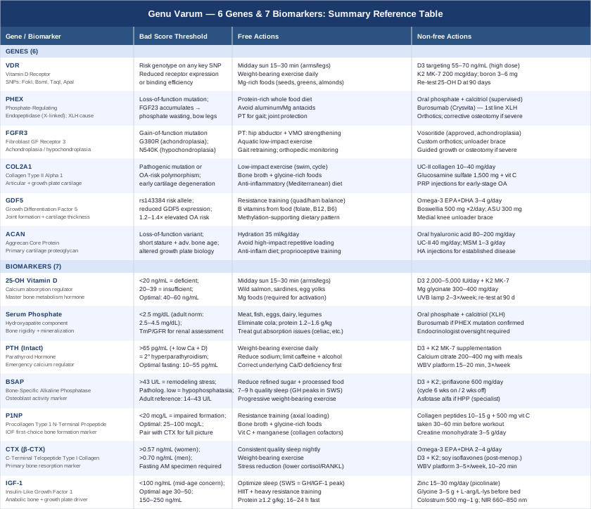
The picture from both biomarkers and genetics is, taken together, actionable. The following table consolidates the key genes and biomarkers into a reference you can return to.
Reference Table: Genes, Biomarkers, and Action Plans
10 Things From the Huberman Lab on Bone Health That Most People Miss
Andrew Huberman has addressed bone and musculoskeletal health across multiple Huberman Lab podcast episodes, particularly in conversations with Dr. Rhonda Patrick and other researchers focused on nutrient science, sleep physiology, and exercise biology. While no single episode focuses exclusively on genu varum, the framework that emerges across this body of work challenges several assumptions in standard bone health advice — and aligns directly with the biomarker and genetic findings above.
1. Vitamin D Behaves as a Hormone, Not a Supplement
The Huberman Lab consistently frames vitamin D as a nuclear hormone that regulates over 1,000 genes across multiple tissues. The implication: taking D3 without addressing receptor function (VDR variants), co-factor availability (magnesium, K2), and baseline deficiency depth is a partial intervention at best. Knowing your 25-OH D level and VDR genotype before choosing a dose is the rational approach.
2. Morning Light Is the First Bone Health Protocol
Huberman's morning sunlight protocol — 10–30 minutes of outdoor light exposure within an hour of waking — serves the circadian system but also directly supports the skin's capacity for UVB-mediated vitamin D synthesis later in the day. The neuroendocrine environment set by appropriate morning light also supports GH pulsatility (and therefore IGF-1), making it relevant to the anabolic bone context.
3. Only Impact-Generating Exercise Builds Bone
Swimming and cycling preserve cardiovascular fitness but provide minimal osteogenic stimulus. Huberman highlights that piezoelectric signaling within bone matrix — triggered by impact, compression, and tension — is what actually stimulates new bone formation. Running, jumping, and loaded resistance work are irreplaceable for this purpose. For genu varum patients with knee pain, low-impact substitution should be strategic, not total.
4. Sleep Architecture Determines Your Anabolic Environment
Growth hormone and the downstream IGF-1 it generates are secreted primarily during slow-wave sleep. Huberman explicitly connects chronic poor sleep to chronically suppressed anabolic bone signaling — an insight rarely included in standard bone health counseling. Optimizing sleep architecture (consistent schedule, temperature, light management) is framed as foundational, not optional.
5. Cortisol Is a Direct Bone Antagonist
Sustained psychological stress elevates cortisol, which suppresses GH/IGF-1 release, upregulates RANKL (the primary osteoclast activator), and impairs intestinal calcium absorption. Huberman argues this makes stress management a clinical bone health intervention — one that operates over years and meaningfully affects the CTX/P1NP balance documented in the biomarker section above.
6. Omega-3 Fatty Acids Are Underweighted in Bone Protocols
In multiple episodes covering anti-inflammatory biochemistry, Huberman and guests highlight omega-3 EPA and DHA as having direct relevance to bone resorption via prostaglandin E2 modulation. Most bone health protocols emphasize calcium and vitamin D; omega-3s are typically absent despite consistent RCT evidence for CTX reduction.
7. Protein Is Rate-Limiting for Bone Matrix, Not Just Muscle
Multiple Huberman Lab discussions push back on the calcium-first bone health narrative, emphasizing that type I collagen — which constitutes approximately 90% of bone organic matrix — requires adequate amino acid availability and specific cofactors for synthesis. At 1.2–1.6 g/kg/day minimum, protein intake directly determines whether P1NP can rise in response to exercise and supplementation.
8. Collagen Timing Relative to Exercise Is Not Trivial
Huberman has referenced research showing that collagen peptide supplementation taken 30–60 minutes before resistance training, with concurrent vitamin C, produces greater increases in collagen synthesis markers than identical supplementation at other times. The mechanism is the coincidence of circulating amino acid availability with the post-exercise anabolic window when synthesis rates are highest.
9. The Gut-Bone Axis Is Emerging and Worth Tracking
In conversations about microbiome science, Rhonda Patrick and Huberman flag that gut bacteria modulate bone metabolism through short-chain fatty acid production, microbial K2 synthesis, and immune signaling that affects osteoclast activity. Prebiotic fiber and fermented food intake are framed as indirect but real bone health variables — consistent with the emerging literature on microbiome-bone crosstalk.
10. Genetic Risk Is About Dose, Not Direction
Huberman invokes gene-environment interaction consistently: SNPs like VDR variants or the GDF5 rs143384 polymorphism do not shut down a pathway — they change how much input is needed to produce the same output. The practical message for anyone with genu varum and a relevant genetic variant is not resignation. It is calibration: higher doses of the right inputs, more consistent application, and closer monitoring of biomarker response.
Complementary Approaches With Evidence for Genu Varum and Its Complications
No complementary modality corrects the angular geometry of established bow legs. What these approaches address are the secondary consequences that drive most of the daily burden in genu varum: medial knee pain, cartilage wear, gait-related muscle imbalances, and joint inflammation. The three modalities below have the most condition-relevant evidence among the available options.
Low-Level Laser Therapy (Photobiomodulation)
Low-level laser therapy (LLLT) uses specific wavelengths of red and near-infrared light (typically 630–900 nm) to penetrate joint and periarticular tissue, stimulate mitochondrial cytochrome c oxidase activity, reduce local inflammatory cytokine expression (IL-1β, TNF-α), and accelerate cellular repair processes without thermal damage. For genu varum, LLLT is most relevant as a non-pharmacological intervention for the medial knee osteoarthritis that is both a common complication of the condition and a driver of progressive structural deterioration. It is non-invasive, well-tolerated, and can be applied consistently at home with consumer-grade devices.
A systematic review and meta-analysis published in BMC Musculoskeletal Disorders (Brosseau et al.) found that LLLT significantly reduced pain and improved functional outcomes in knee osteoarthritis when applied at adequate doses (4–50 J/cm²) in multiple randomized controlled trials. The Osteoarthritis Research Society International (OARSI) has included LLLT as a conditionally recommended therapy for knee osteoarthritis management. It is worth noting that evidence for slowing structural progression (cartilage thickness preservation) remains more limited than evidence for symptomatic benefit.
For practical application: a clinical device at 810 nm or a home near-infrared device (660/850 nm dual-wavelength panel) applied directly to the skin over the medial knee joint line and surrounding periarticular tissue. Sessions of 10–20 minutes per knee, 3 times per week for a minimum 8–12 week course. Expect meaningful pain and mobility improvement within 4–6 weeks of consistent application. For those with active skin conditions or taking photosensitizing medications, consult a physician before beginning. LLLT is most effective when combined with the nutritional and exercise strategies outlined in the biomarker sections above — it does not replace them.
Yoga
Yoga is relevant to genu varum not as a structural correction tool but as a targeted system for addressing the specific muscular imbalances that develop around bow legs and worsen their joint consequences. The characteristic posture associated with genu varum includes external femoral rotation, overactivation of the IT band and lateral hip rotators, and relative weakness of the hip adductors and vastus medialis oblique (VMO). These imbalances increase varus stress at the medial knee and accelerate cartilage wear. Specific yoga postures — warrior II, chair pose, bridge variations, goddess pose, wide-legged forward folds — address these imbalances with relative specificity, targeting both the tightness and weakness patterns simultaneously.
A randomized controlled trial published in the Journal of Alternative and Complementary Medicine examined yoga-based knee rehabilitation in participants with knee osteoarthritis — the primary degenerative complication of genu varum — and found significant improvements in pain, functional mobility, and disease-specific quality of life scores at 8 weeks compared to usual care. The intervention used 60-minute sessions three times per week. No RCT has directly examined yoga for genu varum alignment specifically, but the evidence base for yoga in knee OA and lower limb muscle imbalance is meaningful and consistent enough to recommend it as a conservative adjunct.
A realistic protocol for genu varum: 3–4 sessions per week, 30–45 minutes, structured around hip adductor strengthening (goddess pose, bound angle, wide-legged work), VMO activation (low chair pose, single-leg modifications), and lateral hip and IT band releasing (pigeon pose, figure-four, thread-the-needle). Avoid excessive loaded deep knee flexion early in the protocol — begin with partial ranges and progress. Hot yoga should be avoided when knee inflammation is present. Individuals with significant knee pain should obtain a physiotherapy assessment before beginning an independent yoga practice to rule out structural contraindications.
Massage Therapy
Massage therapy addresses a specific and clinically significant aspect of genu varum: the chronic tension and trigger point patterns that develop in the IT band, lateral gastrocnemius, peroneal muscles, and lateral hip rotators as the body compensates for altered mechanics over years. This lateral chain overrecruitment — which develops in response to medial instability and altered weight-bearing — increases varus knee stress, reduces proprioceptive accuracy, and generates the referred knee, hip, and lower back pain that many people with bow legs experience. Myofascial release and deep tissue work targeting these structures can interrupt this compensatory pattern and reduce secondary mechanical load on medial knee structures.
A Cochrane review of massage therapy for musculoskeletal conditions found consistent short-term benefit for pain reduction and functional improvement, with a favorable safety profile. More specifically, trigger point therapy directed at the lateral quadriceps and IT band in patients with knee osteoarthritis has demonstrated reduced knee pain and improved range of motion in small randomized trials. Evidence is primarily symptomatic and functional — massage does not change bone geometry or directly slow cartilage degradation, and its benefits require maintenance.
For application to genu varum: monthly or biweekly deep tissue sessions with a therapist experienced in musculoskeletal work, targeting the IT band, lateral hip rotators, and peroneal chain specifically. Between professional sessions, foam rolling the IT band (daily, 60–90 seconds per side at a moderate, tolerable pressure) and using a lacrosse ball on the lateral gastrocnemius maintains tissue quality and prolongs session benefits. Trigger point release at the proximal IT band insertion reduces measurable medial knee varus stress when combined with the strengthening strategies described in the yoga section. Massage is most effective as a complement to active rehabilitation — not as a standalone intervention.
Conclusion
Genu varum is rarely a single-cause problem, and it is almost never a no-options problem. Whether the underlying drivers are a childhood nutritional deficiency that has left its structural mark, a phosphate-wasting gene variant running unchecked, a cartilage matrix polymorphism accelerating medial knee wear, or a combination of metabolic imbalances visible in your biomarkers — the biology is largely trackable, and in many respects adjustable.
The seven biomarkers covered here give you a real-time view of your bone metabolism: whether the balance of formation and resorption is running in your favor, whether your vitamin D and phosphate systems are functional, and whether your anabolic environment can support structural maintenance. The six genes provide the explanatory layer — telling you why some individuals are more susceptible and guiding which interventions are most likely to matter for their specific biology.
A practical next step: request a baseline panel that includes 25-OH vitamin D, serum phosphate, calcium, PTH, and alkaline phosphatase. If your physician is amenable, add P1NP and β-CTX for a full bone turnover picture. If there is a family history of bow legs, short stature, or early knee osteoarthritis, a genetics consultation is worth raising. Bring what you find to a metabolic physician, endocrinologist, or functional medicine practitioner who can interpret these markers together rather than in isolation.
Better data does not guarantee a better outcome — but it reliably leads to better decisions. That is a meaningful place to start.
Musculoskeletal Endocrine & Metabolic
Musculoskeletal: Bone Conditions Joint Conditions
Autoimmune: Inflammatory Conditions Connective Tissue Conditions