This article contains AI generated content.
Gerdy's Tubercle Avulsion: 4 Genes and 7 Biomarkers to Track
Introduction
A Gerdy's tubercle avulsion is a specific kind of injury: a small but structurally important piece of bone, torn away by the pull of the iliotibial band at the moment of a hard pivot, jump landing, or sudden deceleration. Most of what gets written about it stops at the mechanics — immobilize, protect, image again in a few weeks, clear for return to sport. That advice is not wrong, but it treats every knee and every healing timeline as identical, when they clearly are not.
Two people with the same X-ray can heal at very different speeds, and recover with very different odds of re-injury, depending on things that never show up on the scan: how efficiently their bone is remodeling, how their collagen is built, how their inflammation is trending, and whether the basic raw materials for repair are actually present in their blood. Generic rehab handouts cannot account for any of that, because they are written for the average patient, not for the person reading them.
This article takes a narrower, more useful approach. It looks at the biomarkers that plausibly track how well an avulsion fracture is healing and how resilient the surrounding tissue is likely to be going forward, alongside the genetic variants that current sports-medicine research associates with connective tissue and bone injury risk. Neither list is a diagnosis. Both are tools for asking better questions during recovery.
None of this replaces an orthopedic surgeon, a physical therapist, or a follow-up X-ray — and nothing here promises faster healing or a guaranteed return to sport. What it offers is a clearer map of what can realistically be measured, what those numbers may mean, and what a sensible, evidence-aware response looks like, with or without supplements or equipment.
Summary
Before going deeper, here is the shape of what follows. The article opens with seven blood-based biomarkers — from vitamin D and calcium to the bone formation and resorption markers P1NP and CTX-I — that give a working picture of whether the biological environment around a Gerdy's tubercle avulsion actually supports repair, and where the gaps might be. Each one comes with realistic testing costs, how often to recheck it, and two concrete plans: one built entirely around food, sunlight, sleep, and loading, and one that adds supplements or equipment, always with frequency, cycling, and side effects spelled out.
From there, the article turns to genetics: four genes — COL5A1, COL1A1, GDF5, and VDR — that current research links to how tendon, ligament, and bone tissue is built and how it responds to load, along with what can and cannot be done about a less favorable variant. There is also a practical breakdown of the ideas from Built to Move by Kelly and Juliet Starrett, a book that reframes joint and tissue health around daily physical habits rather than occasional workouts, and a short section on complementary approaches — massage, photobiomodulation, biofeedback, and mindfulness — that have real, if sometimes modest, human evidence behind them for knee and soft-tissue recovery. Read together, these sections turn a single injury into a much more specific, trackable picture.
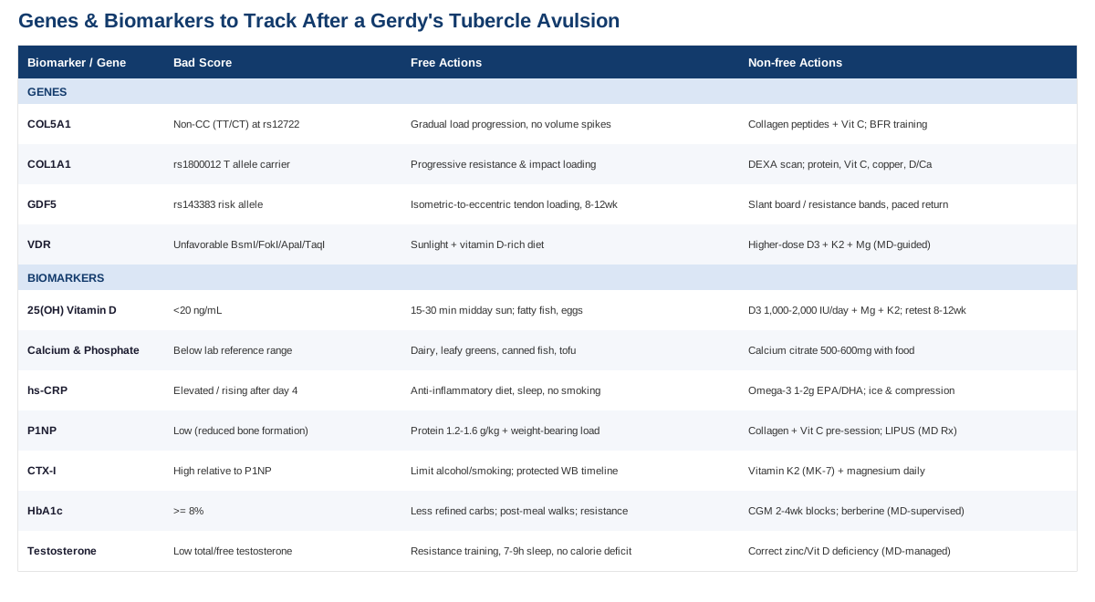
Bone and soft-tissue healing do not happen in isolation from the rest of the body, which is why the biomarker picture below is worth building before assuming a delayed recovery is simply "bad luck."
7 Biomarkers Worth Tracking After a Gerdy's Tubercle Avulsion
An avulsion fracture heals through the same biology as any other bone injury: an inflammatory phase, a soft callus of collagen and cartilage, then mineralization into hard bone, followed by remodeling that can continue for a year or more. Each of these seven biomarkers maps onto a different part of that process, and each is realistically testable through a standard blood draw.
25-Hydroxyvitamin D
Vitamin D governs how efficiently the gut absorbs calcium and phosphate, the two minerals that actually harden the healing callus. Low 25(OH)D has been directly associated with delayed and non-union fractures — one review found that roughly 60% of patients with non-union tibial fractures had markedly low vitamin D levels, compared to about 30% of those who healed normally within three to six months, as summarized in this narrative overview of vitamin D deficiency in orthopaedic trauma. A phase II trial testing vitamin D3 supplementation for acute fracture healing found a possible modest benefit, though the authors were careful to call it preliminary.
How to measure it
A simple serum 25(OH)D blood test, typically $40 to $90 out of pocket, or bundled into a standard metabolic panel through a physician. Retest every 8 to 12 weeks while actively correcting a deficiency, then once or twice a year afterward.If the level is low: the plan without supplements
Fifteen to thirty minutes of midday sun exposure on the arms and legs, several times a week, adjusted for skin tone and latitude, plus dietary sources like fatty fish, egg yolks, and fortified dairy. This raises levels slowly and safely but is genuinely limited in winter months or northern climates.If the level is low: the plan with supplements or equipment
Vitamin D3 (cholecalciferol), typically 1,000 to 2,000 IU daily for maintenance, or a clinician-supervised higher repletion dose for significant deficiency, paired with magnesium and a modest dose of vitamin K2 (see below) to support proper use of the extra calcium. Retest at 8 to 12 weeks and taper to the maintenance dose once levels normalize; there is no need to keep dosing at repletion levels indefinitely. The main side effect risk is hypercalcemia from excessive, unsupervised megadosing, so routine bloodwork should accompany any dose above standard maintenance. The official NIH vitamin D fact sheet is a reasonable reference for safe upper limits.Serum Calcium and Phosphate
These are the literal building blocks of the mineralized callus. Deficiency is uncommon in otherwise healthy people eating a normal diet, but it is cheap and easy to rule out, and worth checking in anyone with restrictive eating patterns, malabsorption, or heavy antacid use.
How to measure it
Part of any basic metabolic panel, usually $20 to $40, often already included in routine post-injury bloodwork.If the level is low: the plan without supplements
Dairy, leafy greens, canned fish with bones, and tofu for calcium; phosphate is abundant in almost any protein-containing diet, so true dietary phosphate deficiency is rare outside of specific medical conditions.If the level is low: the plan with supplements or equipment
Calcium citrate or carbonate, generally in 500 to 600 mg doses taken with food for better absorption, staying under a total daily intake of roughly 2,000 to 2,500 mg from food and supplements combined. There is no need to cycle calcium, but total intake should be reassessed every few months against dietary changes. Excess supplemental calcium (rather than dietary calcium) has been associated in some studies with kidney stone risk and debated cardiovascular effects, which is why food sources are generally preferred as the first option — see the NIH calcium fact sheet for dosing detail.High-Sensitivity C-Reactive Protein (hs-CRP)
CRP rises predictably after any fracture or surgical fixation, and tracking its trajectory can help distinguish normal healing inflammation from a problem. Research on orthopedic patients shows CRP typically peaks within about four days of surgery; a second peak or a level that fails to decline afterward is a recognized warning sign for infection, as described in this analysis of CRP as a marker of postoperative infection in fracture patients.
How to measure it
A standard blood test, $20 to $50, easily added to routine follow-up labs after a surgically fixed avulsion.If the level is high: the plan without supplements
An anti-inflammatory eating pattern built around vegetables, fiber, and fatty fish, consistent sleep, gradual rather than aggressive return to loading, and avoiding smoking, which independently slows bone healing.If the level is high: the plan with supplements or equipment
Omega-3 fish oil, roughly 1 to 2 grams of combined EPA/DHA daily, has modest evidence for lowering hs-CRP over 8 to 12 weeks; a cryotherapy cuff or simple ice and compression can help manage local swelling in the first one to two weeks. Side effects of fish oil are mostly mild gastrointestinal upset, with a bleeding-risk caution for anyone on blood thinners. It's worth noting that some inflammation in the first week or two after injury is a necessary healing signal, not something to aggressively suppress.P1NP (Procollagen Type I N-Terminal Propeptide)
P1NP is released when osteoblasts actively lay down new type I collagen matrix, making it a reasonable proxy for how vigorously new bone is forming at the healing site, as outlined in this review of bone turnover markers and this StatPearls overview of bone biochemical markers.
How to measure it
A specialty lab test, typically $80 to $150, usually ordered through an endocrinologist or metabolic bone specialist rather than routine primary care. It should be drawn fasting in the morning, since levels shift with circadian rhythm.If the level is low: the plan without supplements
Consistent protein intake around 1.2 to 1.6 g/kg of body weight daily to supply collagen-building amino acids, plus progressive, cleared weight-bearing activity, since mechanical loading is one of the strongest known stimuli for osteoblast activity.If the level is low: the plan with supplements or equipment
Fifteen grams of collagen peptides with roughly 50 mg of vitamin C taken 30 to 60 minutes before a rehab or loading session, timed to coincide with collagen synthesis windows. A physician-prescribed low-intensity pulsed ultrasound (LIPUS) bone stimulator, used about 20 minutes daily, is an option specifically for confirmed delayed union, not a general-purpose device. Collagen peptides are generally well tolerated with occasional mild digestive upset; LIPUS should only be used under a treating physician's guidance.CTX-I (C-Terminal Telopeptide)
CTX-I reflects osteoclast activity, the resorption side of bone remodeling. Read alongside P1NP, it helps show whether formation and resorption are reasonably balanced or whether resorption is outpacing new bone formation.
How to measure it
A specialty lab test, $80 to $150, fasting morning draw. Reference ranges are less standardized between labs than for P1NP, so trend over time matters more than a single absolute number.If the level is high relative to P1NP: the plan without supplements
Limiting alcohol and smoking, both of which increase bone resorption, prioritizing sleep, and following the surgeon's protected weight-bearing timeline closely — both prolonged immobilization and rushing weight-bearing too early can skew this balance in the wrong direction.If the level is high relative to P1NP: the plan with supplements or equipment
Vitamin K2 (MK-7 form), 90 to 180 mcg daily, to support proper calcium incorporation into bone matrix, and magnesium, 300 to 400 mg daily, as a cofactor alongside vitamin D and calcium. A supervised, progressive resistance program once cleared by a physical therapist is the strongest lever available. Vitamin K2 interacts with anticoagulant medications like warfarin, so anyone on blood thinners should check with their physician before adding it.HbA1c and Fasting Glucose
Chronic hyperglycemia impairs osteoblast function and increases advanced glycation end-products that stiffen and weaken the collagen matrix, a mechanism well documented in reviews of diabetes and bone fracture healing and diabetes-related fracture risk, showing meaningfully higher fracture risk once HbA1c crosses roughly 8%.
How to measure it
A standard blood test, $15 to $40, often already part of an annual physical.If the level is elevated: the plan without supplements
Reducing refined carbohydrates and added sugar, taking short walks after meals, and adding resistance training to improve insulin sensitivity — all of which also support the loading side of bone healing.If the level is elevated: the plan with supplements or equipment
A continuous glucose monitor, roughly $50 to $100 a month, used in focused 2 to 4 week blocks rather than continuously, can identify which specific foods or meal patterns spike an individual's glucose during rehab. Berberine, 500 mg two to three times daily, has glucose-lowering evidence but can cause GI upset and should not be combined with other glucose-lowering medications without medical supervision, given the real risk of hypoglycemia.Total and Free Testosterone
Gerdy's tubercle avulsions are especially common in adolescent and young adult athletes, which makes hormonal status genuinely relevant — but the evidence here argues for caution rather than optimization. Sex hormones influence collagen turnover, yet studies on testosterone replacement therapy show it is associated with a higher risk of tendon tear, not better healing, a pattern also discussed in this review of sex hormones and tendon physiology.
How to measure it
A morning fasting blood test, $60 to $120, through primary care or endocrinology, given testosterone's diurnal variation.If the level is low: the plan without supplements
Resistance training, 7 to 9 hours of sleep, and avoiding chronic caloric deficits — a real risk in young athletes training hard while restricting food, which suppresses sex hormones and directly impairs bone healing.If the level is low: the plan with supplements or equipment
Correcting a genuine zinc or vitamin D deficiency, if present, can support normal endogenous hormone production. Exogenous testosterone or prohormones should not be self-administered to "speed up" healing from an avulsion fracture; the evidence points toward increased tendon injury risk, not improved bone or ligament repair, and any true clinical deficiency should be managed by a physician, not self-treated.Blood chemistry tells part of the story, but some of the variation in how tissue is built and how it responds to load traces back further, to genetics.
4 Genes That May Influence Injury Risk and Healing
Consumer genomics figures like Ali Torkamani, whose research focuses on polygenic risk and precision health, and Gary Brecka, known for popularizing panels that flag variants in genes like these, have helped bring this kind of testing into the mainstream. It's worth being direct about where the science currently stands: these are population-level associations from sports-genetics research, not deterministic predictions for any one person's knee, and the field is still young compared to something like pharmacogenomics.
COL5A1
Type V collagen helps regulate the diameter and organization of collagen fibrils in tendons and ligaments, including the iliotibial band that inserts at Gerdy's tubercle. Certain variants, particularly the rs12722 CC genotype, have been associated with a reduced risk of tendon-ligament injury in a meta-analysis of Caucasian populations, and a similar protective pattern was found in a study of ACL injury risk in skiers.
If the gene is unfavorable: the plan without supplements
The genotype itself cannot be changed, but the practical response is training-load management: gradual progression rather than sudden spikes in volume or intensity, since tendon and ligament tissue adapts more slowly than muscle, often over many weeks rather than days.If the gene is unfavorable: the plan with supplements or equipment
The same collagen peptide plus vitamin C pre-loading protocol described above, and, where available, supervised blood-flow restriction training equipment, which can stimulate connective tissue adaptation under lower absolute joint loads. There are no meaningful supplement side effects here beyond standard collagen tolerance; the real risk is still training-load errors, not the gene itself.COL1A1
Type I collagen is the main structural protein in both bone and ligament. The rs1800012 polymorphism has been linked in a meta-analysis to sports-related tendon and ligament injury risk, and separately, COL1A1 variants have a longer history of association with bone fragility.
If the gene is unfavorable: the plan without supplements
Progressive bone-loading exercise — resistance training and controlled impact work once cleared — remains the strongest modifiable stimulus for collagen matrix quality, regardless of genotype.If the gene is unfavorable: the plan with supplements or equipment
Adequate protein, vitamin C, copper, and vitamin D/calcium status support collagen cross-linking. A baseline DEXA bone density scan ($75 to $250) is a reasonable one-time check for anyone with a family history of fragility fractures, repeated roughly every one to two years if results warrant monitoring — not a test that needs frequent repetition in an otherwise young, healthy patient.GDF5
Growth differentiation factor 5 is involved in joint and tendon development. The rs143383 variant has been associated with an increased risk of Achilles tendinopathy in more than one population, discussed in this broader review of the genetics of sports injuries and athletic performance, though its link to ACL injury specifically is less consistent across studies.
If the gene is unfavorable: the plan without supplements
Tendon-specific loading protocols — isometric holds progressing to eccentric loading, three times weekly for 8 to 12 weeks — have broad evidence for improving tendon resilience regardless of genotype, alongside a graded, not rushed, return-to-sport timeline.If the gene is unfavorable: the plan with supplements or equipment
No supplement directly corrects GDF5 expression. A slant board or resistance bands for structured eccentric loading, combined with adequate recovery time between high-impact sessions, are the realistic tools here; reassess symptoms after each 8 to 12 week block before progressing further.VDR (Vitamin D Receptor Gene)
VDR polymorphisms (BsmI, FokI, ApaI, TaqI) affect how efficiently cells respond to circulating vitamin D, though a systematic review of VDR variants and bone mineral density found the overall evidence inconsistent — some studies show an effect, others do not, once study quality is accounted for.
If the gene is unfavorable: the plan without supplements
Since receptor sensitivity can't be directly altered, the practical lever is making sure there is more circulating substrate for the receptor to use in the first place — the same sunlight and dietary strategies described in the vitamin D biomarker section.If the gene is unfavorable: the plan with supplements or equipment
The same vitamin D3, magnesium, and K2 approach as above, with a reasonable case for targeting the upper end of the sufficient range (with physician input) rather than just clearing the minimum threshold, since a less responsive receptor may need more substrate to achieve the same effect. Retest 25(OH)D at 8 to 12 weeks; side effects mirror the standard vitamin D3 supplementation cautions already covered.Numbers and genes describe the biology, but recovery is also a matter of daily habits — which is where a recent, widely discussed book offers a useful counterweight.
10 Ideas from Built to Move That Are Worth Applying During Recovery
Built to Move, by Kelly Starrett and Juliet Starrett, argues that most joint and tissue problems are less about a single injury event and more about years of restricted range of motion and sedentary posture that leave tissue unprepared for the loads sport eventually demands. That framing challenges the common clinical habit of treating an avulsion fracture as an isolated event rather than a signal about the surrounding tissue's readiness. The following ten ideas are the ones most relevant to someone recovering from a Gerdy's tubercle avulsion.
1. The sit-to-stand test matters more than it seems
Being able to sit on the floor and stand back up without using hands or support is presented as a rough proxy for hip and knee mobility and strength. Struggling with it after a knee injury is a signal to rebuild that range gradually, not to avoid the movement pattern indefinitely.2. Hip range of motion protects the knee
Restricted hip internal and external rotation forces the knee to absorb rotational forces it wasn't designed for — directly relevant to an injury caused by the iliotibial band pulling on the tibia during a pivot.3. Ankle stiffness travels upward
Limited ankle dorsiflexion changes landing mechanics and can shift stress higher up the kinetic chain, including onto the lateral knee structures near Gerdy's tubercle.4. Movement should be distributed across the whole day
A single workout doesn't offset hours of sitting. Short, frequent movement breaks are framed as more protective for tissue health than one intense session bookended by stillness — a useful principle during a rehab period where full workouts aren't yet possible.5. Walking volume is an underrated recovery tool
A daily step target, once weight-bearing is cleared, is described as one of the simplest ways to maintain general tissue and metabolic health during a period when higher-impact training is off the table.6. Grip strength as a broader signal
Grip strength is used as a rough marker of overall physical resilience. It's not directly about the knee, but it's a low-effort number worth tracking as a general recovery and conditioning barometer.7. Nasal breathing and an aerobic base support tissue repair
Building aerobic capacity through easy, nasal-breathing-paced effort is framed as supportive of general recovery capacity, relevant when higher-intensity conditioning is temporarily restricted after injury.8. Sleep is treated as a non-negotiable, not a bonus
Tissue repair, including bone remodeling, happens disproportionately during sleep. The book treats consistent sleep as equally important as any specific exercise during recovery.9. Minimum viable range of motion, checked regularly
Rather than occasional intense stretching, the book advocates brief, frequent range-of-motion checks and work — a few minutes daily rather than one long session weekly — which fits naturally into a structured knee rehab schedule.10. Pain is information, not identity
The book pushes back on treating persistent discomfort as something to simply push through or ignore, framing it instead as data that should inform load and pacing decisions — a useful mindset for anyone tempted to return to sport before tissue is ready.Alongside these habit-level changes, a handful of adjunct therapies have specific human evidence worth considering during the rehab phase itself.
Complementary Approaches Worth Considering
These are add-ons to standard orthopedic care and physical therapy, not replacements for it, and the evidence for each varies in strength.
Massage Therapy
Massage is relevant here specifically because the iliotibial band itself — the structure whose pull causes this avulsion — is a common site of tightness and friction-related irritation, and manual therapy is one of the more directly studied interventions for it. It can help manage the soft-tissue tightness and compensatory guarding that often develops around the knee during a period of reduced activity and altered gait.
A randomized trial of deep transverse friction massage combined with standard physical therapy for iliotibial band friction syndrome found added benefit over physical therapy alone, and a separate study on percussive massage and IT band tightness in field athletes found meaningful short-term reductions in pain and improved range of motion.
In practice, this means gentle, gradually progressing manual work on the IT band and surrounding quadriceps and hip muscles once a physical therapist confirms the fracture site is stable enough to tolerate it — not deep pressure directly over the healing bone itself, and not before clearance for soft-tissue work.
Photobiomodulation (Low-Level Laser Therapy)
Photobiomodulation uses low-level laser or LED light to influence cellular metabolism, and preclinical research suggests it may support the angiogenesis and osteoblast activity involved in bone repair, making it conceptually relevant to a healing avulsion fracture.
Mechanistic and animal research shows LLLT can promote bone regeneration by coupling angiogenesis and osteogenesis, and a review of its use for acute tissue injury and sport recovery describes benefits for pain and inflammation control, while explicitly noting that human clinical evidence specific to bone healing remains limited compared to the animal literature.
Given that gap, this is best treated as a genuinely optional adjunct rather than a core part of a recovery plan — something to discuss with a treating physician or physical therapist who has access to clinical-grade equipment, rather than a home device purchased on the assumption it will meaningfully speed bone union.
Biofeedback (EMG-Guided Quadriceps Training)
After any knee injury involving immobilization or surgery, the quadriceps — particularly the vastus medialis — tend to shut down neurologically well before they weaken structurally, a phenomenon called arthrogenic muscle inhibition. Electromyographic biofeedback gives real-time visual or auditory feedback on muscle activation, helping retrain that connection.
A randomized controlled trial found EMG biofeedback improved knee extension recovery following ACL reconstruction, and a separate RCT found EMG-guided quadriceps strengthening improved outcomes in patellofemoral pain, both directly relevant to the quadriceps and patellar mechanics surrounding Gerdy's tubercle.
Realistically, this looks like sessions with a physical therapist using surface EMG sensors during isometric quadriceps sets, typically a few times a week during the early-to-mid rehab phase, tapering off once voluntary activation and strength normalize — it's a rehab tool for a defined window, not an indefinite practice.
Mindfulness Meditation and MBSR
Return to sport after a significant knee injury carries a well-documented psychological component — fear of re-injury and pain-related anxiety can slow rehab progress independent of tissue healing itself. Mindfulness-based approaches address that layer directly.
A systematic review and meta-analysis found mindfulness-based interventions reduced postoperative pain after hip and knee procedures, and a study specifically in injured athletes found MBSR improved pain tolerance and mental health during recovery.
In practice, this can be as simple as a guided 10 to 15 minute daily practice through a reputable app or a structured MBSR course, particularly useful in the weeks surrounding return-to-sport testing, when anxiety about re-injury tends to peak.
Conclusion
A Gerdy's tubercle avulsion is a mechanical injury, but healing well from it depends on biology that a cast or a brace alone can't influence: vitamin D and mineral status, inflammation trends, bone formation and resorption balance, glucose control, and hormonal context, layered on top of a genetic backdrop that shapes how tendon, ligament, and bone tissue are built in the first place. None of these factors guarantee a particular outcome on their own, and none of them replace the surgeon's imaging or the physical therapist's clearance to progress load.
What they offer instead is a way to ask sharper questions during recovery — is there an actual reason healing feels slower than expected, is inflammation trending the right direction, is the training load being reintroduced sensibly for this particular tissue. The most useful next step is a practical one: pick two or three biomarkers from this list that are easy to get through an existing physician relationship, get a baseline, and revisit them on the timeline suggested above rather than guessing. Pair that with an honest conversation with a treating orthopedic professional about what the numbers do and don't change about the rehab plan, and let that combination — not any single test or supplement — guide the pace of return to sport.
Musculoskeletal Endocrine & Metabolic
Musculoskeletal: Joint Conditions